
(408) 984-3333 ■ mind@growing.com ■ www.growing.com/mind ■ www.cortexercise.com
Transcranial Direct Current Stimulation (tDCS)
Selected Abstracts
Draft version: 4-13-11
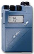
NOTE: Transcranial direct current stimulation protocols generally call for a constant current of 1-2 mA over a period of 20-30 minutes. Such steady currents are produced by Iontophoresis devices, such as the one shown above. Iontophoresis devices are not approved by the FDA for transcranial direct current stimulation. Nonetheless, as these studies show, the constant current made possible by high quality iontophoresis devices has made them a standard all around the world for tDCS research. Widely used models have included IoMed's Phoresor, a unit which is now discontinued in favor of more flexible and portable units, and devices by Germany's NeuroConn.
Biol Psychiatry. 2011 Apr 15;69(8):e23-4. Epub 2011 Feb 18.
Efficacy and safety of transcranial direct current stimulation in major depression.
Dell'osso B, Priori A, Altamura AC.
Department of Psychiatry, Fondazione IRCCS Cà Granda, Ospedale Maggiore Policlinico, University of Milan, Italy.
http://www.ncbi.nlm.nih.gov/pubmed/21310394
Gastrointest Endosc. 2011 Apr 4. [Epub ahead of print]
Feasibility, safety, and effectiveness of transcranial direct current stimulation for decreasing post-ERCP pain: a randomized, sham-controlled, pilot study.
Borckardt JJ, Romagnuolo J, Reeves ST, Madan A, Frohman H, Beam W, George MS.
Department of Psychiatry and Behavioral Sciences (J.J.B., A.M., M.S.G.), Department of Anesthesiology and Perioperative Medicine (J.J.B., S.T.R., H.F., W.B.), Department of Gastroenterology and Hepatology (J.R.), Medical University of South Carolina, Charleston, South Carolina, Ralph H. Johnson Veterans Affairs Medical Center (M.S.G.).
BACKGROUND: Emerging evidence shows that transcranial direct current stimulation (tDCS), a minimally invasive brain stimulation technique, has analgesic effects in chronic pain patients and in healthy volunteers with experimental pain. No studies have examined the analgesic effects of tDCS immediately after surgical/endoscopic procedures. Endoscopy investigating abdominal pain, especially ERCP, can cause significant postprocedural pain. OBJECTIVE: To test the feasibility, efficacy, and safety of tDCS on post-ERCP pain and analgesia use. DESIGN: Randomized, sham-controlled, pilot study. SETTING: Tertiary-care medical center. PATIENTS: This study involved 21 patients who were hospitalized overnight for ERCP for unexplained right upper quadrant pain. INTERVENTION: Twenty minutes of real 2.0 mA tDCS or sham (anode over left prefrontal cortex; cathode over gut-representation of right sensory cortex) immediately after ERCP. MAIN OUTCOME MEASUREMENTS: Pain (visual analogue scale, McGill pain questionnaire, brief pain inventory), patient-controlled analgesia use, adverse events. RESULTS: Real tDCS was associated with 22% less total hydromorphone use, versus sham. The slope of the cumulative patient-controlled analgesia usage curve was significantly steeper in the sham tDCS group (F [2,13] = 15.96; P = .0003). Real tDCS patients reported significantly less pain interference with sleep (t [17] = 3.70; P = .002) and less throbbing pain (t [16] = 2.37; P = .03). Visual analogue scale pain and mood scores (4 hours post-ERCP) suggested a nonsignificant advantage for real tDCS, despite less hydromorphone use. Side effects of tDCS were limited to mild, self-limited tingling, itching, and stinging under electrodes. LIMITATIONS: Small sample size, variability in chronic pain, and chronic opioid use. CONCLUSION: In this pilot study, tDCS appears to be safe, has minimal side effects, and may reduce postprocedural analgesia requirements and subjective pain ratings. Future studies appear warranted.
http://www.ncbi.nlm.nih.gov/pubmed/21470608
Brain Lang. 2011 Apr 1. [Epub ahead of print]
Mechanisms of aphasia recovery after stroke and the role of noninvasive brain stimulation.
Hamilton RH, Chrysikou EG, Coslett B.
University of Pennsylvania, Department of Neurology, Center for Cognitive Neuroscience, Philadelphia, PA, United States; Laboratory for Cognition and Neural Stimulation, University of Pennsylvania, Philadelphia, PA, United States.
One of the most frequent symptoms of unilateral stroke is aphasia, the impairment or loss of language functions. Over the past few years, behavioral and neuroimaging studies have shown that rehabilitation interventions can promote neuroplastic changes in aphasic patients that may be associated with the improvement of language functions. Following left hemisphere strokes, the functional reorganization of language in aphasic patients has been proposed to involve both intrahemispheric interactions between damaged left hemisphere and perilesional sites and transcallosal interhemispheric interactions between the lesioned left hemisphere language areas and homotopic regions in the right hemisphere. A growing body of evidence for such reorganization comes from studies using transcranial magnetic stimulation (TMS) and transcranial direct current stimulation (tDCS), two safe and noninvasive procedures that can be applied clinically to modulate cortical excitability during post-stroke language recovery. We discuss a hierarchical model for the plastic changes in language representation that occur in the setting of dominant hemisphere stroke and aphasia. We further argue that TMS and tDCS are potentially promising tools for enhancing functional recovery of language and for further elucidating mechanisms of plasticity in patients with aphasia.
http://www.ncbi.nlm.nih.gov/pubmed/21459427
Clin Neurophysiol. 2011 Apr;122(4):777-83. Epub 2010 Nov 11.
Transcranial direct current stimulation over the motor association cortex induces plastic changes in ipsilateral primary motor and somatosensory cortices.
Kirimoto H, Ogata K, Onishi H, Oyama M, Goto Y, Tobimatsu S.
Department of Occupational Therapy, Faculty of Rehabilitation, Niigata University of Health and Welfare, Japan.
OBJECTIVE: This study was performed to elucidate whether transcranial direct current stimulation (tDCS) over the motor association cortex modifies the excitability of primary motor (M1) and somatosensory (S1) cortices via neuronal connectivity. METHODS: Anodal, cathodal, and sham tDCS (1 mA) over the left motor association cortex was applied to 10 subjects for 15min using electrodes of two sizes (9 and 18cm(2)). Both motor evoked potentials (MEPs) and somatosensory evoked potentials (SEPs) were recorded before, immediately after, and 15min after tDCS. Electrode positions were confirmed by overlaying them on MRI anatomical surface images of two individuals. RESULTS: After applying anodal tDCS using the large electrode, amplitudes of MEP components significantly decreased, whereas those of early SEP components (N20 and P25) increase. Opposite effects were observed on MEPs and SEPs after cathodal tDCS. However, a small electrode did not significantly influence either MEPs or SEPs, irrespective of polarity. The small electrode covered mainly the dorsal premotor cortex (PMd) while the large electrode involved the supplementary motor area (SMA) in addition to PMd. CONCLUSIONS: These results suggest that anodal tDCS over PMd together with SMA enhanced the inhibitory input to M1 and excitatory input to S1, and that cathodal tDCS might lead to an opposite effect. SIGNIFICANCE: The finding that only the large electrode modulated M1 and S1 implies that activation of PMd together with SMA by tDCS can induce plastic changes in primary sensorimotor areas.
http://www.ncbi.nlm.nih.gov/pubmed/21074492
Clin Neurophysiol. 2011 Apr;122(4):803-7. Epub 2010 Oct 25.
Comparing cutaneous perception induced by electrical stimulation using rectangular and round shaped electrodes.
Ambrus GG, Antal A, Paulus W.
Department of Clinical Neurophysiology, Georg-August University of Göttingen, 37075 Göttingen, Germany.
OBJECTIVE: We have investigated the cutaneous perception differences for anodal and cathodal transcranial direct current stimulation (tDCS) and transcranial random noise stimulation (tRNS) between two electrode configurations: a standard, rectangle-shaped, and a circle-shaped, round geometry with the same surface area, and thus, same nominal current distribution. We have aimed to find whether a smaller perimeter length and the absence of corners in the case of the round configuration would lead to altered skin perception characteristics when compared to the rectangular geometry. METHODS: Twelve subjects were tested for tDCS and tRNS skin perception characteristics in the intensity range of 200-2000μA using round and rectangular electrode configurations. RESULTS: We have not found any substantial differences between detection thresholds, detection rates, false positive rates or consistent alterations in the sites of perceived stimulation. CONCLUSION: We conclude that there is no difference between the round and the rectangular electrode configurations regarding their blinding potentials. SIGNIFICANCE: The results of this investigation indicate that the altering of the electrode geometry to a round configuration is unwarranted for better blinding purposes in future studies using tDCS and tRNS.
http://www.ncbi.nlm.nih.gov/pubmed/20980196
Exp Brain Res. 2011 Apr;210(2):217-27. Epub 2011 Mar 25.
Different resting state brain activity and functional connectivity in patients who respond and not respond to bifrontal tDCS for tinnitus suppression.
Vanneste S, Focquaert F, Van de Heyning P, De Ridder D.
Brai²n, TRI and Department of Neurosurgery, University Hospital Antwerp, Wilrijkstraat 10, 2650, Edegem, Belgium, sven.vanneste@ua.ac.be.
Tinnitus is an ongoing phantom percept. It has been demonstrated that bifrontal transcranial direct current stimulation (tDCS) can reduce tinnitus. In this study, one group of patients reported a substantial improvement in their tinnitus perception, whereas another group described minor or no beneficial effect at all. The objective was to verify whether the activity and connectivity of the resting brain is different for people who will respond to bifrontal tDCS for tinnitus in comparison with non-responders. Higher gamma band activity was demonstrated in right primary and secondary auditory cortex and right parahippocampus for responders. It has been shown that gamma band activity in the auditory cortex is correlated with tinnitus loudness and that the anterior cingulate is involved in tinnitus distress. People who were going to respond to bifrontal tDCS also demonstrated an increased functional connectivity in the gamma band between the right dorsolateral prefrontal cortex (DLPFC) and the right parahippocampus as well as the right DLPFC and subgenual anterior cingulate cortex (sgACC). An analysis revealed that responders to bifrontal tDCS also experienced a larger suppression effect on TMS placed over the right temporal cortex (i.e. auditory cortex) than non-responders. Responders to bifrontal tDCS seem to differ in resting brain activity compared to non-responders in the right auditory cortex and parahippocampal area. They also have a different functional connectivity between DLPFC and, respectively, the sgACC and parahippocampal area. These connectivities might explain the suppression effect for both tinnitus loudness and tinnitus-related distress.
http://www.ncbi.nlm.nih.gov/pubmed/21437634
Int J Neuropsychopharmacol. 2011 Apr;14(3):425-6. Epub 2010 Oct 6.
Avoiding skin burns with transcranial direct current stimulation: preliminary considerations.
Loo CK, Martin DM, Alonzo A, Gandevia S, Mitchell PB, Sachdev P.
School of Psychiatry, University of New South Wales, Sydney, Australia.
http://www.ncbi.nlm.nih.gov/pubmed/20923600
Neurosci Lett. 2011 Apr 1;492(2):105-8. Epub 2011 Feb 1.
The enhanced cortical activation induced by transcranial direct current stimulation during hand movements.
Kwon YH, Jang SH.
Department of Physical Therapy, Yeungnam College of Science & Technology, 1737, Daemyung 7-Dong, Namgu, Daegu 705-703,Republic of Korea.
The aim of this study is to evaluate whether tDCS applied on the primary motor cortex (M1) in company with hand movements could enhance cortical activation, using functional MRI (fMRI). Twelve right-handed normal subjects were recruited. Real tDCS and sham tDCS with hand movements were applied during fMRI scanning. Subjects performed grasp-release hand movements at a metronome-guided frequency of 1Hz, while direct current with 1.0mA was delivered to the primary motor cortex. The averaged cortical map and the intensity index were compared between real tDCS with hand movements and sham tDCS with hand movements. Our result showed that cortical activation on the primary sensorimotor cortex was observed under both of two conditions; real tDCS with hand movements and sham tDCS with hand movements. Voxel count and peak intensity were 365.10±227.23 and 5.66±1.97, respectively, in the left primary sensorimotor cortex during real tDCS with right hand movements; in contrast, those were 182.20±117.88 and 4.12±0.88, respectively, during sham tDCS with right hand movements. Significant differences in voxel count and peak intensity were observed between real tDCS and sham tDCS (p<0.05). We found that anodal tDCS application during motor task enhanced cortical activation on the underlying targeted motor cortex, compared with the same motor task without tDCS. Therefore, it seemed that tDCS induced more cortical activity and modulated brain function when concurrently applied with motor task.
http://www.ncbi.nlm.nih.gov/pubmed/21291959
Pediatr Neurol. 2011 Apr;44(4):239-53.
Care for child development: basic science rationale and effects of interventions.
Holt RL, Mikati MA.
Department of Pediatric Neurology, Children's Health Center, Duke University Medical Center, Durham, North Carolina.
The past few years have witnessed increasing interest in devising programs to enhance early childhood development. We review current understandings of brain development, recent advances in this field, and their implications for clinical interventions. An expanding body of basic science laboratory data demonstrates that several interventions, including environmental enrichment, level of parental interaction, erythropoietin, antidepressants, transcranial magnetic stimulation, transcranial direct current stimulation, hypothermia, nutritional supplements, and stem cells, can enhance cerebral plasticity. Emerging clinical data, using functional magnetic resonance imaging and clinical evaluations, also support the hypothesis that clinical interventions can increase the developmental potential of children, rather than merely allowing the child to achieve an already predetermined potential. Such interventions include early developmental enrichment programs, which have improved cognitive function; high-energy and high-protein diets, which have increased brain growth in infants with perinatal brain damage; constraint-induced movement therapy, which has improved motor function in patients with stroke, cerebral palsy, and cerebral hemispherectomy; and transcranial magnetic stimulation, which has improved motor function in stroke patients.
http://www.ncbi.nlm.nih.gov/pubmed/21397164
Stroke. 2011 Apr;42(4):1035-40. Epub 2011 Mar 24.
Noninvasive brain stimulation may improve stroke-related Dysphagia: a pilot study.
Kumar S, Wagner CW, Frayne C, Zhu L, Selim M, Feng W, Schlaug G.
Beth Israel Deaconess Medical Center and Harvard Medical School, 330 Brookline Avenue, Palmer 127 Boston, MA 02215. skumar@bidmc.harvard.edu.
BACKGROUND AND PURPOSE: Treatment options for stroke-related dysphagia are currently limited. In this study, we investigated whether noninvasive brain stimulation in combination with swallowing maneuvers facilitates swallowing recovery in dysphagic stroke patients during early stroke convalescence. METHODS: Fourteen patients with subacute unilateral hemispheric infarction were randomized to anodal transcranial direct current stimulation (tDCS) versus sham stimulation to the sensorimotor cortical representation of swallowing in the unaffected hemisphere over the course of 5 consecutive days with concurrent standardized swallowing maneuvers. Severity of dysphagia was measured using a validated swallowing scale, Dysphagia Outcome and Severity scale, before the first and after the last session of tDCS or sham. The effect of tDCS was analyzed in a multivariate linear regression model using changes in Dysphagia Outcome and Severity Scale as the outcome variable after adjusting for the effects of other potential confounding variables such as the National Institutes of Health Stroke Scale and Dysphagia Outcome and Severity scale scores at baseline, acute ischemic lesion volumes, patient age, and time from stroke onset to stimulation. RESULTS: Patients who received anodal tDCS gained 2.60 points of improvement in Dysphagia Outcome and Severity scale scores compared to patients in the sham stimulation group who showed an improvement of 1.25 points (P=0.019) after controlling for the effects of other aforementioned variables. Six out 7 (86%) patients in tDCS stimulation group gained at least 2 points of improvement compared with 3 out 7 (43%) patients in the sham group (P=0.107). CONCLUSIONS: Because brain stem swallowing centers have bilateral cortical innervations, measures that enhance cortical input and sensorimotor control of brain stem swallowing may be beneficial for dysphagia recovery.
http://www.ncbi.nlm.nih.gov/pubmed/21441148
J Cogn Neurosci. 2011 Mar 31. [Epub ahead of print]
Activation of Inhibition: Diminishing Impulsive Behavior by Direct Current Stimulation over the Inferior Frontal Gyrus.
Jacobson L, Javitt DC, Lavidor M.
Bar Ilan University, Ramat Gan, Israel.
A common feature of human existence is the ability to reverse decisions after they are made but before they are implemented. This cognitive control process, termed response inhibition, refers to the ability to inhibit an action once initiated and has been localized to the right inferior frontal gyrus (rIFG) based on functional imaging and brain lesion studies. Transcranial direct current stimulation (tDCS) is a brain stimulation technique that can facilitate as well as impair cortical function. To explore whether response inhibition can be improved through rIFG electrical stimulation, we administered focal tDCS before subjects performed the stop signal task (SST), which measures response inhibition. Notably, activation of the rIFG by unilateral anodal stimulation significantly improved response inhibition, relative to a sham condition, whereas the same tDCS protocol did not affect response time in the go trials of the SST and in a control task. Furthermore, the SST was not affected by tDCS at a control site, the right angular gyrus. Our results are the first demonstration of response inhibition improvement with brain stimulation over rIFG and further confirm the rIFG involvement in this task. Although this study was conducted in healthy subjects, present findings with anodal rIFG stimulation support the use of similar paradigms for the treatment of cognitive control impairments in pathological conditions.
http://www.ncbi.nlm.nih.gov/pubmed/21452949
Neuroimage. 2011 Mar 31. [Epub ahead of print]
Modulating inhibitory control with direct current stimulation of the superior medial frontal cortex.
Hsu TY, Tseng LY, Yu JX, Kuo WJ, Hung DL, J L Tzeng O, Walsh V, Muggleton NG, Juan CH.
Institute of Neuroscience, National Yang-Ming University, Taipei 112, Taiwan; Institute of Cognitive Neuroscience, National Central University, Jhongli 320, Taiwan; Laboratories for Cognitive Neuroscience, National Yang-Ming University, Taipei 112, Taiwan.
The executive control of voluntary action involves not only choosing from a range of possible actions but also the inhibition of responses as circumstances demand. Recent studies have demonstrated that many clinical populations, such as people with attention-deficit hyperactivity disorder, exhibit difficulties in inhibitory control. One prefrontal area that has been particularly associated with inhibitory control is the pre-supplementary motor area (Pre-SMA). Here we applied non-invasive transcranial direct current stimulation (tDCS) over Pre-SMA to test its role in this behavior. tDCS allows for current to be applied in two directions to selectively excite or suppress the neural activity of Pre-SMA. Our results showed that anodal tDCS improved efficiency of inhibitory control. Conversely, cathodal tDCS showed a tendency towards impaired inhibitory control. To our knowledge, this is the first demonstration of non-invasive intervention tDCS altering subjects' inhibitory control. These results further our understanding of the neural bases of inhibitory control and suggest a possible therapeutic intervention method for clinical populations.
http://www.ncbi.nlm.nih.gov/pubmed/21459149
Neuropsychologia. 2011 Mar 30. [Epub ahead of print]
The enhancement of cortical excitability over the DLPFC before and during training impairs categorization in the prototype distortion task.
Ambrus GG, Zimmer M, Kincses ZT, Harza I, Kovács G, Paulus W, Antal A.
Department of Clinical Neurophysiology, Georg-August University of Göttingen, Robert Koch Straße 40, 37075 Göttingen, Germany.
The present study investigated the effects of transcranial weak electrical stimulation techniques applied to the right and left dorsolateral prefrontal cortex (DLPFC) on categorization learning measured using a variant of the prototype distortion task. During the training phase of this task subjects saw low- and high distortions of a prototype dot-pattern. 60 participants received 10min of either anodal or cathodal transcranial direct current (tDCS), transcranial random noise (tRNS) or sham stimulation before and during the training. We have assessed the effects of the intervention during a test phase, where the subjects had to decide whether the consecutive high- and low-distortion versions of the prototype or random patterns that were presented belonged to the category established in the training phase. Our results show that the categorization of prototypes is significantly impaired by the application of anodal tDCS and tRNS to the DLPFC. The prototype-effect, observable in the case of the sham stimulation group, was severed in all active stimulation conditions.
http://www.ncbi.nlm.nih.gov/pubmed/21440562
Neurorehabil Neural Repair. 2011 Mar 24. [Epub ahead of print]
Single Session of Transcranial Direct Current Stimulation Transiently Increases Knee Extensor Force in Patients With Hemiparetic Stroke.
Tanaka S, Takeda K, Otaka Y, Kita K, Osu R, Honda M, Sadato N, Hanakawa T, Watanabe K.
BACKGROUND: Transcranial direct current stimulation (tDCS) of the motor cortex can enhance the performance of a paretic upper extremity after stroke. Reported effects on lower limb (LL) function are sparse. OBJECTIVE: The authors examined whether tDCS can increase the force production of the paretic quadriceps. METHODS: In this double-blind, crossover, sham-controlled experimental design, 8 participants with chronic subcortical stroke performed knee extension using their hemiparetic leg before, during, and after anodal or sham tDCS of the LL motor cortex representation in the affected hemisphere. Affected hand-grip force was also recorded. RESULTS: The maximal knee-extension force increased by 21 N (13.2%, P < .01) during anodal tDCS compared with baseline and sham stimulation. The increase persisted less than 30 minutes. Maximal hand-grip force did not change. CONCLUSIONS: Anodal tDCS transiently enhanced knee extensor strength. The modest increase was specific to the LL. Thus, tDCS might augment the rehabilitation of stroke patients when combined with lower extremity strengthening or functional training.
http://www.ncbi.nlm.nih.gov/pubmed/21436391
J Neurophysiol. 2011 Mar 23. [Epub ahead of print]
Transcranial Direct Current Stimulation Effects on I-wave activity in man.
Lang N, Nitsche MA, Dileone M, Mazzone P, De Andrés-Arés J, Diaz-Jara L, Paulus W, Di Lazzaro V, Oliviero A.
1Christian-Albrechts University, Kiel, Germany;
Transcranial direct current stimulation (tDCS) of the human cerebral cortex modulates cortical excitability non-invasively in a polarity-specific manner: anodal tDCS leads to lasting facilitation and cathodal tDCS to inhibition of motor cortex excitability. To further elucidate the underlying physiological mechanisms we recorded corticospinal volleys evoked by single-pulse transcranial magnetic stimulation of the primary motor cortex before and after a 5 min period of anodal or cathodal tDCS in eight conscious patients who had electrodes implanted in the cervical epidural space for the control of pain. The effects of anodal tDCS were evaluated in six subjects, while the effects of cathodal tDCS in five subjects. Three subjects were studied using both polarities. Anodal tDCS increased the excitability of cortical circuits generating I waves in the corticospinal system, including the earliest wave (I1 wave), whereas cathodal tDCS suppressed later I waves. The motor evoked potential (MEP) amplitude changes immediately following tDCS periods were in agreement with the effects produced on intracortical circuitry. The results deliver additional evidence that tDCS changes the excitability of cortical neurons.
http://www.ncbi.nlm.nih.gov/pubmed/21430275
Curr Biol. 2011 Mar 22;21(6):480-4. Epub 2011 Mar 3.
The Role of GABA in Human Motor Learning.
Stagg CJ, Bachtiar V, Johansen-Berg H.
Oxford Centre for Functional Magnetic Resonance Imaging of the Brain (FMRIB), Department of Clinical Neurosciences, University of Oxford, Oxford OX3 9DU, UK.
GABA modification plays an important role in motor cortical plasticity [1-4]. We therefore hypothesized that interindividual variation in the responsiveness of the GABA system to modification influences learning capacity in healthy adults. We assessed GABA responsiveness by transcranial direct current stimulation (tDCS), an intervention known to decrease GABA [5, 6]. The magnitude of M1 GABA decrease induced by anodal tDCS correlated positively with both the degree of motor learning and the degree of fMRI signal change within the left M1 during learning. This study therefore suggests that the responsiveness of the GABAergic system to modification may be relevant to short-term motor learning behavior and learning-related brain activity.
http://www.ncbi.nlm.nih.gov/pubmed/21376596
Soc Cogn Affect Neurosci. 2011 Mar 18. [Epub ahead of print]
When anger leads to aggression: induction of relative left frontal cortical activity with transcranial direct current stimulation increases the anger-aggression relationship.
Hortensius R, Schutter DJ, Harmon-Jones E.
Department of Psychology, Texas A&M University, 4235 TAMU, College Station, TX 77843-4235, USA, eddiehj@gmail.com.
The relationship between anger and aggression is imperfect. Based on work on the neuroscience of anger, we predicted that anger associated with greater relative left frontal cortical activation would be more likely to result in aggression. To test this hypothesis, we combined transcranial direct current stimulation (tDCS) over the frontal cortex with interpersonal provocation. Participants received insulting feedback after 15 min of tDCS and were able to aggress by administering noise blasts to the insulting participant. Individuals who received tDCS to increase relative left frontal cortical activity behaved more aggressively when they were angry. No relation between anger and aggression was observed in the increase relative right frontal cortical activity or sham condition. These results concur with the motivational direction model of frontal asymmetry, in which left frontal activity is associated with anger. We propose that anger with approach motivational tendencies is more likely to result in aggression.
http://www.ncbi.nlm.nih.gov/pubmed/21421731
Neuroimage. 2011 Mar 15;55(2):590-6. Epub 2011 Jan 4.
Transcranial direct current stimulation over the primary motor cortex during fMRI.
Antal A, Polania R, Schmidt-Samoa C, Dechent P, Paulus W.
Department of Clinical Neurophysiology, Georg-August University of Göttingen, Göttingen, Germany. Aantal@gwdg.de
Measurements of motor evoked potentials (MEPs) have shown that anodal and cathodal transcranial direct current stimulations (tDCS) have facilitatory or inhibitory effects on corticospinal excitability in the stimulated area of the primary motor cortex (M1). Here, we investigated the online effects of short periods of anodal and cathodal tDCS on human brain activity of healthy subjects and associated hemodynamics by concurrent blood-oxygenation-level-dependent (BOLD) functional magnetic resonance imaging (fMRI) at 3T. Using a block design, 20s periods of tDCS at 1 mA intensity over the left M1 altered with 20s periods without tDCS. In different fMRI runs, the effect of anodal or cathodal tDCS was assessed at rest or during finger tapping. A control experiment was also performed, in which the electrodes were placed over the left and right occipito-temporo-parietal junction. Neither anodal nor cathodal tDCS over the M1 for 20s stimulation duration induced a detectable BOLD signal change. However, in comparison to a voluntary finger tapping task without stimulation, anodal tDCS during finger tapping resulted in a decrease in the BOLD response in the supplementary motor area (SMA). Cathodal stimulation did not result in significant change in BOLD response in the SMA, however, a tendency toward decreased activity could be seen. In the control experiment neither cathodal nor anodal stimulation resulted in a significant change of BOLD signal during finger tapping in any brain area including SMA, PM, and M1. These findings demonstrate that the well-known polarity-dependent shifts in corticospinal excitability that have previously been demonstrated using measurements of MEPs after M1 stimulation are not paralleled by analogous changes in regional BOLD signal. This difference implies that the BOLD signal and measurements of MEPs probe diverse physiological mechanisms. The MEP amplitude reflects changes in transsynaptic excitability of large pyramidal neurons while the BOLD signal is a measure of net synaptic activity of all cortical neurons.
http://www.ncbi.nlm.nih.gov/pubmed/21211569
Neuroimage. 2011 Mar 15;55(2):644-57. Epub 2010 Dec 10.
Prefrontal direct current stimulation modulates resting EEG and event-related potentials in healthy subjects: a standardized low resolution tomography (sLORETA) study.
Keeser D, Padberg F, Reisinger E, Pogarell O, Kirsch V, Palm U, Karch S, Möller HJ, Nitsche MA, Mulert C.
Department of Psychiatry and Psychotherapy, Ludwig-Maximilian University Munich, Munich, Germany.
Prefrontal transcranial direct current stimulation (tDCS) with the anode placed on the left dorsolateral prefrontal cortex (DLPFC) has been reported to enhance working memory in healthy subjects and to improve mood in major depression. However, its putative antidepressant, cognitive and behavior action is not well understood. Here, we evaluated the distribution of neuronal electrical activity changes after anodal tDCS of the left DLPFC and cathodal tDCS of the right supraorbital region using spectral power analysis and standardized low resolution tomography (sLORETA). Ten healthy subjects underwent real and sham tDCS on separate days in a double-blind, placebo-controlled cross-over trial. Anodal tDCS was applied for 20 min at 2 mA intensity over the left DLPFC, while the cathode was positioned over the contralateral supraorbital region. After tDCS, EEG was recorded during an eyes-closed resting state followed by a working memory (n-back) task. Statistical non-parametric mapping showed reduced left frontal delta activity in the real tDCS condition. Specifically, a significant reduction of mean current densities (sLORETA) for the delta band was detected in the left subgenual PFC, the anterior cingulate and in the left medial frontal gyrus. Moreover, the effect was strongest for the first 5 min (p<0.01). The following n-back task revealed a positive impact of prefrontal tDCS on error rate, accuracy and reaction time. This was accompanied by increased P2- and P3- event-related potentials (ERP) component-amplitudes for the 2-back condition at the electrode Fz. A source localization using sLORETA for the time window 250-450 ms showed enhanced activity in the left parahippocampal gyrus for the 2-back condition. These results suggest that anodal tDCS of the left DLPFC and/or cathodal tDCS of the contralateral supraorbital region may modulate regional electrical activity in the prefrontal and anterior cingulate cortex in addition to improving working memory performance.
http://www.ncbi.nlm.nih.gov/pubmed/21146614
Cephalalgia. 2011 Mar 11. [Epub ahead of print]
Cathodal transcranial direct current stimulation of the visual cortex in the prophylactic treatment of migraine.
Antal A, Kriener N, Lang N, Boros K, Paulus W.
Georg-August University, Germany.
Background: The purpose of this study was to determine whether transcranial direct current stimulation (tDCS) can be an effective prophylactic therapy for migraine and migraine-associated pain. Method: This painless and non-invasive method was applied for 6 weeks over the visual cortex (V1), delivered three times per week. Thirty patients were assigned to cathodal or to sham stimulation, and 26 patients participated in the final analyses (cathodal: n = 13, sham: n = 13). During the first 3 weeks both groups received only placebo stimulation. Measures of attack frequency and duration, intensity of pain and number of migraine-related days were recorded 2 months before, during and 2 months post-treatment. Results: Patients treated by cathodal tDCS showed a significant reduction in the duration of attacks, the intensity of pain and the number of migraine-related days post-treatment as compared to the baseline period, but not in the frequency of the attacks. However, compared to the sham group, only the intensity of the pain was significantly reduced post-stimulation. No patients experienced severe adverse effects. Conclusion: Our results suggest that the application of cathodal stimulation over the V1 might be an effective prophylactic therapy in migraine, at least with regard to pain control.
http://www.ncbi.nlm.nih.gov/pubmed/21398419
Neurosci Lett. 2011 Mar 10;491(1):40-3. Epub 2011 Jan 5.
A rat model for measuring the effectiveness of transcranial direct current stimulation using fMRI.
Takano Y, Yokawa T, Masuda A, Niimi J, Tanaka S, Hironaka N.
NTT Communication Science Laboratories, 3-1, Morinosato Wakamiya, Atsugi-shi, Kanagawa Pref., 243-0198, Japan. ytakano@cs.brl.ntt.co.jp
Transcranial direct current stimulation (tDCS) is one of the noteworthy noninvasive brain stimulation techniques, but the mechanism of its action remains unclear. With the aim of clarifying the mechanism, we developed a rat model and measured its effectiveness using fMRI. Carbon fiber electrodes were placed on the top of the head over the frontal cortex as the anode and on the neck as the cathode. The stimulus was 400- or 40-μA current applied for 10 min after a baseline recording under an anesthetized condition. The 400-μA stimulation significantly increased signal intensities in the frontal cortex and nucleus accumbens. This suggests anodal tDCS over the frontal cortex induces neuronal activation in the frontal cortex and in its connected brain region.
http://www.ncbi.nlm.nih.gov/pubmed/21215288
J Neurophysiol. 2011 Mar 9. [Epub ahead of print]
Cathodal transcranial direct current stimulation suppresses ipsilateral projections to presumed propriospinal neurons of the proximal upper limb.
Bradnam LV, Stinear CM, Byblow WD.The University of Auckland.
This study investigated whether cathodal transcranial direct current stimulation (c-tDCS) of left primary motor cortex (M1) modulates excitability of ipsilateral propriospinal pre-motoneurons (PNs) in healthy humans. Transcranial magnetic stimulation (TMS) of the right motor cortex was used to obtain motor evoked potentials (MEPs) from the left biceps brachii (BB) while participants maintained contraction of the left BB. To examine presumed PN excitability, left BB MEPs were compared to those conditioned by median nerve stimulation (MNS) at the left elbow. Interstimulus intervals between TMS and MNS were set to produce summation at the C3-4 level of the spinal cord. MNS facilitated BB MEPs elicited at TMS intensities near active motor threshold, but inhibited BB MEPs at slightly higher intensities, indicative of putative PN modulation. Cathodal tDCS suppressed the facilitatory and inhibitory effects of MNS. Sham c-tDCS did not alter either component. There was no effect of c-tDCS and sham tDCS on non-conditioned left BB MEPs, or on the ipsilateral silent period of left BB. Right first dorsal interosseous (FDI) MEPs were suppressed by c-tDCS. These results indicate that M1 c-tDCS can be used to modulate excitability of ipsilateral projections to presumed PNs controlling the proximal arm muscle BB. This technique may hold promise for promoting motor recovery of proximal upper limb function after stroke.
http://www.ncbi.nlm.nih.gov/pubmed/21389299
Clin Neurophysiol. 2011 Mar 3. [Epub ahead of print]
Transcranial direct current stimulation modulates the spinal plasticity induced with patterned electrical stimulation.
Fujiwara T, Tsuji T, Honaga K, Hase K, Ushiba J, Liu M.
Department of Rehabilitation Medicine, Keio University School of Medicine, Japan.
OBJECTIVE: Patterned sensory electrical stimulation (PES) has been shown to induce plasticity in spinal reciprocal Ia inhibition of the calf muscles. To study the cortical modulation of spinal plasticity, we examined the effects of giving transcranial direct current stimulation (tDCS) to the motor cortex before PES. METHODS: Seven healthy volunteers participated in this study. PES involved stimulating the left common peroneal nerve at the fibular head with a train of 10 pulses at 100Hz every 1.5s for 20min using an intensity equal to the motor threshold of the tibialis anterior. tDCS was applied for 10min before PES. For anodal stimulation, the electrode was placed over the motor cortex, and the cathodal electrode over the contralateral supraorbital area. For cathodal stimulation, the electrodes were reversed. Reciprocal inhibition was assessed using a soleus H reflex conditioning-test paradigm. RESULTS: PES increased disynaptic reciprocal inhibition from the peroneal nerve to the soleus H reflex. When cathodal tDCS was applied before PES, PES no longer increased reciprocal inhibition. CONCLUSIONS: Applying tDCS before PES modulated the effects of PES on spinal reciprocal inhibition in a polarity specific manner. SIGNIFICANCE: We suggest that the motor cortex may play a role in spinal plasticity.
http://www.ncbi.nlm.nih.gov/pubmed/21377414
Exp Brain Res. 2011 Mar;209(1):9-17. Epub 2010 Dec 19.
Non-invasive brain stimulation enhances fine motor control of the hemiparetic ankle: implications for rehabilitation.
Madhavan S, Weber KA 2nd, Stinear JW.
Department of Physical Therapy, University of Illinois, 1919 W Taylor St, Chicago, IL 60612, USA. smadhava@uic.edu
We set out to answer two questions with this study: 1. Can stroke patients improve voluntary control of their paretic ankle by practising a visuo-motor ankle-tracking task? 2. Are practice effects enhanced with non-invasive brain stimulation? A carefully selected sample of chronic stroke patients able to perform the experimental task attended three data collection sessions. Facilitatory transcranial direct current stimulation (tDCS) was applied in a random order over the lower limb primary motor cortex of the lesioned hemisphere or the non-lesioned hemisphere or sham stimulation was delivered over the lesioned hemisphere. In each session, tDCS was applied as patients practiced tracking a sinusoidal waveform for 15 min using dorsiflexion-plantarflexion movements of their paretic ankle. The difference in tracking error prior to, and after, the 15 min of practice was calculated. A practice effect was revealed following sham stimulation, and this effect was enhanced with tDCS applied over the lesioned hemisphere. The practice effect observed following sham stimulation was eliminated by tDCS applied over the non-lesioned hemisphere. The study provides the first evidence that non-invasive brain stimulation applied to the lesioned motor cortex of moderate- to well-recovered stroke patients enhances voluntary control of the paretic ankle. The results provide a basis for examining whether this enhanced ankle control can be induced in patients with greater impairments and whether enhanced control of a single or multiple lower limb joints improves hemiparetic gait patterns.
http://www.ncbi.nlm.nih.gov/pubmed/21170708
J Neurophysiol. 2011 Mar;105(3):1141-9. Epub 2010 Dec 22.
Time course of the induction of homeostatic plasticity generated by repeated transcranial direct current stimulation of the human motor cortex.
Fricke K, Seeber AA, Thirugnanasambandam N, Paulus W, Nitsche MA, Rothwell JC.
Dept. of Clinical Neurophysiology, Georg-August-Univ., Robert Koch Strasse 40, D-37075 Göttingen, Germany. mnitsch1@gwdg.de.
Several mechanisms have been proposed that control the amount of plasticity in neuronal circuits and guarantee dynamic stability of neuronal networks. Homeostatic plasticity suggests that the ease with which a synaptic connection is facilitated/suppressed depends on the previous amount of network activity. We describe how such homeostatic-like interactions depend on the time interval between two conditioning protocols and on the duration of the preconditioning protocol. We used transcranial direct current stimulation (tDCS) to produce short-lasting plasticity in the motor cortex of healthy humans. In the main experiment, we compared the aftereffect of a single 5-min session of anodal or cathodal tDCS with the effect of a 5-min tDCS session preceded by an identical 5-min conditioning session administered 30, 3, or 0 min beforehand. Five-minute anodal tDCS increases excitability for about 5 min. The same duration of cathodal tDCS reduces excitability. Increasing the duration of tDCS to 10 min prolongs the duration of the effects. If two 5-min periods of tDCS are applied with a 30-min break between them, the effect of the second period of tDCS is identical to that of 5-min stimulation alone. If the break is only 3 min, then the second session has the opposite effect to 5-min tDCS given alone. Control experiments show that these shifts in the direction of plasticity evolve during the 10 min after the first tDCS session and depend on the duration of the first tDCS but not on intracortical inhibition and facilitation. The results are compatible with a time-dependent "homeostatic-like" rule governing the response of the human motor cortex to plasticity probing protocols.
http://www.ncbi.nlm.nih.gov/pubmed/21177994
Neuropsychopharmacology. 2011 Mar;36(4):879-86. Epub 2010 Dec 15.
Nicotinergic impact on focal and non-focal neuroplasticity induced by non-invasive brain stimulation in non-smoking humans.
Thirugnanasambandam N, Grundey J, Adam K, Drees A, Skwirba AC, Lang N, Paulus W, Nitsche MA.
Department of Clinical Neurophysiology, Georg-August-University Goettingen, Goettingen, Germany.
Nicotine improves cognitive performance and modulates neuroplasticity in brain networks. The neurophysiological mechanisms underlying nicotine-induced behavioral changes have been sparsely studied, especially in humans. Global cholinergic activation focuses on plasticity in humans. However, the specific contribution of nicotinic receptors to these effects is unclear. Henceforth, we explored the impact of nicotine on non-focal neuroplasticity induced by transcranial direct current stimulation (tDCS) and focal, synapse-specific plasticity induced by paired associative stimulation (PAS) in healthy non-smoking individuals. Forty-eight subjects participated in the study. Each subject received placebo and nicotine patches combined with one of the stimulation protocols to the primary motor cortex in different sessions. Transcranial magnetic stimulation (TMS)-elicited motor-evoked potential (MEP) amplitudes were recorded as a measure of corticospinal excitability until the evening of the second day following the stimulation. Nicotine abolished or reduced both PAS- and tDCS-induced inhibitory neuroplasticity. Non-focal facilitatory plasticity was also abolished, whereas focal facilitatory plasticity was slightly prolonged by nicotine. Thus, nicotinergic influence on facilitatory, but not inhibitory plasticity mimics that of global cholinergic enhancement. Therefore, activating nicotinic receptors has clearly discernable effects from global cholinergic activation. These nicotine-generated plasticity alterations might be important for the effects of the drug on cognitive function.
http://www.ncbi.nlm.nih.gov/pubmed/21160466
Stroke. 2011 Mar;42(3):819-21. Epub 2011 Jan 13.
Transcranial direct current stimulation improves naming reaction time in fluent aphasia: a double-blind, sham-controlled study.
Fridriksson J, Richardson JD, Baker JM, Rorden C.
Department of Communication Sciences & Disorders, University of South Carolina, 915 Greene Street, Columbia, SC 29201, USA. jfridrik@sc.edu
BACKGROUND AND PURPOSE: Previous evidence suggests that anodal transcranial direct current stimulation (A-tDCS) applied to the left hemisphere can improve aphasic participants' ability to name common objects. The current study further examined this issue in a more tightly controlled experiment in participants with fluent aphasia. METHODS: We examined the effect of A-tDCS on reaction time during overt picture naming in 8 chronic stroke participants. Anode electrode placement targeted perilesional brain regions that showed the greatest activation on a pretreatment functional MRI scan administered during overt picture naming with the reference cathode electrode placed on the contralateral forehead. A-tDCS (1 mA; 20-minute) was compared with sham tDCS (S-tDCS) in a crossover design. Participants received 10 sessions of computerized anomia treatment; 5 sessions included A-tDCS and 5 included S-tDCS. RESULTS: Coupling A-tDCS with behavioral language treatment reduced reaction time during naming of trained items immediately posttreatment (Z=1.96, P=0.025) and at subsequent testing 3 weeks later (Z=2.52, P=0.006). CONCLUSIONS: A-tDCS administered during language treatment decreased processing time during picture naming by fluent aphasic participants. Additional studies combining A-tDCS, an inexpensive method with no reported serious side effects, with behavioral language therapy are recommended.
http://www.ncbi.nlm.nih.gov/pubmed/21233468
Appetite. 2011 Feb 23;56(3):741-746. [Epub ahead of print]
Prefrontal cortex transcranial direct current stimulation (tDCS) temporarily reduces food cravings and increases the self-reported ability to resist food in adults with frequent food craving.
Goldman RL, Borckardt JJ, Frohman HA, O'Neil PM, Madan A, Campbell LK, Budak A, George MS.
Department of Psychiatry and Behavioral Sciences, Medical University of South Carolina, 1-South, 67 President Street, Charleston, SC 29425, United States.
This study examined whether a 20-min session of prefrontal transcranial direct current stimulation (tDCS) (anode over the right prefrontal cortex and cathode over the left prefrontal cortex) would reduce food cravings and increase the self-reported ability to resist foods in 19 healthy individuals who reported frequent food cravings. Participants viewed computerized images of food and used computerized visual analogue scales to rate food cravings and inability to resist foods before, during, and after receiving either real or sham tDCS. This study employed a randomized within-subject crossover design; participants received both real and sham tDCS and were blind to the condition. Food cravings ratings were reduced in both conditions, however, the percent change in cravings ratings from pre- to post-stimulation was significantly greater for real stimulation than for sham. The percent change in inability to resist food from pre- to post-stimulation also showed a greater decrease in the real condition than for sham. Post hoc analyses suggest that active prefrontal tDCS acutely and significantly decreased food cravings ratings for sweet foods and carbohydrates more so than sham tDCS. No significant differences were seen in the amount of food ingested between real and sham tDCS. These findings in healthy subjects indicate that tDCS is able to temporarily reduce food cravings and improve the self-reported ability to resist foods.
http://www.ncbi.nlm.nih.gov/pubmed/21352881
IEEE Trans Biomed Eng. 2011 Feb 17. [Epub ahead of print]
Transcranial Direct Current Stimulation: Estimation of the Electric Field and of the Current Density in an Anatomical Human Head Model.
Parazzini M, Fiocchi S, Rossi E, Paglialonga A, Ravazzani P.
This paper investigates the spatial distribution of the electric field and of the current density in the brain tissues induced by transcranial direct current stimulation (tDCS) of the primary motor cortex. A numerical method was applied on a realistic human head model to calculate these field distributions in different brain structures, such as the cortex, the white matter, the cerebellum, the hippocampus, the medulla oblongata, the pons, the midbrain and the thalamus. The influence of varying the anode area, the cathode area, and the injected current was also investigated. An electrode area as the one typically used in clinical practice (i.e. both electrodes equal to 35 cm2) resulted into complex and diffuse amplitude distributions over all the examined brain structures, with the region of maximum induced field being below or close to the anode. Variations in either the anode or cathode area corresponded to changes in the field amplitude distribution in all the brain tissues, with the former variation producing more diffuse effects. Variations in the injected current resulted, as could be expected, in linearly correlated changes in the field amplitudes.
http://www.ncbi.nlm.nih.gov/pubmed/21335303
Neuropsychologia. 2011 Feb 16. [Epub ahead of print]
Polarity and timing-dependent effects of transcranial direct current stimulation in explicit motor learning.
Stagg CJ, Jayaram G, Pastor D, Kincses ZT, Matthews PM, Johansen-Berg H.
Centre for Functional Magnetic Resonance Imaging of the Brain (FMRIB), Nuffield Department of Clinical Neurosciences, University of Oxford, Oxford, UK.
Transcranial direct current stimulation (tDCS) is attracting increasing interest as a therapeutic tool for neurorehabilitation, particularly after stroke, because of its potential to modulate local excitability and therefore promote functional plasticity. Previous studies suggest that timing is important in determining the behavioural effects of brain stimulation. Regulatory metaplastic mechanisms exist to modulate the effects of a stimulation intervention in a manner dependent on prior cortical excitability, thereby preventing destabilization of existing cortical networks. The importance of such timing dependence has not yet been fully explored for tDCS. Here, we describe the results of a series of behavioural experiments in healthy controls to determine the importance of the relative timing of tDCS for motor performance. Application of tDCS during an explicit sequence-learning task led to modulation of behaviour in a polarity specific manner: relative to sham stimulation, anodal tDCS was associated with faster learning and cathodal tDCS with slower learning. Application of tDCS prior to performance of the sequence-learning task led to slower learning after both anodal and cathodal tDCS. By contrast, regardless of the polarity of stimulation, tDCS had no significant effect on performance of a simple reaction time task. These results are consistent with the idea that anodal tDCS interacts with subsequent motor learning in a metaplastic manner and suggest that anodal stimulation modulates cortical excitability in a manner similar to motor learning.
http://www.ncbi.nlm.nih.gov/pubmed/21335013
Int J Neuropsychopharmacol. 2011 Feb 15:1-13. [Epub ahead of print]
A systematic review on reporting and assessment of adverse effects associated with transcranial direct current stimulation.
Brunoni AR, Amadera J, Berbel B, Volz MS, Rizzerio BG, Fregni F.
Laboratory of Neuromodulation, Department of Physical Medicine & Rehabilitation, Spaulding Rehabilitation Hospital and Massachusetts General Hospital, Harvard Medical School, Boston, MA, USA.
Transcranial direct current stimulation (tDCS) is a non-invasive method of brain stimulation that has been intensively investigated in clinical and cognitive neuroscience. Although the general impression is that tDCS is a safe technique with mild and transient adverse effects (AEs), human data on safety and tolerability are largely provided from single-session studies in healthy volunteers. In addition the frequency of AEs and its relationship with clinical variables is unknown. With the aim of assessing tDCS safety in different conditions and study designs, we performed a systematic review and meta-analysis of tDCS clinical trials. We assessed Medline and other databases and reference lists from retrieved articles, searching for articles from 1998 (first trial with contemporary tDCS parameters) to August 2010. Animal studies, review articles and studies assessing other neuromodulatory techniques were excluded. According to our eligibility criteria, 209 studies (from 172 articles) were identified. One hundred and seventeen studies (56%) mentioned AEs in the report. Of these studies, 74 (63%) reported at least one AE and only eight studies quantified AEs systematically. In the subsample reporting AEs, the most common were, for active vs. sham tDCS group, itching (39.3% vs. 32.9%, p>0.05), tingling (22.2% vs. 18.3%, p>0.05), headache (14.8% vs. 16.2%, p>0.05), burning sensation (8.7% vs. 10%, p>0.05) and discomfort (10.4% vs. 13.4%, p>0.05). Meta-analytical techniques could be applied in only eight studies for itching, but no definite results could be obtained due to between-study heterogeneity and low number of studies. Our results suggested that some AEs such as itching and tingling were more frequent in the tDCS active group, although this was not statistically significant. Although results suggest that tDCS is associated with mild AEs only, we identified a selective reporting bias for reporting, assessing and publishing AEs of tDCS that hinders further conclusions. Based on our findings, we propose a revised adverse effects questionnaire to be applied in tDCS studies in order to improve systematic reporting of tDCS-related AEs.
http://www.ncbi.nlm.nih.gov/pubmed/21320389
PLoS One. 2011 Feb 14;6(2):e16905.
Transcranial electrical currents to probe EEG brain rhythms and memory consolidation during sleep in humans.
Marshall L, Kirov R, Brade J, Mölle M, Born J.
Department of Neuroendocrinology, University of Lübeck, Lübeck, Germany. marshall@kfg.uni-luebeck.de
Previously the application of a weak electric anodal current oscillating with a frequency of the sleep slow oscillation (∼0.75 Hz) during non-rapid eye movement sleep (NonREM) sleep boosted endogenous slow oscillation activity and enhanced sleep-associated memory consolidation. The slow oscillations occurring during NonREM sleep and theta oscillations present during REM sleep have been considered of critical relevance for memory formation. Here transcranial direct current stimulation (tDCS) oscillating at 5 Hz, i.e., within the theta frequency range (theta-tDCS) is applied during NonREM and REM sleep. Theta-tDCS during NonREM sleep produced a global decrease in slow oscillatory activity conjoint with a local reduction of frontal slow EEG spindle power (8-12 Hz) and a decrement in consolidation of declarative memory, underlining the relevance of these cortical oscillations for sleep-dependent memory consolidation. In contrast, during REM sleep theta-tDCS appears to increase global gamma (25-45 Hz) activity, indicating a clear brain state-dependency of theta-tDCS. More generally, results demonstrate the suitability of oscillating-tDCS as a tool to analyze functions of endogenous EEG rhythms and underlying endogenous electric fields as well as the interactions between EEG rhythms of different frequencies.
http://www.ncbi.nlm.nih.gov/pubmed/21340034
Behav Brain Res. 2011 Feb 2;217(1):99-103. Epub 2010 Sep 6.
Transcranial direct current stimulation decreases convulsions and spatial memory deficits following pilocarpine-induced status epilepticus in immature rats.
Kamida T, Kong S, Eshima N, Abe T, Fujiki M, Kobayashi H.
Department of Neurosurgery, Oita University Faculty of Medicine, Hasama-machi, Oita 879-5593, Japan. kamida@med.oita-u.ac.jp
PURPOSE: Transcranial direct current stimulation (tDCS) is a recently available, noninvasive brain stimulation technique. The effects of cathodal tDCS on convulsions and spatial memory after status epilepticus (SE) in immature animals were investigated. METHODS: Rats underwent lithium-pilocarpine-induced SE at postnatal day (P) 20-21 and received daily 30-min cathodal tDCS for 2 weeks at P23-36 through a unilateral epicranial electrode at 200μA. After tDCS, convulsions over 2 weeks were estimated by 20-h/day video monitoring. The rats were tested in a water maze for spatial learning at P50-53 and the brains were examined for cell loss and mossy fiber sprouting. RESULTS: Long-term treatment with weak cathodal tDCS reduced SE-induced hippocampal cell loss, supragranular and CA3 mossy fiber sprouting, and convulsions (reduction of 21%) in immature rats. The tDCS treatment also rescued cognitive impairment following SE. CONCLUSIONS: These findings suggested that cathodal tDCS has neuroprotective effects on the immature rat hippocampus after pilocarpine-induced SE, including reduced sprouting and subsequent improvements in cognitive performance. Such treatment might also have an antiepileptic effect.
http://www.ncbi.nlm.nih.gov/pubmed/20826186
PLoS One. 2011 Feb 2;6(2):e16655.
Facilitate insight by non-invasive brain stimulation.
Chi RP, Snyder AW.
Centre for the Mind, University of Sydney, Sydney, Australia.
Our experiences can blind us. Once we have learned to solve problems by one method, we often have difficulties in generating solutions involving a different kind of insight. Yet there is evidence that people with brain lesions are sometimes more resistant to this so-called mental set effect. This inspired us to investigate whether the mental set effect can be reduced by non-invasive brain stimulation. 60 healthy right-handed participants were asked to take an insight problem solving task while receiving transcranial direct current stimulation (tDCS) to the anterior temporal lobes (ATL). Only 20% of participants solved an insight problem with sham stimulation (control), whereas 3 times as many participants did so (p = 0.011) with cathodal stimulation (decreased excitability) of the left ATL together with anodal stimulation (increased excitability) of the right ATL. We found hemispheric differences in that a stimulation montage involving the opposite polarities did not facilitate performance. Our findings are consistent with the theory that inhibition to the left ATL can lead to a cognitive style that is less influenced by mental templates and that the right ATL may be associated with insight or novel meaning. Further studies including neurophysiological imaging are needed to elucidate the specific mechanisms leading to the enhancement.
http://www.ncbi.nlm.nih.gov/pubmed/21311746
Exp Neurol. 2011 Feb;227(2):322-7. Epub 2010 Dec 11.
Transcranial direct current stimulation induces polarity-specific changes of cortical blood perfusion in the rat.
Wachter D, Wrede A, Schulz-Schaeffer W, Taghizadeh-Waghefi A, Nitsche MA, Kutschenko A, Rohde V, Liebetanz D.
Department of Neurosurgery, University Medical Center Göttingen, Germany. dorothee.wachter@gmx.de
OBJECTIVE: Transcranial direct current stimulation (tDCS) induces changes in cortical excitability and improves hand-motor function in chronic stroke. These effects depend on polarity, duration of stimulation and current intensity applied. Towards evaluating the therapeutic potential of tDCS in acute stroke, we investigated tDCS-effects on cerebral blood flow (CBF) in a tDCS rat model adapted for this purpose. METHODS: In a randomised crossover design eight Sprague-Dawley rats received three single cathodal and anodal tDCS for 15 min every other day. At each polarity, current intensities of 25, 50 and 100 μA were applied. CBF was measured prior and after tDCS for at least 30 min with laser Doppler flowmetry (LDF). RESULTS: At higher intensities (50 and 100 μA) anodal tDCS increased CBF up to 30 min. At 100 μA CBF was increased by about 25%, at 50 μA by about 18%. In contrast, cathodal tDCS led to a decrease of CBF, likewise depending on the current intensity applied. At 100 μA the effects were about 25% of baseline levels and persisted for at least 30 min. At 25 and 50 μA, baseline-levels were mostly re-established within 30 min. CONCLUSIONS: tDCS modulates CBF in a polarity specific way, the extent of modulation depending on the stimulation parameters applied. Because of its polarity-specificity, we assume that CBF-alterations are causally related to tDCS-induced alterations in cortical excitability via neuro-vascular coupling. tDCS may constitute a therapeutic option in acute stroke patients or in patients at risk for vasospasm-induced ischemia after subarachnoid hemorrhage.
http://www.ncbi.nlm.nih.gov/pubmed/21147105
Neuroimage. 2011 Feb 1;54(3):2287-96. Epub 2010 Oct 13.
Introducing graph theory to track for neuroplastic alterations in the resting human brain: a transcranial direct current stimulation study.
Polanía R, Paulus W, Antal A, Nitsche MA.
Department of Clinical Neurophysiology, Georg-August University of Göttingen, Göttingen, Germany. rafael.polania@med.uni-goettingen.de
Transcranial direct current stimulation (tDCS) is a non-invasive brain stimulation technique that alters cortical excitability and activity in a polarity-dependent way. Stimulation for a few minutes has been shown to induce plastic alterations of cortical excitability and to improve cognitive performance. These effects might be related to stimulation-induced alterations of functional cortical network connectivity. We aimed to investigate the impact of tDCS on cortical network function by functional connectivity and graph theoretical analysis of the BOLD fMRI spontaneous activity. fMRI resting-state datasets were acquired immediately before and after 10-min bipolar tDCS during rest, with the anode placed over the left primary motor cortex (M1) and the cathode over the contralateral frontopolar cortex. For each dataset, grey matter voxel-based synchronization matrices were calculated and thresholded to construct undirected graphs. Nodal connectivity degree and minimum path length maps were calculated and compared before and after tDCS. Nodal minimum path lengths significantly increased in the left somatomotor (SM1) cortex after anodal tDCS, which means that the number of direct functional connections from the left SM1 to topologically distant grey matter voxels significantly decreased. In contrast, functional coupling between premotor and superior parietal areas with the left SM1 significantly increased. Additionally, the nodal connectivity degree in the left posterior cingulate cortex (PCC) area as well as in the right dorsolateral prefrontal cortex (right DLPFC) significantly increased. In summary, we provide initial support that tDCS-induced neuroplastic alterations might be related to functional connectivity changes in the human brain. Additionally, we propose our approach as a powerful method to track for neuroplastic changes in the human brain.
http://www.ncbi.nlm.nih.gov/pubmed/20932916
Neuroscientist. 2011 Feb;17(1):37-53.
Physiological basis of transcranial direct current stimulation.
Stagg CJ, Nitsche MA.
Centre for Functional Magnetic Resonance Imaging of the Brain, University of Oxford, John Radcliffe Hospital, Oxford, UK. cstagg@fmrib.ox.ac.uk
Since the rediscovery of transcranial direct current stimulation (tDCS) about 10 years ago, interest in tDCS has grown exponentially. A noninvasive stimulation technique that induces robust excitability changes within the stimulated cortex, tDCS is increasingly being used in proof-of-principle and stage IIa clinical trials in a wide range of neurological and psychiatric disorders. Alongside these clinical studies, detailed work has been performed to elucidate the mechanisms underlying the observed effects. In this review, the authors bring together the results from these pharmacological, neurophysiological, and imaging studies to describe their current knowledge of the physiological effects of tDCS. In addition, the theoretical framework for how tDCS affects motor learning is proposed.
http://www.ncbi.nlm.nih.gov/pubmed/21343407
Prog Neuropsychopharmacol Biol Psychiatry. 2011 Jan 15;35(1):96-101. Epub 2010 Sep 18.
Transcranial direct current stimulation (tDCS) in unipolar vs. bipolar depressive disorder.
Brunoni AR, Ferrucci R, Bortolomasi M, Vergari M, Tadini L, Boggio PS, Giacopuzzi M, Barbieri S, Priori A.
Centro Clinico per la Neurostimolazione, le Neurotecnologie ed i Disordini del Movimento, Fondazione IRCCS Ca' Granda Ospedale Maggiore Policlinico, Milan, Italy.
Transcranial direct current stimulation (tDCS) is a non-invasive method for brain stimulation. Although pilot trials have shown that tDCS yields promising results for major depressive disorder (MDD), its efficacy for bipolar depressive disorder (BDD), a condition with high prevalence and poor treatment outcomes, is unknown. In a previous study we explored the effectiveness of tDCS for MDD. Here, we expanded our research, recruiting patients with MDD and BDD. We enrolled 31 hospitalized patients (24 women) aged 30-70 years 17 with MDD and 14 with BDD (n = 14). All patients received stable drug regimens for at least two weeks before enrollment and drug dosages remained unchanged throughout the study. We applied tDCS over the dorsolateral prefrontal cortex (anodal electrode on the left and cathodal on the right) using a 2 mA-current for 20 min, twice-daily, for 5 consecutive days. Depression was measured at baseline, after 5 tDCS sessions, one week later, and one month after treatment onset. We used the scales of Beck (BDI) and Hamilton-21 items (HDRS). All patients tolerated treatment well without adverse effects. After the fifth tDCS session, depressive symptoms in both study groups diminished, and the beneficial effect persisted at one week and one month. In conclusion, our preliminary study suggests that tDCS is a promising treatment for patients with MDD and BDD.
http://www.ncbi.nlm.nih.gov/pubmed/20854868
Cephalalgia. 2011 Jan 13. [Epub ahead of print]
Modulation of human trigeminal and extracranial nociceptive processing by transcranial direct current stimulation of the motor cortex.
Hansen N, Obermann M, Poitz F, Holle D, Diener HC, Antal A, Paulus W, Katsarava Z.
University Duisburg-Essen and Julius-Maximilians-University, Germany.
Objective: The study was conducted to investigate the after-effect of transcranial direct current stimulation (tDCS) applied over the human primary motor cortex (M1) on trigeminal and extracranial nociceptive processing. Basic procedures: Nineteen healthy volunteers were stimulated using cathodal, anodal (both 1 mA) or sham tDCS for 20 minutes. Pain processing was assessed by recording trigeminal and extracranial pain-related evoked potentials (PREPs) following electrical stimulation of the contralateral forehead and hand at baseline, 0, 20 and 50 minutes post-tDCS. Main findings: Cathodal tDCS resulted in decreased peak-to-peak amplitudes (PPAs) by 18% while anodal tDCS lead to increased PPAs of PREPs by 35% (p < .05). Principal conclusions: The decreased PPAs suggest an inhibition and the increased PPAs of PREPs suggest an excitation of trigeminal and extracranial pain processing induced by tDCS of the M1. These results may provide evidence for the effectiveness of tDCS as a therapeutic instrument in treating headache disorders.
http://www.ncbi.nlm.nih.gov/pubmed/21233281
Neurology. 2011 Jan 11;76(2):187-93.
Rethinking the thinking cap: ethics of neural enhancement using noninvasive brain stimulation.
Hamilton R, Messing S, Chatterjee A.
Department of Neurology, University of Pennsylvania, Philadelphia, USA. royhhamilton@yahoo.com
Although a growing body of evidence suggests that noninvasive brain stimulation techniques such as transcranial magnetic stimulation and transcranial direct current stimulation have the capacity to enhance neural function in both brain-injured and neurally intact individuals, the implications of their potential use for cosmetic self-enhancement have not been fully explored. We review 3 areas in which noninvasive brain stimulation has the potential to enhance neurologic function: cognitive skills, mood, and social cognition. We then characterize the ethical problems that affect the practice of cosmetic neurology, including safety, character, justice, and autonomy, and discuss how these problems may apply to the use of noninvasive brain stimulation for self-enhancement.
http://www.ncbi.nlm.nih.gov/pubmed/21220723
BMC Neurosci. 2011 Jan 6;12:2.
Transcranial direct current stimulation of the prefrontal cortex modulates working memory performance: combined behavioural and electrophysiological evidence.
Zaehle T, Sandmann P, Thorne JD, Jäncke L, Herrmann CS.
Department of Neurology, Otto v, Guericke University Magdeburg, Germany. tino.zaehle@DZNE.de
BACKGROUND: Transcranial direct current stimulation (tDCS) is a technique that can systematically modify behaviour by inducing changes in the underlying brain function. In order to better understand the neuromodulatory effect of tDCS, the present study examined the impact of tDCS on performance in a working memory (WM) task and its underlying neural activity. In two experimental sessions, participants performed a letter two-back WM task after sham and either anodal or cathodal tDCS over the left dorsolateral prefrontal cortex (DLPFC). RESULTS: Results showed that tDCS modulated WM performance by altering the underlying oscillatory brain activity in a polarity-specific way. We observed an increase in WM performance and amplified oscillatory power in the theta and alpha bands after anodal tDCS whereas cathodal tDCS interfered with WM performance and decreased oscillatory power in the theta and alpha bands under posterior electrode sides. CONCLUSIONS: The present study demonstrates that tDCS can alter WM performance by modulating the underlying neural oscillations. This result can be considered an important step towards a better understanding of the mechanisms involved in tDCS-induced modulations of WM performance, which is of particular importance, given the proposal to use electrical brain stimulation for the therapeutic treatment of memory deficits in clinical settings.
http://www.ncbi.nlm.nih.gov/pubmed/21211016
Neurocase. 2011 Jan 6:1-5. [Epub ahead of print]
Mood and cognitive effects of transcranial direct current stimulation in post-stroke depression.
Bueno VF, Brunoni AR, Boggio PS, Bensenor IM, Fregni F.
Centro de Pesquisas Clinicas, Hospital Universitario, Universidade de Sao Paulo, Brazil.
Depression following stroke (PSD) affects up to 33% of patients and is associated with increased mortality. Antidepressant drugs have several side effects; therefore novel treatments are needed. Transcranial direct current stimulation (tDCS) has induced mood and cognitive gain in several neuropsychiatric conditions but has not been tested for PSD to date. Here, we report a patient with significant mood and cognitive impairment who showed marked amelioration of these symptoms following anodal stimulation (2 mA per 30 minutes per 10 days) over the left dorsolateral prefrontal cortex. We discuss the possible mechanisms of tDCS in improving PSD. This initial preliminary data is useful to encourage further controlled trials on the field.
http://www.ncbi.nlm.nih.gov/pubmed/21213180
J ECT. 2011 Jan 4. [Epub ahead of print]
Hypomania Induction in a Patient With Bipolar II Disorder by Transcranial Direct Current Stimulation (tDCS).
Gálvez V, Alonzo A, Martin D, Mitchell PB, Sachdev P, Loo CK.
From the School of Psychiatry, University of New South Wales, Sydney, Australia; Black Dog Institute, Sydney, Australia; Mood Disorders Clinical and Research Unit, Psychiatry Department, Bellvitge University Hospital, L'Hospitalet de Llobregat, Barcelona, Spain; Neuroscience Group, Bellvitge Biomedical Research Institute (IDIBELL), Barcelona, Spain; Neuropsychiatric Institute, Sydney, Australia; and St. George Hospital, Sydney, Australia.
OBJECTIVES:: To report a case of hypomania induced by transcranial direct current stimulation (tDCS) given with an extracephalic reference electrode. Transcranial direct current stimulation is a noninvasive brain stimulation technique in which a weak current is applied through the scalp to produce changes in neuronal excitability in the underlying cerebral tissue. Recent clinical trials have shown promising results with left anodal prefrontal tDCS in treating depression. When the reference cathodal electrode in tDCS is moved from the cranium to an extracephalic position, larger areas of both cerebral hemispheres are stimulated, with potential implications for both efficacy and safety. METHODS:: We report the case of a 33-year-old female with bipolar II disorder, on mood stabilizer medication, who had previously participated in a clinical trial of tDCS given with a bifrontal electrode montage for the treatment of major depression without incident, but became hypomanic when she received a later course of tDCS given with a frontoextracephalic configuration. Factors contributing to the development of hypomania in the second course of tDCS are examined. RESULTS:: No substantial differences were found in the patient's clinical presentation between the 2 tDCS courses to explain the emergence of hypomania only after the second course. The different montage used in the second course appeared to be the main contributory factor in the induction of hypomania. CONCLUSIONS:: The reported case suggests that frontoextracephalic tDCS has antidepressant properties and the potential to induce hypomanic symptoms. In particular, it raises the question of whether frontoextracephalic tDCS requires additional precautions when administered to bipolar patients compared to bifrontal tDCS.
http://www.ncbi.nlm.nih.gov/pubmed/21206371
Brain Stimul. 2011 Jan;4(1):38-42. Epub 2010 Jun 17.
Reducing procedural pain and discomfort associated with transcranial direct current stimulation.
McFadden JL, Borckardt JJ, George MS, Beam W.
Brain Stimulation Laboratory, Medical University of South Carolina, Charleston, South Carolina, USA.
BACKGROUND: Transcranial direct current stimulation (tDCS) appears to have modulatory effects on the excitability of cortical brain tissue. Though tDCS as presently applied causes no apparent harm to brain structure or function, a number of uncomfortable sensations can occur beneath the electrodes during stimulation, including tingling, pain, itching, and burning sensations. Therefore, we investigated the effect of topically applied Eutectic mixture of local anesthetics (EMLA) on tDCS-related discomfort. METHODS: Nine healthy adults received both anodal and cathodal 2.0 mA tDCS for 5 minutes over the prefrontal cortex with the skin pretreated for 20 minutes with either EMLA or placebo cream. Participants rated procedural discomfort six times across eight dimensions of sensation. RESULTS: On average, the mean sensation ratings for EMLA-associated tDCS stimulation were significantly lower than placebo-associated stimulation for every cutaneous sensation evaluated. Cathodal stimulation was associated with higher ratings of "sharpness" and intolerability than anodal stimulation. CONCLUSIONS: Topical EMLA may reduce tDCS-related discomfort.
http://www.ncbi.nlm.nih.gov/pubmed/21255753
Brain Stimul. 2011 Jan;4(1):17-27. Epub 2010 Feb 11.
Neuropsychologic effects of neuromodulation techniques for treatment-resistant depression: a review.
Moreines JL, McClintock SM, Holtzheimer PE.
Emory College, Emory University, Atlanta, Georgia, USA.
Electroconvulsive therapy (ECT) and ablative neurosurgical procedures are established interventions for treatment-resistant depression (TRD), but their use may be limited in part by neuropsychological adverse effects. Additional neuromodulation strategies are being developed that aim to match or exceed the efficacy of ECT/ablative surgery with a better neurocognitive side effect profile. In this review, we briefly discuss the neurocognitive effects of ECT and ablative neurosurgical procedures, then synthesize the available neurocognitive information for emerging neuromodulation therapies, including repetitive transcranial magnetic stimulation, magnetic seizure therapy, transcranial direct current stimulation, vagus nerve stimulation, and deep brain stimulation. The available evidence suggests these procedures may be more cognitively benign relative to ECT or ablative neurosurgical procedures, though further research is clearly needed to fully evaluate the neurocognitive effects, both positive and negative, of these novel neuromodulation interventions.
http://www.ncbi.nlm.nih.gov/pubmed/21255751
Contemp Clin Trials. 2011 Jan;32(1):90-8. Epub 2010 Sep 18.
Sertraline vs. Electrical Current Therapy for Treating Depression Clinical Trial--SELECT TDCS: design, rationale and objectives.
Brunoni AR, Valiengo L, Baccaro A, Zanao TA, de Oliveira JF, Vieira GP, Bueno VF, Goulart AC, Boggio PS, Lotufo PA, Bensenor IM, Fregni F.
Clinical Research Center, University Hospital, University of São Paulo, Brazil. brunoni@usp.br
BACKGROUND: Despite significant advancements in psychopharmacology, treating major depressive disorder (MDD) is still a challenge considering the efficacy, tolerability, safety, and economical costs of most antidepressant drugs. One approach that has been increasingly investigated is modulation of cortical activity with tools of non-invasive brain stimulation - such as transcranial magnetic stimulation and transcranial direct current stimulation (tDCS). Due to its profile, tDCS seems to be a safe and affordable approach. METHODS AND DESIGN: The SELECT TDCS trial aims to compare sertraline vs. tDCS in a double-blinded, randomized, factorial trial enrolling 120 participants to be allocated to four groups to receive sertraline+tDCS, sertraline, tDCS or placebo. Eligibility criteria are moderate-to-severe unipolar depression (Hamilton Depression Rating Scale >17) not currently on sertraline treatment. Treatment will last 6weeks and the primary outcome is depression change in the Montgomery-Asberg Depression Rating Score (MADRS). Potential biological markers that mediate response, such as BDNF serum levels, Val66Met BDNF polymorphism, and heart rate variability will also be examined. A neuropsychological battery with a focus on executive functioning will be administered. DISCUSSION: With this design we will be able to investigate whether tDCS is more effective than placebo in a sample of patients free of antidepressants and in addition, we will be able to secondarily compare the effect sizes of sertraline vs. tDCS and also the comparison between tDCS and combination of tDCS and sertraline.
http://www.ncbi.nlm.nih.gov/pubmed/20854930
Epilepsy Behav. 2011 Jan;20(1):126-31. Epub 2010 Dec 17.
Transcranial direct current stimulation in adolescent and adult Rasmussen's encephalitis.
San-Juan D, Calcáneo Jde D, González-Aragón MF, Bermúdez Maldonado L, Avellán AM, Argumosa EV, Fregni F.
Neurophysiology Service, National Institute of Neurology, Mexico. pegaso31@yahoo.com
Rasmussen's encephalitis is a rare, progressive inflammatory disease that typically affects one cerebral hemisphere and causes intractable partial-onset seizures. Currently, the only effective therapy is hemispherectomy; however, this procedure is associated with irreversible neurological deficits. Novel therapeutic approaches to this condition are therefore necessary. One possible option that has not yet been extensively studied is electrical cathodal transcranial direct current stimulation (cTDCS). We describe the cases of two patients with atypical-onset Rasmussen's encephalitis who underwent cTDCS at 1- and 2-mA intensity for 60 minutes in four sessions (on days 0, 7, 30, and 60). No complications were recorded during their therapy. At follow-up evaluations 6 and 12 months later, one patient had a significant reduction in seizure frequency and one was seizure free. Additionally, both patients had improved levels of alertness and language. This is the first time that cTDCS has been applied in serial sessions to treat Rasmussen's encephalitis to avoid or delay surgical treatment. A constant current of 1-mA intensity was applied to the scalp (C3 [–/cathode]/ and contralateral supraorbital area [+/anode]).
http://www.ncbi.nlm.nih.gov/pubmed/21167786
Neurology. 2010 Dec 14;75(24):2176-84. Epub 2010 Nov 10.
Bihemispheric brain stimulation facilitates motor recovery in chronic stroke patients.
Lindenberg R, Renga V, Zhu LL, Nair D, Schlaug G.
Department of Neurology, Beth Israel Deaconess Medical Center and Harvard Medical School, Boston, MA 02215, USA. gschlaug@bidmc.harvard.edu
Comment in: Neurology. 2010 Dec 14;75(24):2146-7.
OBJECTIVE: Motor recovery after stroke depends on the integrity of ipsilesional motor circuits and interactions between the ipsilesional and contralesional hemispheres. In this sham-controlled randomized trial, we investigated whether noninvasive modulation of regional excitability of bilateral motor cortices in combination with physical and occupational therapy improves motor outcome after stroke. METHODS: Twenty chronic stroke patients were randomly assigned to receive 5 consecutive sessions of either 1) bihemispheric transcranial direct current stimulation (tDCS) (anodal tDCS to upregulate excitability of ipsilesional motor cortex and cathodal tDCS to downregulate excitability of contralesional motor cortex) with simultaneous physical/occupational therapy or 2) sham stimulation with simultaneous physical/occupational therapy. Changes in motor impairment (Upper Extremity Fugl-Meyer) and motor activity (Wolf Motor Function Test) assessments were outcome measures while functional imaging parameters were used to identify neural correlates of motor improvement. RESULTS: The improvement of motor function was significantly greater in the real stimulation group (20.7% in Fugl-Meyer and 19.1% in Wolf Motor Function Test scores) when compared to the sham group (3.2% in Fugl-Meyer and 6.0% in Wolf Motor Function Test scores). The effects outlasted the stimulation by at least 1 week. In the real-stimulation group, stronger activation of intact ipsilesional motor regions during paced movements of the affected limb were found postintervention whereas no significant activation changes were seen in the control group. CONCLUSIONS: The combination of bihemispheric tDCS and peripheral sensorimotor activities improved motor functions in chronic stroke patients that outlasted the intervention period. This novel approach may potentiate cerebral adaptive processes that facilitate motor recovery after stroke. Classification of evidence: This study provides Class I evidence that for adult patients with ischemic stroke treated at least 5 months after their first and only stroke, bihemispheric tDCS and simultaneous physical/occupational therapy given over 5 consecutive sessions significantly improves motor function as measured by the Upper Extremity Fugl-Meyer assessment (raw change treated 6.1 ± 3.4, sham 1.2 ± 1.0).
http://www.ncbi.nlm.nih.gov/pubmed/21068427
Neuroreport. 2010 Dec 10. [Epub ahead of print]
Enhancement of precise hand movement by transcranial direct current stimulation.
Matsuo A, Maeoka H, Hiyamizu M, Shomoto K, Morioka S, Seki K.
Department of Physical Therapy, Faculty of Health Science, Kio University, Kobe University Graduate School of Health Sciences, Japan.
The effect of transcranial direct current stimulation (tDCS) on the precise nondominant hand movement was investigated by applying anodal stimulation over the right primary motor cortex. We recruited 14 healthy participants for this single-blind, sham-controlled crossover trial. A circle-drawing task was performed before, immediately after, and at 30 min after 20 min of 1 mA anodal or sham tDCS. Anodal tDCS, compared with sham stimulation, significantly improved the circle-drawing task compared with sham stimulation. The deviation area and path length of the task were significantly decreased after anodal tDCS application and were further enhanced at 30 min after stimulation. These results suggest that anodal tDCS over the primary motor cortex enhances the precise movement of the nondominant hand for 30 min in healthy participants.
http://www.ncbi.nlm.nih.gov/pubmed/21150805
Cereb Cortex. 2010 Dec 9. [Epub ahead of print]
Dissociating the Roles of the Cerebellum and Motor Cortex during Adaptive Learning: The Motor Cortex Retains What the Cerebellum Learns.
Galea JM, Vazquez A, Pasricha N, Orban de Xivry JJ, Celnik P.
Department of Physical Medicine and Rehabilitation, Johns Hopkins Medical Institution, Baltimore, MD 21231, USA.
Adaptation to a novel visuomotor transformation has revealed important principles regarding learning and memory. Computational and behavioral studies have suggested that acquisition and retention of a new visuomotor transformation are distinct processes. However, this dissociation has never been clearly shown. Here, participants made fast reaching movements while unexpectedly a 30-degree visuomotor transformation was introduced. During visuomotor adaptation, subjects received cerebellar, primary motor cortex (M1) or sham anodal transcranial direct current stimulation (tDCS), a noninvasive form of brain stimulation known to increase excitability. We found that cerebellar tDCS caused faster adaptation to the visuomotor transformation, as shown by a rapid reduction of movement errors. These findings were not present with similar modulation of visual cortex excitability. In contrast, tDCS over M1 did not affect adaptation, but resulted in a marked increase in retention of the newly learnt visuomotor transformation. These results show a clear dissociation in the processes of acquisition and retention during adaptive motor learning and demonstrate that the cerebellum and primary motor cortex have distinct functional roles. Furthermore, they show that it is possible to enhance cerebellar function using tDCS.
http://www.ncbi.nlm.nih.gov/pubmed/21139077
Clin Neurophysiol. 2010 Dec;121(12):2083-9. Epub 2010 Jun 8.
Transcranial direct current stimulation of the motor cortex induces distinct changes in thermal and mechanical sensory percepts.
Bachmann CG, Muschinsky S, Nitsche MA, Rolke R, Magerl W, Treede RD, Paulus W, Happe S.
Department of Clinical Neurophysiology, Georg August-University, Goettingen, Germany. cbachma@gwdg.de
OBJECTIVE: The aim of this single-blinded, complete crossover study was to evaluate the effects of tDCS on thermal and mechanical perception, as assessed by quantitative sensory testing (QST). METHODS: QST was performed upon the radial part of both hands of eight healthy subjects (3 female, 5 male, 25-41years of age). These subjects were examined before and after cathodal, anodal or sham tDCS, applied in a random order. TDCS was administered for 15min at a 1mA current intensity, with the active electrode placed over the left primary motor cortex and the reference electrode above the right orbit. RESULTS: After cathodal tDCS, cold detection thresholds (CDT), mechanical detection thresholds (MDT), and mechanical pain thresholds (MPT) significantly increased in the contralateral hand, when compared to the baseline condition. CONCLUSIONS: Cathodal tDCS temporarily reduced the sensitivity to A-fiber mediated somatosensory inputs. SIGNIFICANCE: Impairment of these somatosensory percepts suggests a short-term suppression of lemniscal or suprathalamic sensory pathways following motor cortex stimulation by cathodal tDCS.
http://www.ncbi.nlm.nih.gov/pubmed/20570558
Drug Alcohol Depend. 2010 Dec 1;112(3):220-5. Epub 2010 Aug 21.
Modulation of risk-taking in marijuana users by transcranial direct current stimulation (tDCS) of the dorsolateral prefrontal cortex (DLPFC).
Boggio PS, Zaghi S, Villani AB, Fecteau S, Pascual-Leone A, Fregni F.
Cognitive Neuroscience Laboratory and Developmental Disorders Program, Center for Health and Biological Sciences, Mackenzie Presbyterian University, Rua Piaui, 181, 10 Andar, Sao Paulo, SP 01241-001, Brazil. boggio@mackenzie.br
Cognitive deficits that are reported in heavy marijuana users (attention, memory, affect perception, decision-making) appear to be completely reversible after a prolonged abstinence period of about 28 days. However, it remains unclear whether the reversibility of these cognitive deficits indicates that (1) chronic marijuana use is not associated with long-lasting changes in cortical networks or (2) that such changes occur but the brain adapts to and compensates for the drug-induced changes. Therefore, we examined whether chronic marijuana smokers would demonstrate a differential pattern of response in comparison to healthy volunteers on a decision-making paradigm (Risk Task) while undergoing sham or active transcranial direct current stimulation (tDCS) of the dorsolateral prefrontal cortex (DLPFC). Twenty-five chronic marijuana users who were abstinent for at least 24h were randomly assigned to receive left anodal/right cathodal tDCS of DLPFC (n=8), right anodal/left cathodal tDCS of DLPFC (n=9), or sham stimulation (n=8); results on Risk Task during sham/active tDCS were compared to healthy volunteers from a previously published dataset. Chronic marijuana users demonstrated more conservative (i.e. less risky) decision-making during sham stimulation. While right anodal stimulation of the DLPFC enhanced conservative decision-making in healthy volunteers, both right anodal and left anodal DLPFC stimulation increased the propensity for risk-taking in marijuana users. These findings reveal alterations in the decision-making neural networks among chronic marijuana users. Finally, we also assessed the effects of tDCS on marijuana craving and observed that right anodal/left cathodal tDCS of DLPFC is significantly associated with a diminished craving for marijuana. Setup - F3 & F4, 2mA for 10 minutes.
http://www.ncbi.nlm.nih.gov/pubmed/20729009
Exp Brain Res. 2010 Dec;207(3-4):283-90. Epub 2010 Nov 3.
Transcranial direct current stimulation affects visual perception measured by threshold perimetry.
Kraft A, Roehmel J, Olma MC, Schmidt S, Irlbacher K, Brandt SA.
Department of Neurology, Universitätsmedizin Charité Berlin, Charitéplatz 1, 10117, Berlin, Germany.
In this study, we aimed to characterize the effect of anodal and cathodal direct current stimulation (tDCS) on contrast sensitivity inside the central 10 degrees of the visual field in healthy subjects. Distinct eccentricities were investigated separately, since at the cortical level, more central regions of the visual field are represented closer to the occipital pole, i.e. closer to the polarizing electrodes, than are the more peripheral regions. Using a double-blind and sham-controlled within-subject design, we measured the effect of stimulation and potential learning effect separately across testing days. Anodal stimulation of the visual cortex compared to sham stimulation yielded a significant increase in contrast sensitivity within 8° of the visual field. A significant increase in contrast sensitivity between the conditions "pre" and "post" anodal stimulation was only obtained for the central positions at eccentricities smaller than 2°. Cathodal stimulation of the visual cortex did not affect contrast sensitivity at either eccentricity. Perceptual learning across testing days was only observed for threshold perimetry before stimulation. Measuring contrast sensitivity changes after tDCS with a standard clinical tool such as threshold perimetry may provide an interesting perspective in assessing therapeutic effects of tDCS in ophthalmological or neurological defects (e.g. with foveal sparing vs. foveal splitting).
http://www.ncbi.nlm.nih.gov/pubmed/21046369
Front Aging Neurosci. 2010 Dec 1;2:149.
Non-invasive brain stimulation: enhancing motor and cognitive functions in healthy old subjects.
Zimerman M, Hummel FC.
Brain Imaging and Neurostimulation Laboratory, Abteilung für Neurologie, Universitätsklinikum Hamburg-Eppendorf Hamburg, Germany.
Healthy aging is accompanied by changes in cognitive and motor functions that result in impairment of activities of daily living. This process involves a number of modifications in the brain and is associated with metabolic, structural, and physiological changes; some of these serving as adaptive responses to the functional declines. Up to date there are no universally accepted strategies to ameliorate declining functions in this population. An essential basis to develop such strategies is a better understanding of neuroplastic changes during healthy aging. In this context, non-invasive brain stimulation techniques, such as transcranial direct current or transcranial magnetic stimulation, provide an attractive option to modulate cortical neuronal assemblies, even with subsequent changes in neuroplasticity. Thus, in the present review we discuss the use of these techniques as a tool to study underlying cortical mechanisms during healthy aging and as an interventional strategy to enhance declining functions and learning abilities in aged subjects.
http://www.ncbi.nlm.nih.gov/pubmed/21151809
PM R. 2010 Dec;2(12 Suppl 2):S269-78.
Cortical stimulation as an adjuvant to upper limb rehabilitation after stroke.
Harvey RL, Stinear JW.
Department of Physical Medicine and Rehabilitation, Northwestern University Feinberg School of Medicine, Chicago, IL 60611, USA. rharvey@ric.org
Recovery of upper limb function after stroke remains a clinical challenge in rehabilitation. New insights into the role of activity-dependent motor recovery have guided clinicians to develop novel task-oriented therapies that are effective in reducing functional limitations in hand use after stroke. A number of brain-stimulation techniques have been examined as therapeutic adjuvants applied to enhance functional outcomes. Cortical stimulation with the use of either noninvasive techniques or implanted technology has shown some promise as an adjuvant therapy but has yet to be supported in well-designed clinical trials. In this article, we review the physiology of neural plasticity and of cortical stimulation. Laboratory studies and early clinical trials of repetitive transcranial magnetic stimulation, transcranial direct current stimulation, and epidural cortical stimulation are reported. Cortical stimulation may have a role in facilitating motor recovery after stroke, but a better understanding of the physics of cortical stimulation, biological response to stimulation, effective stimulation protocols, and proper patient selection is needed.
http://www.ncbi.nlm.nih.gov/pubmed/21172688
Disabil Rehabil. 2010 Nov 26. [Epub ahead of print]
Transcranial direct current stimulation: electrode montage in stroke.
Mahmoudi H, Haghighi AB, Petramfar P, Jahanshahi S, Salehi Z, Fregni F.
Acquired Brain Injury Rehabilitation Centre, Shiraz, Islamic Republic of Iran.
Neurophysiological and computer modelling studies have shown that electrode montage is a critical parameter to determine the neuromodulatory effects of transcranial direct current stimulation (tDCS). We tested these results clinically by systematically investigating optimal tDCS electrode montage in stroke. Ten patients received in a counterbalanced and randomised order the following conditions of stimulation (i) anodal stimulation of affected M1 (primary motor cortex) and cathodal stimulation of unaffected M1 ('bilateral tDCS'); (ii) anodal stimulation of affected M1 and cathodal stimulation of contralateral supraorbital area ('anodal tDCS'); (iii) cathodal stimulation of unaffected M1 and anodal stimulation of contralateral supraorbital area ('cathodal tDCS'); (iv) anodal stimulation of affected M1 and cathodal stimulation of contralateral deltoid muscle ('extra-cephalic tDCS') and (v) sham stimulation. We used the Jebsen-Taylor Test (JTT) as a widely accepted measure of upper limb function. Bilateral tDCS, anodal tDCS and cathodal tDCS were shown to be associated with significant improvements on the JTT. Placing the reference electrode in an extracephalic position and use of sham stimulation did not induce any significant effects. This small sham controlled cross-over clinical trial is important to provide additional data on the clinical effects of tDCS in stroke and for planning and designing future large tDCS trials in patients with stroke.
http://www.ncbi.nlm.nih.gov/pubmed/21110732
Curr Biol. 2010 Nov 23;20(22):R975-7.
Numerical processing: stimulating numbers.
Lepage JF, Théoret H.
Département de psychologie and Centre Hospitalier Universitaire Sainte-Justine, CP 6128, Succ. Centre-Ville, Montréal, QC, H3C3J7, Canada.
A new study using transcranial direct current stimulation shows that modulating parietal cortex activity during the learning of abstract numerical material can enhance numerical competency for up to six months.
http://www.ncbi.nlm.nih.gov/pubmed/21093789
Curr Biol. 2010 Nov 23;20(22):2016-20. Epub 2010 Nov 4.
Modulating neuronal activity produces specific and long-lasting changes in numerical competence.
Cohen Kadosh R, Soskic S, Iuculano T, Kanai R, Walsh V.
Department of Experimental Psychology and Oxford Centre for Functional MRI of the Brain, University of Oxford, Oxford OX1 3UD, UK. roi.cohenkadosh@psy.ox.ac.uk
Around 20% of the population exhibits moderate to severe numerical disabilities [1-3], and a further percentage loses its numerical competence during the lifespan as a result of stroke or degenerative diseases [4]. In this work, we investigated the feasibility of using noninvasive stimulation to the parietal lobe during numerical learning to selectively improve numerical abilities. We used transcranial direct current stimulation (TDCS), a method that can selectively inhibit or excitate neuronal populations by modulating GABAergic (anodal stimulation) and glutamatergic (cathodal stimulation) activity [5, 6]. We trained subjects for 6 days with artificial numerical symbols, during which we applied concurrent TDCS to the parietal lobes. The polarity of the brain stimulation specifically enhanced or impaired the acquisition of automatic number processing and the mapping of number into space, both important indices of numerical proficiency [7-9]. The improvement was still present 6 months after the training. Control tasks revealed that the effect of brain stimulation was specific to the representation of artificial numerical symbols. The specificity and longevity of TDCS on numerical abilities establishes TDCS as a realistic tool for intervention in cases of atypical numerical development or loss of numerical abilities because of stroke or degenerative illnesses.
http://www.ncbi.nlm.nih.gov/pubmed/21055945
Neuroimage. 2010 Nov 19. [Epub ahead of print]
TDCS guided using fMRI significantly accelerates learning to identify concealed objects.
Clark VP, Coffman BA, Mayer AR, Weisend MP, Lane TD, Calhoun VD, Raybourn EM, Garcia CM, Wassermann EM.
Mind Research Network, Albuquerque, NM 87106, USA; Department of Psychology, University of New Mexico, Albuquerque, NM 87131, USA.
The accurate identification of obscured and concealed objects in complex environments was an important skill required for survival during human evolution, and is required today for many forms of expertise. Here we used transcranial direct current stimulation (tDCS) guided using neuroimaging to increase learning rate in a novel, minimally guided discovery-learning paradigm. Ninety-six subjects identified threat-related objects concealed in naturalistic virtual surroundings used in real-world training. A variety of brain networks were found using functional magnetic resonance imaging (fMRI) data collected at different stages of learning, with two of these networks focused in right inferior frontal and right parietal cortex. Anodal 2.0mA tDCS performed for 30min over these regions in a series of single-blind, randomized studies resulted in significant improvements in learning and performance compared with 0.1mA tDCS. This difference in performance increased to a factor of two after a one-hour delay. A dose-response effect of current strength on learning was also found. Taken together, these brain imaging and stimulation studies suggest that right frontal and parietal cortex are involved in learning to identify concealed objects in naturalistic surroundings. Furthermore, they suggest that the application of anodal tDCS over these regions can greatly increase learning, resulting in one of the largest effects on learning yet reported. The methods developed here may be useful to decrease the time required to attain expertise in a variety of settings.
http://www.ncbi.nlm.nih.gov/pubmed/21094258
Dev Psychobiol. 2010 Nov 17. [Epub ahead of print]
Recovery of motor function after stroke.
Sharma N, Cohen LG.
Human Cortical Physiology and Stroke Neurorehabilitation Section, NINDS, NIH, Bethesda, Maryland.
The human brain possesses a remarkable ability to adapt in response to changing anatomical (e.g., aging) or environmental modifications. This form of neuroplasticity is important at all stages of life but is critical in neurological disorders such as amblyopia and stroke. This review focuses upon our new understanding of possible mechanisms underlying functional deficits evidenced after adult-onset stroke. We review the functional interactions between different brain regions that may contribute to motor disability after stroke and, based on this information, possible interventional approaches to motor stroke disability. New information now points to the involvement of non-primary motor areas and their interaction with the primary motor cortex as areas of interest. The emergence of this new information is likely to impact new efforts to develop more effective neurorehabilitative interventions using transcranial magnetic stimulation (TMS) and transcranial direct current stimulation (tDCS) that may be relevant to other neurological disorders such as amblyopia. http://www.ncbi.nlm.nih.gov/pubmed/21086514
Front Psychol. 2010 Nov 11;1(193). pii: 00193.
Non-invasive brain stimulation applied to Heschl's gyrus modulates pitch discrimination.
Mathys C, Loui P, Zheng X, Schlaug G.
Music and Neuroimaging Laboratory, Department of Neurology, Beth Israel Deaconess Medical Center and Harvard Medical School, Boston, MA, USA.
The neural basis of the human brain's ability to discriminate pitch has been investigated by functional neuroimaging and the study of lesioned brains, indicating the critical importance of right and left Heschl's gyrus (HG) in pitch perception. Nonetheless, there remains some uncertainty with regard to localization and lateralization of pitch discrimination, partly because neuroimaging results do not allow us to draw inferences about the causality. To address the problem of causality in pitch discrimination functions, we used transcranial direct current stimulation to downregulate (via cathodal stimulation) and upregulate (via anodal stimulation) excitability in either left or right auditory cortex and measured the effect on performance in a pitch discrimination task in comparison with sham stimulation. Cathodal stimulation of HG on the left and on the right hemispheres adversely affected pitch discrimination in comparison to sham stimulation, with the effect on the right being significantly stronger than on the left. Anodal stimulation on either side had no effect on performance in comparison to sham. Our results indicate that both left and right HG are causally involved in pitch discrimination, although the right auditory cortex might be a stronger contributor.
http://www.ncbi.nlm.nih.gov/pubmed/21286253
Am J Phys Med Rehabil. 2010 Nov;89(11):879-86.
Effect of transcranial direct current stimulation on motor recovery in patients with subacute stroke.
Kim DY, Lim JY, Kang EK, You DS, Oh MK, Oh BM, Paik NJ.
Department of Rehabilitation Medicine, Asan Medical Center, University of Ulsan College of Medicine, Seoul, Republic of Korea.
OBJECTIVE: To test the hypothesis that 10 sessions of transcranial direct current stimulation combined with occupational therapy elicit more improvement in motor function of the paretic upper limb than sham stimulation in patients with subacute stroke. DESIGN: Eighteen patients with subacute stroke with hand motor impairment were randomly assigned to one of the three 10-day sessions of (a) anodal transcranial direct current stimulation over the affected motor cortex, (b) cathodal transcranial direct current stimulation over the unaffected motor cortex, or (c) sham stimulation. Blinded evaluators assessed upper limb motor impairment and global functional state with the Fugl-Meyer Assessment score and the Modified Barthel Index at baseline, 1 day after stimulation, and 6 mos after stimulation. RESULTS: Baseline scores for Fugl-Meyer Assessment and Modified Barthel Index were comparable in all groups (P > 0.05). At 6-mo follow-up, cathodal transcranial direct current stimulation led to a greater improvement in Fugl-Meyer Assessment than the sham procedure (P < 0.05). There was a significant inverse correlation between baseline Fugl-Meyer Assessment and Fugl-Meyer Assessment increase at 6 mos (r = -0.846; P < 0.01). CONCLUSIONS: Our results suggest a potentially beneficial effect of noninvasive cortical stimulation during rehabilitative motor training of patients who have suffered from subacute strokes.
http://www.ncbi.nlm.nih.gov/pubmed/20962598
Clin Neurophysiol. 2010 Nov;121(11):1908-14. Epub 2010 May 14.
Cutaneous perception thresholds of electrical stimulation methods: comparison of tDCS and tRNS.
Ambrus GG, Paulus W, Antal A.
Department of Clinical Neurophysiology, Georg-August University, 37075 Göttingen, Germany. g.ambrus@gmail.com
OBJECTIVE: Controlled blinded studies using transcranial electrical stimulation (tES) paradigms need a validated sham stimulation paradigm since an itching or tingling sensation on the skin surface under the electrode can be associated with current flow. METHODS: Here we investigated the skin perception thresholds of transcranial direct current stimulation (tDCS) and transcranial random noise stimulation (tRNS) for current intensities ranging from 200 to 2000μA and additional non-stimulation trials using a motor cortex-contralateral orbit montage in three different healthy subject groups: subjects naïve to tES methods, subjects with previous experience with these techniques and investigators, who use these methods in their research. RESULTS: Taking the whole sample into consideration the 50% perception threshold for both tDCS conditions was at 400μA while this threshold was at 1200μA in the case of tRNS. Anodal and cathodal tDCS are indistinguishable regarding sites of perception. Experienced investigators show a significantly higher anodal stimulation detection rate when compared to the naïve group, furthermore investigators performed significantly better than naïve subjects in non-stimulation discrimination. CONCLUSIONS: tRNS has the advantage of higher cutaneous perception thresholds and lower response rates in when compared with tDCS. Further investigation in blinding methods (such as placebo itching) is warranted in order to improve sham control. SIGNIFICANCE: As tRNS has been shown to have similar aftereffects as anodal tDCS, this finding points to the application of tRNS as a possible alternative with a better blinding control.
http://www.ncbi.nlm.nih.gov/pubmed/20471313
J Cogn Neurosci. 2010 Nov;22(11):2427-36.
Electrical stimulation of Broca's area enhances implicit learning of an artificial grammar.
de Vries MH, Barth AC, Maiworm S, Knecht S, Zwitserlood P, Flöel A.
University of Münster, Germany. meinou.devries@mpi.nl
Artificial grammar learning constitutes a well-established model for the acquisition of grammatical knowledge in a natural setting. Previous neuroimaging studies demonstrated that Broca's area (left BA 44/45) is similarly activated by natural syntactic processing and artificial grammar learning. The current study was conducted to investigate the causal relationship between Broca's area and learning of an artificial grammar by means of transcranial direct current stimulation (tDCS). Thirty-eight healthy subjects participated in a between-subject design, with either anodal tDCS (20 min, 1 mA) or sham stimulation, over Broca's area during the acquisition of an artificial grammar. Performance during the acquisition phase, presented as a working memory task, was comparable between groups. In the subsequent classification task, detecting syntactic violations, and specifically, those where no cues to superficial similarity were available, improved significantly after anodal tDCS, resulting in an overall better performance. A control experiment where 10 subjects received anodal tDCS over an area unrelated to artificial grammar learning further supported the specificity of these effects to Broca's area. We conclude that Broca's area is specifically involved in rule-based knowledge, and here, in an improved ability to detect syntactic violations. The results cannot be explained by better tDCS-induced working memory performance during the acquisition phase. This is the first study that demonstrates that tDCS may facilitate acquisition of grammatical knowledge, a finding of potential interest for rehabilitation of aphasia.
http://www.ncbi.nlm.nih.gov/pubmed/19925194
Neurol Neurochir Pol. 2010 Nov-Dec;44(6):580-90.
[Influence of transcranial direct current stimulation on cognitive functioning of patients with brain injury].
[Article in Polish]
Polanowska K, Seniów J.
II Klinika Neurologiczna, Instytut Psychiatrii i Neurologii, ul. Sobieskiego 9, 02-957 Warszawa. kpolanow@ipin.edu.pl
Clinical consequences of brain injuries are not simply the result of the initial insult, but also reflect dynamic changes of activity in disrupted neural networks, some of which might be maladaptive. Transcranial direct current stimulation (tDCS), which delivers weak polarizing direct currents to the cortex, is used to modulate cortical excitability. The nature of neuromodulation depends on the stimulation polarity: anodal stimulation increases cortical excitability while cathodal stimulation reduces it. It has been demonstrated that tDCS-induced brain modulations are associated with cognitive changes. In most paradigms tested, excitability-enhancing anodal tDCS proved beneficial to learning and memory processes, attention, and linguistic skills. In this context, tDCS appears to be a promising method to improve cognitive functions in patients with various neurological disorders, including stroke and neurodegenerative diseases. Exposure to brain polarization may help in specific and selective enhancement of adaptive patterns of activity, suppression of non-adaptive activation patterns, and balancing interhemispheric interactions.
http://www.ncbi.nlm.nih.gov/pubmed/21225521
J Cogn Neurosci. 2010 Oct 14. [Epub ahead of print]
Transcranial Direct Current Stimulation Improves Word Retrieval in Healthy and Nonfluent Aphasic Subjects.
Fiori V, Coccia M, Marinelli CV, Vecchi V, Bonifazi S, Ceravolo MG, Provinciali L, Tomaiuolo F, Marangolo P.
University of Rome, Italy.
A number of studies have shown that modulating cortical activity by means of transcranial direct current stimulation (tDCS) affects performances of both healthy and brain-damaged subjects. In this study, we investigated the potential of tDCS to enhance associative verbal learning in 10 healthy individuals and to improve word retrieval deficits in three patients with stroke-induced aphasia. In healthy individuals, tDCS (20 min, 1 mA) was applied over Wernicke's area (position CP5 of the International 10-20 EEG System) while they learned 20 new "words" (legal nonwords arbitrarily assigned to 20 different pictures). The healthy subjects participated in a randomized counterbalanced double-blind procedure in which they were subjected to one session of anodic tDCS over left Wernicke's area, one sham session over this location and one session of anodic tDCS stimulating the right occipito-parietal area. Each experimental session was performed during a different week (over three consecutive weeks) with 6 days of intersession interval. Over 2 weeks, three aphasic subjects participated in a randomized double-blind experiment involving intensive language training for their anomic difficulties in two tDCS conditions. Each subject participated in five consecutive daily sessions of anodic tDCS (20 min, 1 mA) and sham stimulation over Wernicke's area while they performed a picture-naming task. By the end of each week, anodic tDCS had significantly improved their accuracy on the picture-naming task. Both normal subjects and aphasic patients also had shorter naming latencies during anodic tDCS than during sham condition. At two follow-ups (1 and 3 weeks after the end of treatment), performed only in two aphasic subjects, response accuracy and reaction times were still significantly better in the anodic than in the sham condition, suggesting a long-term effect on recovery of their anomic disturbances.
http://www.ncbi.nlm.nih.gov/pubmed/20946060
Curr Biol. 2010 Oct 12;20(19):1745-51. Epub 2010 Sep 30.
The involvement of the left motor cortex in learning of a novel action word lexicon.
Liuzzi G, Freundlieb N, Ridder V, Hoppe J, Heise K, Zimerman M, Dobel C, Enriquez-Geppert S, Gerloff C, Zwitserlood P, Hummel FC.
University Hospital Hamburg-Eppendorf, Department of Neurology, 20246 Hamburg, Germany.
Current theoretical positions assume that action-related word meanings are established by functional connections between perisylvian language areas and the motor cortex (MC) according to Hebb's associative learning principle. To test this assumption, we probed the functional relevance of the left MC for learning of a novel action word vocabulary by disturbing neural plasticity in the MC with transcranial direct current stimulation (tDCS). In combination with tDCS, subjects learned a novel vocabulary of 76 concrete, body-related actions by means of an associative learning paradigm. Compared with a control condition with "sham" stimulation, cathodal tDCS reduced success rates in vocabulary acquisition, as shown by tests of novel action word translation into the native language. The analysis of learning behavior revealed a specific effect of cathodal tDCS on the ability to associatively couple actions with novel words. In contrast, we did not find these effects in control experiments, when tDCS was applied to the prefrontal cortex or when subjects learned object-related words. The present study lends direct evidence to the proposition that the left MC is causally involved in the acquisition of novel action-related words.
http://www.ncbi.nlm.nih.gov/pubmed/20888226
J ECT. 2010 Oct 5. [Epub ahead of print]
Cognitive, Mood, and Electroencephalographic Effects of Noninvasive Cortical Stimulation With Weak Electrical Currents.
Tadini L, El-Nazer R, Brunoni AR, Williams J, Carvas M, Boggio P, Priori A, Pascual-Leone A, Fregni F.
From the *Laboratory of Neuromodulation, Spaulding Rehabilitation Hospital, Harvard Medical School, Boston, MA; †Centro Clinico per le Neuronanotecnologie e la Neurostimolazione, ‡Unità Operativa di Neurofisiologia Clinica, Fondazione IRCCS Ospedale Maggiore Policlinico, Mangiagalli e Regina Elena; §Dipartimento di Scienze Neurologiche, Università di Milano, Milan, Italy; ∥Department of Neurology, Berenson-Allen Center for Noninvasive Brain Stimulation, Beth Israel Deaconess Medical Center, Harvard Medical School, Boston, MA; ¶Department of Neurosciences and Behavior, Institute of Psychology, University of São Paulo; and **Laboratório de Neurociência Cognitiva e Programa de Transtorno do Desenvolvimento, Centro de Saúde e Ciências Biológicas, Universidade Presbiteriana Mackenzie, Sao Paulo, Brazil.
OBJECTIVES:: The use of noninvasive cortical electrical stimulation with weak currents has significantly increased in basic and clinical human studies. Initial, preliminary studies with this technique have shown encouraging results; however, the safety and tolerability of this method of brain stimulation have not been sufficiently explored yet. The purpose of our study was to assess the effects of direct current (DC) and alternating current (AC) stimulation at different intensities in order to measure their effects on cognition, mood, and electroencephalogram. METHODS:: Eighty-two healthy, right-handed subjects received active and sham stimulation in a randomized order. We conducted 164 ninety-minute sessions of electrical stimulation in 4 different protocols to assess safety of (1) anodal DC of the dorsolateral prefrontal cortex (DLPFC); (2) cathodal DC of the DLPFC; (3) intermittent anodal DC of the DLPFC and; (4) AC on the zygomatic process. We used weak currents of 1 to 2 mA (for DC experiments) or 0.1 to 0.2 mA (for AC experiment). RESULTS:: We found no significant changes in electroencephalogram, cognition, mood, and pain between groups and a low prevalence of mild adverse effects (0.11% and 0.08% in the active and sham stimulation groups, respectively), mainly, sleepiness and mild headache that were equally distributed between groups. CONCLUSIONS:: Here, we show no neurophysiological or behavioral signs that transcranial DC stimulation or AC stimulation with weak currents induce deleterious changes when comparing active and sham groups. This study provides therefore additional information for researchers and ethics committees, adding important results to the safety pool of studies assessing the effects of cortical stimulation using weak electrical currents. Further studies in patients with neuropsychiatric disorders are warranted.
http://www.ncbi.nlm.nih.gov/pubmed/20938352
Brain Stimul. 2010 Oct;3(4):230-7. Epub 2010 Jan 14.
Brain-derived neurotrophic factor (BDNF) gene polymorphisms shape cortical plasticity in humans.
Antal A, Chaieb L, Moliadze V, Monte-Silva K, Poreisz C, Thirugnanasambandam N, Nitsche MA, Shoukier M, Ludwig H, Paulus W.
Department of Clinical Neurophysiology, Georg-August University of Göttingen, Göttingen, Germany. Aantal@gwdg.de
BACKGROUND: The brain-derived neurotrophic factor (BDNF) gene is involved in mechanisms of synaptic plasticity in the adult brain. It has been demonstrated that BDNF also plays a significant role in shaping externally induced human brain plasticity. Plasticity induced in the human motor cortex by intermittent theta-burst stimulation (iTBS) was impaired in individuals expressing the Val66Met polymorphism. METHODS: To explore whether this polymorphism is also important for other neuroplasticity-inducing tools in humans with modes of action differing from that of iTBS, namely, transcranial direct current (tDCS) and random noise stimulation (tRNS), we retrospectively analyzed the data of 64 subjects studied in our laboratory with regard to BDNF genotype. RESULTS: Fifteen subjects with the Val66Met allele, 46 subjects with the Val66Val allele, and 3 Met66Met carriers were identified. The response of the Val66Met allele carriers to stimulation differed in two protocols compared with the response of Val66Val individuals. For iTBS (15 subjects, 5 heterozygotes), plasticity could be only induced in the Val66Val allele carriers. However, for facilitatory tDCS (24 subjects, 10 heterozygotes), as well as for inhibitory tDCS, (19 subjects, 8 heterozygotes), carriers of the Val66Met allele displayed enhanced plasticity, whereas for transcranial random noise stimulation (29 subjects, 8 heterozygotes), the difference between groups was not so pronounced. CONCLUSIONS: BDNF polymorphism has a definite impact on plasticity in humans, which might differ according to the mechanism of plasticity induction. This impact of BDNF on plasticity should be taken into account for future studies, as well as having wider ranging implications for the treatment of neuropsychiatric disorders with transcranial stimulation tools, as it may predetermine their efficacy for the treatment of disease and rehabilitation.
http://www.ncbi.nlm.nih.gov/pubmed/20965453
J Korean Med Sci. 2010 Oct;25(10):1499-505. Epub 2010 Sep 20.
Functional and histologic changes after repeated transcranial direct current stimulation in rat stroke model.
Kim SJ, Kim BK, Ko YJ, Bang MS, Kim MH, Han TR.
Department of Rehabilitation Medicine, Seoul National University, College of Medicine, Seoul, Korea.
Transcranial direct current stimulation (tDCS) is associated with enhancement or weakening of the NMDA receptor activity and change of the cortical blood flow. Therefore, repeated tDCS of the brain with cerebrovascular injury will induce the functional and histologic changes. Sixty-one Sprague-Dawley rats with cerebrovascular injury were used. Twenty rats died during the experimental course. The 41 rats that survived were allocated to the exercise group, the anodal stimulation group, the cathodal stimulation group, or the control group according to the initial motor function. Two-week treatment schedules started from 2 days postoperatively. Garcia, modified foot fault, and rota-rod performance scores were checked at 2, 9, and 16 days postoperatively. After the experiments, rats were sacrificed for the evaluation of histologic changes (changes of the white matter axon and infarct volume). The anodal stimulation and exercise groups showed improvement of Garcia's and modified foot fault scores at 16 days postoperatively. No significant change of the infarct volume happened after exercise and tDCS. Neuronal axons at the internal capsule of infarct hemispheres showed better preserved axons in the anodal stimulation group. From these results, repeated tDCS might have a neuroprotective effect on neuronal axons in rat stroke model.
http://www.ncbi.nlm.nih.gov/pubmed/20890433
J Neurol Neurosurg Psychiatry. 2010 Oct;81(10):1105-11.
Transcranial direct current stimulation for the treatment of Parkinson's disease.
Benninger DH, Lomarev M, Lopez G, Wassermann EM, Li X, Considine E, Hallett M.
Medical Neurology Branch, National Institute of Neurological Disorders and Stroke, National Institutes of Health, Bethesda, Maryland 20892, USA. benningerd@ninds.nih.gov
Erratum in: J Neurol Neurosurg Psychiatry. 2011 Mar;82(3):354.
Comment in: J Neurol Neurosurg Psychiatry. 2010 Oct;81(10):1061.
BACKGROUND: Progression of Parkinson's disease (PD) is characterised by motor deficits which eventually respond less to dopaminergic therapy and thus pose a therapeutic challenge. Deep brain stimulation has proven efficacy but carries risks and is not possible in all patients. Non-invasive brain stimulation has shown promising results and may provide a therapeutic alternative. OBJECTIVE: To investigate the efficacy of transcranial direct current stimulation (tDCS) in the treatment of PD. DESIGN: Randomised, double blind, sham controlled study. SETTING: Research institution. METHODS: The efficacy of anodal tDCS applied to the motor and prefrontal cortices was investigated in eight sessions over 2.5 weeks. Assessment over a 3 month period included timed tests of gait (primary outcome measure) and bradykinesia in the upper extremities, Unified Parkinson's Disease Rating Scale (UPDRS), Serial Reaction Time Task, Beck Depression Inventory, Health Survey and self-assessment of mobility. RESULTS: Twenty-five PD patients were investigated, 13 receiving tDCS and 12 sham stimulation. tDCS improved gait by some measures for a short time and improved bradykinesia in both the on and off states for longer than 3 months. Changes in UPDRS, reaction time, physical and mental well being, and self-assessed mobility did not differ between the tDCS and sham interventions. CONCLUSION: tDCS of the motor and prefrontal cortices may have therapeutic potential in PD but better stimulation parameters need to be established to make the technique clinically viable. This study was publicly registered (clinicaltrials.org: NCT00082342).
http://www.ncbi.nlm.nih.gov/pubmed/20870863
J Neurol Neurosurg Psychiatry. 2010 Oct;81(10):1061. Epub 2010 Jun 19.
Transcranial direct current stimulation as a treatment for Parkinson's disease--interesting, but not ready for prime time.
Chen R.
Comment on: J Neurol Neurosurg Psychiatry. 2010 Oct;81(10):1105-11.
http://www.ncbi.nlm.nih.gov/pubmed/20562431
Neuroimage. 2010 Oct 1;52(4):1268-78. Epub 2010 May 7.
Transcranial direct current stimulation in patients with skull defects and skull plates: high-resolution computational FEM study of factors altering cortical current flow.
Datta A, Bikson M, Fregni F.
Department of Biomedical Engineering, The City College of New York of CUNY, New York, NY 10031, USA. abhishek.datta@gmail.com
Preliminary positive results of transcranial direct current stimulation (tDCS) in enhancing the effects of cognitive and motor training indicate that this technique might also be beneficial in traumatic brain injury or patients who had decompressive craniectomy for trauma and cerebrovascular disease. One perceived hurdle is the presence of skull defects or skull plates in these patients that would hypothetically alter the intensity and location of current flow through the brain. We aimed to model tDCS using a magnetic resonance imaging (MRI)-derived finite element head model with several conceptualized skull injuries. Cortical electric field (current density) peak intensities and distributions were compared with the healthy (skull intact) case. The factors of electrode position (C3-supraorbital or O1-supraorbital), electrode size skull defect size, skull defect state (acute and chronic) or skull plate (titanium and acrylic) were analyzed. If and how electric current through the brain was modulated by defects was found to depend on a specific combination of factors. For example, the condition that led to largest increase in peak cortical electric field was when one electrode was placed directly over a moderate sized skull defect. In contrast, small defects midway between electrodes did not significantly change cortical currents. As the conductivity of large skull defects/plates was increased (chronic to acute to titanium), current was shunted away from directly underlying cortex and concentrated in cortex underlying the defect perimeter. The predictions of this study are the first step to assess safety of transcranial electrical therapy in subjects with skull injuries and skull plates.
http://www.ncbi.nlm.nih.gov/pubmed/20435146
Neuropsychologia. 2010 Oct;48(12):3671-4. Epub 2010 Jul 24.
Improved proper name recall by electrical stimulation of the anterior temporal lobes.
Ross LA, McCoy D, Wolk DA, Coslett HB, Olson IR.
Department of Psychology, Temple University, Philadelphia, PA 19112, USA. larsross21@gmail.com
People's names have an embarrassing propensity to be forgotten. This problem is exacerbated by normal aging and by some kinds of dementia. As evidence from neuroimaging and neuropsychology suggest that portions of the anterior temporal lobes play a role in proper name retrieval, we hypothesized that transcranial direct current stimulation (tDCS), a technique that modulates neural transmission, to the anterior temporal lobes would alter the retrieval of proper names. Fifteen young adults received left anodal, right anodal, or sham stimulation of the anterior temporal lobes while naming pictures of famous individuals and landmarks. Right anterior temporal lobe stimulation significantly improved naming for people but not landmarks. These findings are consistent with the notion that the anterior temporal lobes are critically involved in the retrieval of people's names.
http://www.ncbi.nlm.nih.gov/pubmed/20659489
Brain Res. 2010 Sep 24;1353:168-75. Epub 2010 Aug 2.
Visual memory improved by non-invasive brain stimulation.
Chi RP, Fregni F, Snyder AW.
Centre for the Mind, Main Quadrangle (A14), University of Sydney, NSW 2006, Australia.
Our visual memories are susceptible to errors, but less so in people who have a more literal cognitive style. This inspired us to attempt to improve visual memory with non-invasive brain stimulation. We applied 13 min of bilateral transcranial direct current stimulation (tDCS) to the anterior temporal lobes. Our stimulation protocol included 3 conditions, each with 12 neurotypical participants: (i) left cathodal stimulation together with right anodal stimulation, (ii) left anodal stimulation together with right cathodal stimulation, and (iii) sham (control) stimulation. Only participants who received left cathodal stimulation (decrease in excitability) together with right anodal stimulation (increase in excitability) showed an improvement in visual memory. This 110% improvement in visual memory was similar to the advantage people with autism, who are known to be more literal, show over normal people in the identical visual task. Importantly, participants receiving stimulation of the opposite polarity (left anodal together with right cathodal stimulation) failed to show any change in memory performance. This is the first demonstration that visual memory can be enhanced in healthy people using non-invasive brain stimulation.
http://www.ncbi.nlm.nih.gov/pubmed/20682299
J Affect Disord. 2010 Sep 20. [Epub ahead of print]
Physical treatments for bipolar disorder: A review of electroconvulsive therapy, stereotactic surgery and other brain stimulation techniques.
Loo C, Katalinic N, Mitchell PB, Greenberg B.
School of Psychiatry, University of New South Wales, Sydney, Australia; St. George Hospital, Sydney, Australia; Bipolar Disorders Clinic, Black Dog Institute, Sydney, Australia.
BACKGROUND: Despite pharmacological advances, bipolar disorder continues to be difficult to treat. This article reviews the evidence base for the use of electroconvulsive therapy (ECT) and other brain stimulation therapies in bipolar disorder. METHODS: The evidence base for the efficacy of ECT and transcranial magnetic stimulation in the treatment of mania, bipolar depression and mixed affective states was reviewed. Reports on the use of vagus nerve stimulation, stereotaxic surgery, deep brain stimulation, magnetic seizure therapy and transcranial direct current stimulation in treating depression, as well as bipolar disorder were also reviewed. Studies were identified from Medline and Embase database searches. RESULTS: There are a few randomized controlled trials of ECT in mania and bipolar depression, and none in mixed affective states. Nevertheless, such studies consistently reported clinically meaningful efficacy, with a majority of pharmacotherapy resistant patients responding to ECT. Evidence for the use of other brain stimulation therapies in treating bipolar mood states is preliminary and limited. CONCLUSIONS: ECT is an effective treatment for acute mania, bipolar depression and mixed affective states and has useful efficacy even in pharmacotherapy-resistant patients. Other brain stimulation techniques may have potential for the treatment of bipolar disorder and should be further researched.
http://www.ncbi.nlm.nih.gov/pubmed/20858566
J Physiol. 2010 Sep 15;588(Pt 18):3415-24. Epub 2010 Jul 26.
Dosage-dependent non-linear effect of L-dopa on human motor cortex plasticity.
Monte-Silva K, Liebetanz D, Grundey J, Paulus W, Nitsche MA.
Department of Clinical Neurophysiology, Georg- August-University Göttingen, Robert-Koch-Strasse 40, 37099 Göttingen, Germany.
The neuromodulator dopamine affects learning and memory formation and their likely physiological correlates, long-term depression and potentiation, in animals and humans. It is known from animal experiments that dopamine exerts a dosage-dependent, inverted U-shaped effect on these functions. However, this has not been explored in humans so far. In order to reveal a non-linear dose-dependent effect of dopamine on cortical plasticity in humans, we explored the impact of 25, 100 and 200 mg of L-dopa on transcranial direct current (tDCS)-induced plasticity in twelve healthy human subjects. The primary motor cortex served as a model system, and plasticity was monitored by motor evoked potential amplitudes elicited by transcranial magnetic stimulation. As compared to placebo medication, low and high dosages of L-dopa abolished facilitatory as well as inhibitory plasticity, whereas the medium dosage prolonged inhibitory plasticity, and turned facilitatory plasticity into inhibition. Thus the results show clear non-linear, dosage-dependent effects of dopamine on both facilitatory and inhibitory plasticity, and support the assumption of the importance of a specific dosage of dopamine optimally suited to improve plasticity. This might be important for the therapeutic application of dopaminergic agents, especially for rehabilitative purposes, and explain some opposing results in former studies.
http://www.ncbi.nlm.nih.gov/pubmed/20660568
Cochrane Database Syst Rev. 2010 Sep 8;(9):CD008208.
Non-invasive brain stimulation techniques for chronic pain.
O'Connell NE, Wand BM, Marston L, Spencer S, Desouza LH.
Centre for Research in Rehabilitation, School of Health Sciences and Social Care, Brunel University, Kingston Lane, Uxbridge, Middlesex, UK, UB8 3PH.
BACKGROUND: Non-invasive brain stimulation techniques aim to induce an electrical stimulation of the brain in an attempt to reduce chronic pain by directly altering brain activity. They include repetitive transcranial magnetic stimulation (rTMS), cranial electrotherapy stimulation (CES) and transcranial direct current stimulation (tDCS). OBJECTIVES: To evaluate the efficacy of non-invasive brain stimulation techniques in chronic pain. SEARCH STRATEGY: We searched CENTRAL, MEDLINE, EMBASE, CINAHL, PsycINFO, LILACS, the Cochrane PaPaS Group Trials Register and clinical trials registers. SELECTION CRITERIA: Randomised and quasi-randomised studies of rTMS, CES or tDCS if they employed a sham stimulation control group, recruited patients over the age of 18 with pain of three months duration or more and measured pain as a primary outcome. DATA COLLECTION AND ANALYSIS: Two authors independently extracted and verified data. Where possible we entered data into meta-analyses. We excluded studies judged as being at high risk of bias from the analysis. MAIN RESULTS: We included 33 trials in the review (involving 937 people)(19 rTMS, eight CES and six tDCS). Only one study was judged as being at low risk of bias.Studies of rTMS (involving 368 participants ) demonstrated significant heterogeneity. Pre-specified subgroup analyses suggest that low-frequency stimulation is ineffective. A short-term effect on pain of active high-frequency stimulation of the motor cortex in single-dose studies was suggested (standardised mean difference (SMD) -0.40, 95% confidence interval (CI) -0.26 to -0.54, P < 0.00001). This equates to a 15% (95% CI 10% to 20%) reduction in pain which does not clearly exceed the pre-established criteria for a minimally clinically important difference (> 15%).For CES (four studies, 133 participants) no statistically significant difference was found between active stimulation and sham. Analysis of tDCS studies (five studies, 83 people) demonstrated significant heterogeneity and did not find a significant difference between active and sham stimulation. Pre-specified subgroup analysis of tDCS applied to the motor cortex suggested superiority of active stimulation over sham (SMD -0.59, 95% CI -1.10 to -0.08).Non-invasive brain stimulation appears to be associated with minor and transient side effects. AUTHORS' CONCLUSIONS: Single doses of high-frequency rTMS of the motor cortex may have small short-term effects on chronic pain. The effects do not clearly exceed the predetermined threshold of minimal clinical significance. Low-frequency rTMS is not effective in the treatment of chronic pain. There is insufficient evidence from which to draw firm conclusions regarding the efficacy of CES or tDCS. The available evidence suggests that tDCS applied to the motor cortex may have short-term effects on chronic pain and that CES may be ineffective. There is a need for further, rigorously designed studies of all types of stimulation.
http://www.ncbi.nlm.nih.gov/pubmed/20824873
Brain. 2010 Sep;133(9):2565-77. Epub 2010 Aug 4.
Effectiveness of transcranial direct current stimulation and visual illusion on neuropathic pain in spinal cord injury.
Soler MD, Kumru H, Pelayo R, Vidal J, Tormos JM, Fregni F, Navarro X, Pascual-Leone A.
Hospital de Neurorehabilitació, Institut Guttmann, Camí Can Ruti s/n. Barcelona, 08916 Badalona, Spain. dsoler@guttmann.com
The aim of this study was to evaluate the analgesic effect of transcranial direct current stimulation of the motor cortex and techniques of visual illusion, applied isolated or combined, in patients with neuropathic pain following spinal cord injury. In a sham controlled, double-blind, parallel group design, 39 patients were randomized into four groups receiving transcranial direct current stimulation with walking visual illusion or with control illusion and sham stimulation with visual illusion or with control illusion. For transcranial direct current stimulation, the anode was placed over the primary motor cortex. Each patient received ten treatment sessions during two consecutive weeks. Clinical assessment was performed before, after the last day of treatment, after 2 and 4 weeks follow-up and after 12 weeks. Clinical assessment included overall pain intensity perception, Neuropathic Pain Symptom Inventory and Brief Pain Inventory. The combination of transcranial direct current stimulation and visual illusion reduced the intensity of neuropathic pain significantly more than any of the single interventions. Patients receiving transcranial direct current stimulation and visual illusion experienced a significant improvement in all pain subtypes, while patients in the transcranial direct current stimulation group showed improvement in continuous and paroxysmal pain, and those in the visual illusion group improved only in continuous pain and dysaesthesias. At 12 weeks after treatment, the combined treatment group still presented significant improvement on the overall pain intensity perception, whereas no improvements were reported in the other three groups. Our results demonstrate that transcranial direct current stimulation and visual illusion can be effective in the management of neuropathic pain following spinal cord injury, with minimal side effects and with good tolerability.
http://www.ncbi.nlm.nih.gov/pubmed/20685806
Stroke. 2010 Sep;41(9):2087-90. Epub 2010 Jul 29.
Cortical neuromodulation modifies cerebral vasomotor reactivity.
Vernieri F, Assenza G, Maggio P, Tibuzzi F, Zappasodi F, Altamura C, Corbetto M, Trotta L, Palazzo P, Ercolani M, Tecchio F, Rossini PM.
Neurologia Clinica, Università Campus Bio-Medico, Via Alvaro del Portillo 200, Roma 00128, Italy. f.vernieri@unicampus.it
BACKGROUND AND PURPOSE: Cerebral vasomotor reactivity (VMR) is a capability of cerebral vessels to dilate in response to hypercapnia. Transcranial direct current stimulation (tDCS) effects on cerebral hemodynamics have been poorly studied. METHODS: Ten healthy subjects underwent anodal/cathodal tDCS on the left motor cortex. Before and after tDCS, VMR assessment by transcranial Doppler and an electrocardiogram were performed. Normalized low-frequency band power of heart rate variability and its reactivity from basal to VMR condition (LFN(react)) were estimated as relative markers of sympathetic activation. tDCS exerted a polarity-specific effect on both VMR (P=0.0001) and LFN(react) (P=0.001). Anodal tDCS decreased VMR by 3.4%/mm Hg CO(2) bilaterally and increased LFN(react), whereas cathodal tDCS increased VMR by 0.8%/mm Hg CO(2) bilaterally and reduced LFN(react). CONCLUSIONS: Cerebral VMR is modified by tDCS. Based on the consensual changes with heart rate variability, we can hypothesize that the sympathetic nervous system could modulate the bihemispheric modification of VMR. Further studies are needed to confirm this hypothesis.
http://www.ncbi.nlm.nih.gov/pubmed/20671257
Subst Use Misuse. 2010 Sep;45(11):1766-86.
Neuromodulation of decision-making in the addictive brain.
Fecteau S, Fregni F, Boggio PS, Camprodon JA, Pascual-Leone A.
Berenson-Allen Center for Noninvasive Brain Stimulation, Beth Israel Deaconess Medical Center, Harvard Medical School, Boston, MA 02215, USA. sfecteau@bidmc.harvard.edu
Noninvasive brain stimulation of the dorsolateral prefrontal cortex with repetitive transcranial magnetic stimulation and transcranial direct current stimulation can modify decision-making behaviors in healthy subjects. The same type of noninvasive brain stimulation can suppress drug craving in substance user patients, who often display impaired decision-making behaviors. We discuss the implications of these studies for the cognitive neurosciences and their translational applications to the treatment of addictions. We propose a neurocognitive model that can account for our findings and suggests a promising therapeutic role of brain stimulation in the treatment of substance abuse and addictive behavior disorders.
http://www.ncbi.nlm.nih.gov/pubmed/20590399
Behav Brain Res. 2010 Aug 25;211(2):164-8. Epub 2010 Mar 20.
Dorsolateral prefrontal cortex specifically processes general - but not personal - knowledge deception: Multiple brain networks for lying.
Mameli F, Mrakic-Sposta S, Vergari M, Fumagalli M, Macis M, Ferrucci R, Nordio F, Consonni D, Sartori G, Priori A.
Centro Clinico per le Neuronanotecnologie e la Neurostimolazione, Fondazione IRCCS Cà Granda-Ospedale Maggiore Policlinico, 20122 Milan, Italy.
Despite intensive research into ways of detecting deception in legal, moral and clinical contexts, few experimental data are available on the neural substrate for the different types of lies. We used transcranial direct current stimulation (tDCS) to modulate dorsolateral prefrontal cortex (DLPFC) function and to assess its influence on various types of lies. Twenty healthy volunteers were tested before and after tDCS (anodal and sham). In each session the Guilty Knowledge Task and Visual Attention Task were administered at baseline and immediately after tDCS ended. A computer-controlled task was used to evaluate truthful responses and lie responses to questions referring to personal information and general knowledge. Dependent variables collected were reaction times (RTs) and accuracy. At baseline the RTs were significantly longer for lies than for truthful responses. After sham stimulation, lie responses remained unchanged (p = 0.24) but after anodal tDCS, RTs decreased significantly only for lies involving general knowledge (p = 0.02). tDCS left the Visual Attention Task unaffected. These findings show that manipulating DLPFC function with tDCS specifically modulates deceptive responses for general information leaving those on personal information unaffected. Multiple cortical networks intervene in deception involving general and personal knowledge. Deception referring to general and personal knowledge probably involves multiple cortical networks.
http://www.ncbi.nlm.nih.gov/pubmed/20307584
Brain Res. 2010 Aug 19;1349:76-89. Epub 2010 Jul 1.
Brain polarization of parietal cortex augments training-induced improvement of visual exploratory and attentional skills.
Bolognini N, Fregni F, Casati C, Olgiati E, Vallar G.
Department of Psychology, University of Milano-Bicocca, Milano, Italy. nadia.bolognini@unimib.it
Recent evidence suggests that behavioural gains induced by behavioural training are maximized when combined with techniques of cortical neuromodulation, such as transcranial Direct Current Stimulation (tDCS). Here we address the validity of this appealing approach by investigating the effect of coupling a multisensory visual field exploration training with tDCS of the posterior parietal cortex (PPC). The multisensory visual field exploration training consisted in the practice of visual search through the systematic audio-visual stimulation of the visual field. Neurologically unimpaired participants performed a bimodal exploration training for 30 min, while simultaneously receiving anodal-excitatory PPC tDCS or sham tDCS. In two different experiments, the left and the right hemisphere were stimulated. Outcome measures included visual exploration speed at different time intervals during the training, and the post-training effects on tests assessing visual scanning and visuo-spatial orienting. Results show that PPC tDCS applied to the right, but not to the left, hemisphere increases the training-induced behavioural improvement of visual exploration, as compared to sham tDCS. In addition, right PPC tDCS brings about an improvement of covert visual orienting, in a task different from the visual search practice. In an additional experiment, we confirm that right parietal tDCS by itself, even without the associated training, can lead to enhancement of visual search. Overall, anodal PPC tDCS is a promising technique to enhance visuo-spatial abilities, when combined to a visual field exploration training task.
http://www.ncbi.nlm.nih.gov/pubmed/20599813
Mov Disord. 2010 Aug 15;25(11):1758-60.
Retraining and transcranial direct current stimulation in musician's dystonia - a case report.
Buttkus F, Baur V, Jabusch HC, Paulus W, Nitsche MA, Altenmüller E.
http://www.ncbi.nlm.nih.gov/pubmed/20645404
Neurosci Lett. 2010 Aug 2;479(3):312-6. Epub 2010 Jun 4.
A selective working memory impairment after transcranial direct current stimulation to the right parietal lobe.
Berryhill ME, Wencil EB, Branch Coslett H, Olson IR.
Department of Psychology, Temple University, Philadelphia, PA, USA; Center for Cognitive Neuroscience, University of Pennsylvania, Philadelphia, PA, USA. berryhil@psych.upenn.edu
The role of the posterior parietal cortex in working memory (WM) is poorly understood. We previously found that patients with parietal lobe damage exhibited a selective WM impairment on recognition but not recall tasks. We hypothesized that this dissociation reflected strategic differences in the utilization of attention. One concern was that these findings, and our subsequent interpretation, would not generalize to normal populations because of the patients' older age, progressive disease processes, and/or possible brain reorganization following injury. To test whether our findings extended to a normal population we applied transcranial direct current stimulation (tDCS) to right inferior parietal cortex. tDCS is a technique by which low electric current applied to the scalp modulates the resting potentials of underlying neural populations and can be used to test structure-function relationships. Eleven normal young adults received cathodal, anodal, or sham stimulation over right inferior posterior parietal cortex and then performed separate blocks of an object WM task probed by recall or recognition. The results showed that cathodal stimulation selectively impaired WM on recognition trials. These data replicate and extend our previous findings of preserved WM recall and impaired WM recognition in patients with parietal lobe lesions.
http://www.ncbi.nlm.nih.gov/pubmed/20570713
J Neurophysiol. 2010 Aug;104(2):1134-40. Epub 2010 Jun 10.
Anodal transcranial direct current stimulation enhances procedural consolidation.
Tecchio F, Zappasodi F, Assenza G, Tombini M, Vollaro S, Barbati G, Rossini PM.
Laboratory of Electrophysiology for Translations neuroScience-LET'S, Istituto di Scienze e Tecnologie della Cognizione, Consiglio Nazionale delle Ricerche, Rome, Italy. franca.tecchio@istc.cnr.it
The primary motor cortex (M1) area recruitment enlarges while learning a finger tapping sequence. Also M1 excitability increases during procedural consolidation. Our aim was to investigate whether increasing M1 excitability by anodal transcranial DC stimulation (AtDCS) when procedural consolidation occurs was able to induce an early consolidation improvement. Forty-seven right-handed healthy participants were trained in a nine-element serial finger tapping task (SFTT) executed with the left hand. Random series blocks were interspersed with training series blocks. Anodal or sham tDCS was administered over the right M1 after the end of the training session. After stimulation, the motor skills of both trained and a new untrained sequential series blocks were tested again. For each block, performance was estimated as the median execution time of correct series. Early consolidation of the trained series, assessed by the performance difference between the first block after and the last block before stimulation normalized by the random, was enhanced by anodal and not by sham tDCS. Stimulation did not affect random series execution. No stimulation effect was found on the on-line learning of the trained and new untrained series. Our results suggest that AtDCS applied on M1 soon after training improves early consolidation of procedural learning. Our data highlight the importance of neuromodulation procedures for understanding learning processes and support their use in the motor rehabilitation setting, focusing on the timing of the application.
http://www.ncbi.nlm.nih.gov/pubmed/20538777
Neuropsychologia. 2010 Aug;48(10):2789-810. Epub 2010 Jun 11.
Electrified minds: transcranial direct current stimulation (tDCS) and galvanic vestibular stimulation (GVS) as methods of non-invasive brain stimulation in neuropsychology--a review of current data and future implications.
Utz KS, Dimova V, Oppenländer K, Kerkhoff G.
Clinical Neuropsychology Unit, Saarland University, Saarbruecken, Germany. k.utz@mx.uni-saarland.de
Transcranial direct current stimulation (tDCS) is a noninvasive, low-cost and easy-to-use technique that can be applied to modify cerebral excitability. This is achieved by weak direct currents to shift the resting potential of cortical neurons. These currents are applied by attaching two electrodes (usually one anode and one cathode) to distinct areas of the skull. Galvanic Vestibular Stimulation (GVS) is a variant of tDCS where the electrodes are attached to the mastoids behind the ears in order to stimulate the vestibular system. tDCS and GVS are safe when standard procedures are used. We describe the basic physiological mechanisms and application of these procedures. We also review current data on the effects of tDCS and GVS in healthy subjects as well as clinical populations. Significant effects of such stimulation have been reported for motor, visual, somatosensory, attentional, vestibular and cognitive/emotional function as well as for a range of neurological and psychiatric disorders. Moreover, both techniques may induce neuroplastic changes which make them promising techniques in the field of neurorehabilitation. A number of open research questions that could be addressed with tDCS or GVS are formulated in the domains of sensory and motor processing, spatial and nonspatial attention including neglect, spatial cognition and body cognition disorders, as well as novel treatments for various neuropsychological disorders. We conclude that the literature suggests that tDCS and GVS are exciting and easily applicable research tools for neuropsychological as well as clinical-therapeutic investigations.
http://www.ncbi.nlm.nih.gov/pubmed/20542047
J Neurosci Methods. 2010 Jul 15;190(2):188-97. Epub 2010 May 19.
Electrodes for high-definition transcutaneous DC stimulation for applications in drug delivery and electrotherapy, including tDCS.
Minhas P, Bansal V, Patel J, Ho JS, Diaz J, Datta A, Bikson M.
Department of Biomedical Engineering, The City College of New York of CUNY, NY 10031, USA.
Transcutaneous electrical stimulation is applied in a range of biomedical applications including transcranial direct current stimulation (tDCS). tDCS is a non-invasive procedure where a weak direct current (<2 mA) is applied across the scalp to modulate brain function. High-definition tDCS (HD-tDCS) is a technique used to increase the spatial focality of tDCS by passing current across the scalp using <12 mm diameter electrodes. The purpose of this study was to design and optimize "high-definition" electrode-gel parameters for electrode durability, skin safety and subjective pain. Anode and cathode electrode potential, temperature, pH and subjective sensation over time were assessed during application of 2 mA direct current, for up to 22 min on agar gel or subject forearms. A selection of five types of solid-conductors (Ag pellet, Ag/AgCl pellet, rubber pellet, Ag/AgCl ring and Ag/AgCl disc) and seven conductive gels (Signa, Spectra, Tensive, Redux, BioGel, Lectron and CCNY-4) were investigated. The Ag/AgCl ring in combination with CCNY-4 gel resulted in the most favorable outcomes. Under anode stimulations, electrode potential and temperature rises were generally observed in all electrode-gel combinations except for Ag/AgCl ring and disc electrodes. pH remained constant for all solid-conductors except for both Ag and rubber pellet electrodes with Signa and CCNY-4 gels. Sensation ratings were independent of stimulation polarity. Ag/AgCl ring electrodes were found to be the most comfortable followed by Ag, rubber and Ag/AgCl pellet electrodes across all gels.
http://www.ncbi.nlm.nih.gov/pubmed/20488204
Neuroimage. 2010 Jul 15;51(4):1310-8. Epub 2010 Mar 27.
Transcranial direct current stimulation (tDCS) in a realistic head model.
Sadleir RJ, Vannorsdall TD, Schretlen DJ, Gordon B.
J. Crayton Pruitt Department of Biomedical Engineering, University of Florida, Box 116131, Gainesville, FL 32611-6131, USA. sadleir@ufl.edu
Distributions of current produced by transcranial direct current stimulation (tDCS) in humans were predicted by a finite-element model representing several individual and collective refinements over prior efforts. A model of the entire human head and brain was made using a finely meshed (1.1x1.1x1.4mm(3) voxel) tissue dataset derived from the MRI data set of a normal human brain. The conductivities of ten tissues were simulated (bone, scalp, blood, CSF, muscle, white matter, gray matter, sclera, fat, and cartilage). We then modeled the effect of placing a "stimulating" electrode with a saline-like conductivity over F3, and a similar "reference" electrode over a right supraorbital (RS) location, as well as the complements of these locations, to compare expectations derived from the simulation with experimental data also using these locations in terms of the presence or absence of subjective and objective effects. The sensitivity of the results to changes in conductivity values were examined by varying white matter conductivity over a factor of ten. Our simulations established that high current densities were found directly under the stimulating and reference electrodes, but values of the same order of magnitude occurred in other structures, and many areas of the brain that might be behaviorally active were also subjected to what may be substantial amounts of current. The modeling also suggests that more targeted stimulations might be achieved by different electrode topologies.
http://www.ncbi.nlm.nih.gov/pubmed/20350607
CNS Neurosci Ther. 2010 Jul 8. [Epub ahead of print]
Brain Stimulation: New Vistas for the Exploration and Treatment of Tinnitus.
Plewnia C.
Department of Psychiatry and Psychotherapy, Neurophysiology and Interventional Psychiatry, University of Tübingen Medical School, Osianderstrasse 24, D-72076 Tübingen, Germany.
SUMMARY Aims: Tinnitus, the perception of sounds or noise in the absence of auditory stimuli, is a frequent and often severely disabling symptom of different disorders of the auditory system. Attempts to develop evidence-based therapies have been thwarted by a poor understanding of the underlying pathophysiology. However, recent work points toward a pivotal role of maladaptive cortical reorganization in the generation and perpetuation of tinnitus. Changes in the representation of sounds, abnormalities of oscillatory activity, and hyperactivity in higher order areas of auditory processing have been linked with the perception of tinnitus. Brain stimulation techniques have entered the field and have opened exciting new perspectives for the modulation of dysfunctional brain activity. In this review, a comprehensive overview on the use of brain-stimulation techniques in the exploration and experimental treatment of tinnitus is provided. Discussions: Noninvasive and invasive brain stimulation techniques, for example, transcranial magnetic stimulation (TMS), direct current stimulation (tDCS), and direct electrical cortical stimulation gave rise to a new line of investigation in tinnitus research. First, it has been shown that focal interference with presumably pathological cortical function can reduce tinnitus at least transiently. Second, the reduction of tinnitus-associated enhancement of cortical activity by neuronavigated TMS has been demonstrated to ameliorate tinnitus. Third, preliminary data suggest that repeated application of TMS or continuous cortical stimulation may lead to a longer lasting suppression of tinnitus. Conclusions: These proof of principle studies point toward a new option for the investigation and neurophysiology based treatment of tinnitus. Based on these findings, larger scale randomized clinical trials are needed to explore the efficacy of different brain stimulation techniques and parameters as well as the optimal target sites and treatment schedules. Particularly, a careful evaluation of clinical relevance under consideration of an adequate sham control and attention to possible unwanted side effects of these new interventions are indispensable.
http://www.ncbi.nlm.nih.gov/pubmed/20626436
Hum Brain Mapp. 2010 Jul 6. [Epub ahead of print]
Modulating functional connectivity patterns and topological functional organization of the human brain with transcranial direct current stimulation.
Polanía R, Nitsche MA, Paulus W.
Department of Clinical Neurophysiology, Georg-August University of Göttingen, 37075 Göttingen, Germany.
Transcranial direct current stimulation (tDCS) is a noninvasive brain stimulation technique that alters cortical excitability and activity in a polarity-dependent way. Stimulation for few minutes has been shown to induce plastic alterations of cortical excitability and to improve cognitive performance. These effects might be caused by stimulation-induced alterations of functional cortical network connectivity. We aimed to investigate the impact of tDCS on cortical network function through functional connectivity and graph theoretical analysis. Single recordings in healthy volunteers with 62 electroencephalography channels were acquired before and after 10 min of facilitatory anodal tDCS over the primary motor cortex (M1), combined with inhibitory cathodal tDCS of the contralateral frontopolar cortex, in resting state and during voluntary hand movements. Correlation matrices containing all 62 pairwise electrode combinations were calculated with the synchronization likelihood (SL) method and thresholded to construct undirected graphs for the theta, alpha, beta, low-gamma and high-gamma frequency bands. SL matrices and undirected graphs were compared before and after tDCS. Functional connectivity patterns significantly increased within premotor, motor, and sensorimotor areas of the stimulated hemisphere during motor activity in the 60-90 Hz frequency range. Additionally, tDCS-induced significant intrahemispheric and interhemispheric connectivity changes in all the studied frequency bands. In summary, we show for the first time evidence for tDCS-induced changes in brain synchronization and topological functional organization. Hum Brain Mapp, 2010. (c) 2010 Wiley-Liss, Inc.
http://www.ncbi.nlm.nih.gov/pubmed/20607750
Psychiatr Pol. 2010 Jul-Aug;44(4):505-18.
[Transcranial direct current stimulation and related techniques in treatment of psychiatric disorders].
[Article in Polish]
Zyss T.
Oddział Kliniczny Kliniki Psychiatrii Dorosłych Szpitala Uniwersyteckiego w Krakowie.
Transcranial Direct Current Stimulation tDCS is one from many techniques of electrical head stimulation, which were or are subjected to clinical investigations for testing their mainly antidepressive efficacy, and which do not evoke the excessive stimulation of brain neurones with eliciting of the paroxysmal discharge. Despite the proven effectiveness of the convulsive techniques, amongst them electroconvulsive therapy ECT, investigations over sub(non) convulsive methods were continued. The paper describes and contains a systematisation trial of the sub(non)convulsive techniques applied in the period of past decades in the therapy of psychical disorders. Their clinical effectiveness is low, and investigations over them have mainly a cognitive value.
http://www.ncbi.nlm.nih.gov/pubmed/20919502
Front Synaptic Neurosci. 2010 Jun 28;2:26.
Transcranial magnetic stimulation provides means to assess cortical plasticity and excitability in humans with fragile x syndrome and autism spectrum disorder.
Oberman L, Ifert-Miller F, Najib U, Bashir S, Woollacott I, Gonzalez-Heydrich J, Picker J, Rotenberg A, Pascual-Leone A.
Department of Neurology, Berenson-Allen Center for Noninvasive Brain Stimulation, Beth Israel Deaconess Medical Center, Harvard University Medical School Boston, MA, USA.
Fragile X Syndrome (FXS) is the most common heritable cause of intellectual disability. In vitro electrophysiologic data from mouse models of FXS suggest that loss of fragile X mental retardation protein affects intracortical excitability and synaptic plasticity. Specifically, the cortex appears hyperexcitable, and use-dependent long-term potentiation (LTP) and long-term depression (LTD) of synaptic strength are abnormal. Though animal models provide important information, FXS and other neurodevelopmental disorders are human diseases and as such translational research to evaluate cortical excitability and plasticity must be applied in the human. Transcranial magnetic stimulation paradigms have recently been developed to non-invasively investigate cortical excitability using paired pulse stimulation, as well as LTP- and LTD-like synaptic plasticity in response to theta burst stimulation (TBS) in vivo in the human. TBS applied on consecutive days can be used to measure metaplasticity (the ability of the synapse to undergo a second plastic change following a recent induction of plasticity). The current study investigated intracortical inhibition, plasticity and metaplasticity in full mutation females with FXS, participants with autism spectrum disorders (ASD), and neurotypical controls. Results suggest that intracortical inhibition is normal in participants with FXS, while plasticity and metaplasticity appear abnormal. ASD participants showed abnormalities in plasticity and metaplasticity, as well as heterogeneity in intracortical inhibition. Our findings highlight the utility of non-invasive neurophysiological measures to translate insights from animal models to humans with neurodevelopmental disorders, and thus provide direct confirmation of cortical dysfunction in patients with FXS and ASD.
http://www.ncbi.nlm.nih.gov/pubmed/21423512
J Neuroeng Rehabil. 2010 Jun 11;7:27.
Modulation of mu rhythm desynchronization during motor imagery by transcranial direct current stimulation.
Matsumoto J, Fujiwara T, Takahashi O, Liu M, Kimura A, Ushiba J.
School of Fundamental Science and Technology, Graduate School of Keio University, Kanagawa, Japan.
BACKGROUND: The mu event-related desynchronization (ERD) is supposed to reflect motor preparation and appear during motor imagery. The aim of this study is to examine the modulation of ERD with transcranial direct current stimulation (tDCS). METHODS: Six healthy subjects were asked to imagine their right hand grasping something after receiving a visual cue. Electroencephalograms (EEGs) were recorded near the left M1. ERD of the mu rhythm (mu ERD) by right hand motor imagery was measured. tDCS (10 min, 1 mA) was used to modulate the cortical excitability of M1. Anodal, cathodal, and sham tDCS were tested in each subject with a randomized sequence on different days. Each condition was separated from the preceding one by more than 1 week in the same subject. Before and after tDCS, mu ERD was assessed. The motor thresholds (MT) of the left M1 were also measured with transcranial magnetic stimulation. RESULTS: Mu ERD significantly increased after anodal stimulation, whereas it significantly decreased after cathodal stimulation. There was a significant correlation between mu ERD and MT. CONCLUSIONS: Opposing effects on mu ERD based on the orientation of the stimulation suggest that mu ERD is affected by cortical excitability.
http://www.ncbi.nlm.nih.gov/pubmed/20540721
Arq Neuropsiquiatr. 2010 Jun;68(3):433-51.
Neuromodulation approaches for the treatment of major depression: challenges and recommendations from a working group meeting.
Brunoni AR, Teng CT, Correa C, Imamura M, Brasil-Neto JP, Boechat R, Rosa M, Caramelli P, Cohen R, Del Porto JA, Boggio PS, Fregni F.
Department and Institute of Psychiatry, University of São Paulo, São Paulo, SP, Brazil.
The use of neuromodulation as a treatment for major depressive disorder (MDD) has recently attracted renewed interest due to development of other non-pharmacological therapies besides electroconvulsive therapy (ECT) such as transcranial magnetic stimulation (TMS), transcranial direct current stimulation (tDCS), deep brain stimulation (DBS), and vagus nerve stimulation (VNS).METHOD: We convened a working group of researchers to discuss the updates and key challenges of neuromodulation use for the treatment of MDD. RESULTS: The state-of-art of neuromodulation techniques was reviewed and discussed in four sections: [1] epidemiology and pathophysiology of MDD; [2] a comprehensive overview of the neuromodulation techniques; [3] using neuromodulation techniques in MDD associated with non-psychiatric conditions; [4] the main challenges of neuromodulation research and alternatives to overcome them. DISCUSSION: ECT is the first-line treatment for severe depression. TMS and tDCS are strategies with a relative benign profile of side effects; however, while TMS effects are comparable to antidepressant drugs for treating MDD; further research is needed to establish the role of tDCS. DBS and VNS are invasive strategies with a possible role in treatment-resistant depression. In summary, MDD is a chronic and incapacitating condition with a high prevalence; therefore clinicians should consider all the treatment options including invasive and non-invasive neuromodulation approaches.
http://www.ncbi.nlm.nih.gov/pubmed/20602051
Clin Neurophysiol. 2010 Jun;121(6):957-61. Epub 2010 Feb 11.
Spinal DC stimulation in humans modulates post-activation depression of the H-reflex depending on current polarity.
Winkler T, Hering P, Straube A.
Department of Neurology, University of Munich, Munich, Germany. Tobias.Winkler@med.uni-muenchen.de
OBJECTIVE: Transcranial direct current stimulation induces long-lasting changes in cortical excitability in humans depending on the current used. Further, transcutaneous spinal application of direct current (tsDCS) induces plastic changes in spinal conduction properties, tested by somatosensory evoked potentials. To verify this thesis on plastic changes in spinal circuitry, we investigated the effects of tsDCS on H-reflex size and post-activation depression. METHODS: Ten healthy subjects participated in the study. The H(max)/M(max) ratio and H-reflex post-activation depression were evaluated before, at current offset, and 15 min after anodal, cathodal or sham tsDCS. Stimulation of the spinal cord (2.5 mA, 0.063 mA/cm(2), 0.056 C/cm(2)) was applied for 15 min at Th11 level. RESULTS: Anodal tsDCS induced a lasting decrease in H-reflex post-activation depression, while cathodal stimulation resulted in a sustained increase. Sham stimulation had no significant effects. The H(max)/M(max) ratio remained unchanged throughout all conditions. CONCLUSION: Anodal and cathodal tsDCS is a non-invasive and painless method that is able to induce lasting changes in the efficacy of the Ia fibre-motoneurone synapse. SIGNIFICANCE: Transcutaneous spinal DC stimulation might be a valuable new tool in modulating spinal motor pathways.
http://www.ncbi.nlm.nih.gov/pubmed/20153248
Med Hypotheses. 2010 Jun;74(6):1044-7. Epub 2010 Jan 21.
Transcranial direct current stimulation in the treatment of anorexia.
Hecht D.
Institute of Cognitive Neuroscience, University College London, London WC1N 3AR, UK. DavidH.Research@gmail.com
Transcranial direct current stimulation (tDCS) is a non-invasive technique for brain stimulation and it increasingly being used in the treatments of some neurological/psychiatric conditions (e.g. chronic pain, epilepsy, depression, motor rehabilitation after stroke and Parkinson's disease). With tDCS, cortical neurons excitability increases in the vicinity of the anodal electrode and suppressed near the cathodal electrode. There is evidence that anorexia is associated with hyperactivity in right-hemisphere frontal regions. tDCS, therefore has a promising potential in facilitating inter-hemispheric balance. A tDCS protocol is proposed: the anode electrode placed over the left prefrontal cortex and the cathode electrode located, either on the right homotopic region for non-SSRI-medicated anorexics, or on a non-cephalic site for SSRI-medicated anorexics. Together with nutritional supplements, psychotherapy and other treatments, tDCS have a good potential, as a complementary tool, in the treatment of anorexia.
http://www.ncbi.nlm.nih.gov/pubmed/20096507
Neuroscientist. 2010 Jun;16(3):285-307. Epub 2009 Dec 29.
Noninvasive brain stimulation with low-intensity electrical currents: putative mechanisms of action for direct and alternating current stimulation.
Zaghi S, Acar M, Hultgren B, Boggio PS, Fregni F.
Berenson-Allen Center for Noninvasive Brain Stimulation, Beth Israel Deaconess Medical Center, Harvard Medical School, Boston, MA 02215, USA.
Transcranial stimulation with weak direct current (DC) has been valuable in exploring the effect of cortical modulation on various neural networks. Less attention has been given, however, to cranial stimulation with low-intensity alternating current (AC). Reviewing and discussing these methods simultaneously with special attention to what is known about their mechanisms of action may provide new insights for the field of noninvasive brain stimulation. Direct current appears to modulate spontaneous neuronal activity in a polarity-dependent fashion with site-specific effects that are perpetuated throughout the brain via networks of interneuronal circuits, inducing significant effects on high-order cortical processes implicated in decision making, language, memory, sensory perception, and pain. AC stimulation has also been associated with a significant behavioral and clinical impact, but the mechanism of AC stimulation has been underinvestigated in comparison with DC stimulation. Even so, preliminary studies show that although AC stimulation has only modest effects on cortical excitability, it has been shown to induce synchronous changes in brain activity as measured by EEG activity. Thus, cranial AC stimulation may render its effects not by polarizing brain tissue, but rather via rhythmic stimulation that synchronizes and enhances the efficacy of endogenous neurophysiologic activity. Alternatively, secondary nonspecific central and peripheral effects may explain the clinical outcomes of DC or AC stimulation. Here the authors review what is known about DC and AC stimulation, and they discuss features that remain to be investigated.
http://www.ncbi.nlm.nih.gov/pubmed/20040569
Stroke. 2010 Jun;41(6):1229-36. Epub 2010 Apr 15.
Using transcranial direct-current stimulation to treat stroke patients with aphasia.
Baker JM, Rorden C, Fridriksson J.
Department of Communication Sciences and Disorders, Williams Brice Building, 1621 Greene St, University of South Carolina, Columbia, SC 29208, USA. bakerjm6@mailbox.sc.edu
BACKGROUND AND PURPOSE: Recent research suggests that increased left hemisphere cortical activity, primarily of the left frontal cortex, is associated with improved naming performance in stroke patients with aphasia. Our aim was to determine whether anodal transcranial direct-current stimulation (tDCS), a method thought to increase cortical excitability, would improve naming accuracy in stroke patients with aphasia when applied to the scalp overlying the left frontal cortex. METHODS: Ten patients with chronic stroke-induced aphasia received 5 days of anodal tDCS (1 mA for 20 minutes) and 5 days of sham tDCS (for 20 minutes, order randomized) while performing a computerized anomia treatment. tDCS positioning was guided by a priori functional magnetic resonance imaging results for each individual during an overt naming task to ensure that the active electrode was placed over structurally intact cortex. RESULTS: Results revealed significantly improved naming accuracy of treated items (F[1,9]=5.72, P<0.040) after anodal tDCS compared with sham tDCS. Patients who demonstrated the most improvement were those with perilesional areas closest to the stimulation site. Crucially, this treatment effect persisted at least 1 week after treatment. CONCLUSIONS: Our findings suggest that anodal tDCS over the left frontal cortex can lead to enhanced naming accuracy in stroke patients with aphasia and, if proved to be effective in larger studies, may provide a supplementary treatment approach for anomia.
http://www.ncbi.nlm.nih.gov/pubmed/20395612
PLoS One. 2010 May 13;5(5):e10623.
Effects of Transcranial Direct Current Stimulation on episodic memory related to emotional visual stimuli.
Penolazzi B, Di Domenico A, Marzoli D, Mammarella N, Fairfield B, Franciotti R, Brancucci A, Tommasi L.
Department of Psychology, University Alma Mater Studiorum of Bologna, Bologna, Italy.
The present study investigated emotional memory following bilateral transcranial electrical stimulation (direct current of 1 mA, for 20 minutes) over fronto-temporal cortical areas of healthy participants during the encoding of images that differed in affective arousal and valence. The main result was a significant interaction between the side of anodal stimulation and image emotional valence. Specifically, right anodal/left cathodal stimulation selectively facilitated the recall of pleasant images with respect to both unpleasant and neutral images whereas left anodal/right cathodal stimulation selectively facilitated the recall of unpleasant images with respect to both pleasant and neutral images. From a theoretical perspective, this double dissociation between the side of anodal stimulation and the advantage in the memory performance for a specific type of stimulus depending on its pleasantness supported the specific-valence hypothesis of emotional processes, which assumes a specialization of the right hemisphere in processing unpleasant stimuli and a specialization of the left hemisphere in processing pleasant stimuli. From a methodological point of view, first we found tDCS effects strictly dependent on the stimulus category, and second a pattern of results in line with an interfering and inhibitory account of anodal stimulation on memory performance. These findings need to be carefully considered in applied contexts, such as the rehabilitation of altered emotional processing or eye-witness memory, and deserve to be further investigated in order to understand their underlying mechanisms of action.
http://www.ncbi.nlm.nih.gov/pubmed/20498700
Eur J Neurosci. 2010 May;31(10):1800-6.
Enhancing multisensory spatial orienting by brain polarization of the parietal cortex.
Bolognini N, Olgiati E, Rossetti A, Maravita A.
Department of Psychology, University of Milano-Bicocca, Viale dell'Innovazione 10, 20126 Milano, Italy. nadia.bolognini@unimib.it
Transcranial direct current stimulation (tDCS) is a noninvasive brain stimulation technique that induces polarity-specific excitability changes in the human brain, therefore altering physiological, perceptual and higher-order cognitive processes. Here we investigated the possibility of enhancing attentional orienting within and across different sensory modalities, namely visual and auditory, by polarization of the posterior parietal cortex (PPC), given the putative involvement of this area in both unisensory and multisensory spatial processing. In different experiments, we applied anodal or sham tDCS to the right PPC and, for control, anodal stimulation of the right occipital cortex. Using a redundant signal effect (RSE) task, we found that anodal tDCS over the right PPC significantly speeded up responses to contralateral targets, regardless of the stimulus modality. Furthermore, the effect was dependant on the nature of the audiovisual enhancement, being stronger when subserved by a probabilistic mechanism induced by blue visual stimuli, which probably involves processing in the PPC. Hence, up-regulating the level of excitability in the PPC by tDCS appears a successful approach for enhancing spatial orienting to unisensory and crossmodal stimuli. Moreover, audiovisual interactions mostly occurring at a cortical level can be selectively enhanced by anodal PPC tDCS, whereas multisensory integration of stimuli, which is also largely mediated at a subcortical level, appears less susceptible to polarization of the cortex.
http://www.ncbi.nlm.nih.gov/pubmed/20584184
Exp Brain Res. 2010 May;202(4):779-85. Epub 2010 Feb 26.
Bilateral dorsolateral prefrontal cortex modulation for tinnitus by transcranial direct current stimulation: a preliminary clinical study.
Vanneste S, Plazier M, Ost J, van der Loo E, Van de Heyning P, De Ridder D.
Brai2n, TRI, Department of Neurosurgery, University Hospital Antwerp, Wilrijkstraat 10, 2650, Edegem, Belgium. sven.vanneste@ua.ac.be
Tinnitus is considered as an auditory phantom percept. Preliminary evidence indicates that transcranial direct current stimulation (tDCS) of the temporo-parietal area might reduce tinnitus. tDCS studies of the prefrontal cortex have been successful in reducing depression, impulsiveness and pain. Recently, it was shown that the prefrontal cortex is important for the integration of sensory and emotional aspects of tinnitus. As such, frontal tDCS might suppress tinnitus as well. In an open label study, a total of 478 tinnitus patients received bilateral tDCS on dorsolateral prefrontal cortex (448 patients anode right, cathode left and 30 anode left, cathode right) for 20 min. Treatment effects were assessed with visual analogue scale for tinnitus intensity and distress. No tinnitus-suppressing effect was found for tDCS with left anode and right cathode. Analyses show that tDCS with right anode and left cathode modulates tinnitus perception in 29.9% of the tinnitus patients. For these responders a significant reduction was found for both tinnitus-related distress and tinnitus intensity. In addition, the amount of suppression for tinnitus-related distress is moderated by an interaction between tinnitus type and tinnitus laterality. This was, however, not the case for tinnitus intensity. Our study supports the involvement of the prefrontal cortex in the pathophysiology of tinnitus.
http://www.ncbi.nlm.nih.gov/pubmed/20186404
J Neurophysiol. 2010 May;103(5):2382-9. Epub 2010 Mar 10.
Task-dependent modulation of inputs to proximal upper limb following transcranial direct current stimulation of primary motor cortex.
Bradnam LV, Stinear CM, Lewis GN, Byblow WD.
Movement Neuroscience Laboratory, University of Auckland, Auckland, New Zealand.
Cathodal transcranial DC stimulation (c-tDCS) suppresses excitability of primary motor cortex (M1) controlling contralateral hand muscles. This study assessed whether c-tDCS would have similar effects on ipsi- and contralateral M1 projections to a proximal upper limb muscle. Transcranial magnetic stimulation (TMS) of left M1 was used to elicit motor evoked potentials (MEPs) in the left and right infraspinatus (INF) muscle immediately before and after c-tDCS of left M1, and at 20 and 40 min, post-c-tDCS. TMS was delivered as participants preactivated each INF in isolation (left, right) or both INF together (bilateral). After c-tDCS, ipsilateral MEPs in left INF and contralateral MEPs in right INF were suppressed in the left task but not in the bilateral or right tasks, indicative of task-dependent modulation. Ipsilateral silent period duration in the left INF was reduced after c-tDCS, indicative of altered transcallosal inhibition. These findings may have implications for the use of tDCS as an adjunct to therapy for the proximal upper limb after stroke.
http://www.ncbi.nlm.nih.gov/pubmed/20220073
J Pain. 2010 May;11(5):436-42. Epub 2009 Dec 16.
Effects of anodal transcranial direct current stimulation on chronic neuropathic pain in patients with multiple sclerosis.
Mori F, Codecà C, Kusayanagi H, Monteleone F, Buttari F, Fiore S, Bernardi G, Koch G, Centonze D.
Clinica Neurologica, Dipartimento di Neuroscienze, Università Tor Vergata, Rome, Italy. francesco808@virgilio.it
Neuropathic pain in patients with MS is frequent and is associated with a great interference with daily life activities. In the present study, we investigated whether anodal transcranial direct current stimulation (tDCS) may be effective in reducing central chronic pain in MS patients. Patients received sham tDCS or real tDCS in a 5-day period of treatment in a randomized, double blind, sham-controlled study. Pain was measured using visual analog scale (VAS) for pain and the short form McGill questionnaire (SF-MPQ). Quality of life was measured using the Multiple Sclerosis Quality of Life-54 scale (MSQoL-54). Depressive symptoms and anxiety were also evaluated as confounding factors using the Beck Depression Inventory (BDI) and VAS for anxiety. Evaluations were performed at baseline, immediately after the end of treatment, and once a week during a 3-week follow-up period. Following anodal but not sham tDCS over the motor cortex, there was a significant pain improvement as assessed by VAS for pain and McGill questionnaire, and of overall quality of life. No depression or anxiety changes were observed. Our results show that anodal tDCS is able to reduce pain-scale scores in MS patients with central chronic pain and that this effect outlasts the period of stimulation, leading to long-lasting clinical effects. PERSPECTIVE: This article presents a new, noninvasive therapeutic approach to chronic, central neuropathic pain in multiple sclerosis, poorly responsive to current conventional medications. tDCS is known to cause long-lasting changes of neuronal excitability at the site of stimulation and in the connected areas in healthy subjects. This led us to hypothesize that pain decrease may be the result of functional plastic changes in brain structures involved in the pathogenesis of chronic neuropathic pain.
http://www.ncbi.nlm.nih.gov/pubmed/20018567
J Pain Symptom Manage. 2010 May;39(5):890-903.
Anodal transcranial direct current stimulation of the motor cortex ameliorates chronic pain and reduces short intracortical inhibition.
Antal A, Terney D, Kühnl S, Paulus W.
Department of Clinical Neurophysiology, Georg-August University, 37075 Göttingen, Germany. Aantal@gwdg.de
CONTEXT: Consecutive sessions of transcranial direct current stimulation (tDCS) over the primary motor cortex (M1) may be a suitable therapy to treat chronic pain, as it can modulate neural activities in the stimulated and interconnected regions. OBJECTIVES: The present study investigated the analgesic effect of five consecutive days of anodal/sham tDCS using subjective (visual analog scale [VAS]) and objective (cortical excitability measured by transcranial magnetic stimulation [TMS]) measurements. METHODS: Patients with therapy-resistant chronic pain syndromes (trigeminal neuralgia, poststroke pain syndrome, back pain, fibromyalgia) participated. As this clinical trial was an exploratory study, statistical analyses implemented exploratory methods. Twelve patients, who underwent both anodal and sham tDCS, were analyzed using a crossover design. An additional nine patients had only anodal or sham stimulation. tDCS was applied over the hand area of the M1 for 20 minutes, at 1mA for five consecutive days, using a randomized, double-blind design. Pain was assessed daily using a VAS rating for one month before, during, and one month post-stimulation. M1 excitability was determined using paired-pulse TMS. RESULTS: Anodal tDCS led to a greater improvement in VAS ratings than sham tDCS, evident even three to four weeks post-treatment. Decreased intracortical inhibition was demonstrated after anodal stimulation, indicating changes in cortico-cortical excitability. No patient experienced severe adverse effects; seven patients suffered from light headache after anodal and six after sham stimulation. CONCLUSION: Results confirm that five daily sessions of tDCS over the hand area of the M1 can produce long-lasting pain relief in patients with chronic pain.
http://www.ncbi.nlm.nih.gov/pubmed/20471549
Schizophr Res. 2010 May;118(1-3):201-10. Epub 2010 Feb 1.
Quality assessment and comparison of evidence for electroconvulsive therapy and repetitive transcranial magnetic stimulation for schizophrenia: a systematic meta-review.
Matheson SL, Green MJ, Loo C, Carr VJ.
Schizophrenia Research Institute, 405 Liverpool St, Darlinghurst, NSW 2031, Australia. s.matheson@schizophreniaresearch.org.au
BACKGROUND: Randomized studies directly comparing the effects of electroconvulsive therapy (ECT) and repetitive transcranial magnetic stimulation (rTMS) for depression generally favour ECT. ECT and rTMS have also been investigated for chronic symptoms of schizophrenia although there are no direct comparisons available. AIMS: We sought to determine the relative benefits and adverse outcomes of ECT and rTMS by comparing effect sizes reported in systematic reviews and to quality assess this evidence using GRADE and QUOROM guidelines. METHOD: Included are systematic reviews with meta-analysis published since 2000, reporting results for people with a diagnosis of schizophrenia, schizoaffective disorder, schizophreniform disorder or first episode schizophrenia. Medline, Embase, CINAHL, Current Contents, PsycINFO and the Cochrane library were searched and hand searching was conducted. Data extraction and quality assessment were completed by two independent reviewers. RESULTS: Fifty-three of 58 reviews were excluded as they did not meet inclusion criteria. The remaining five have a low probability of reporting bias and show that high quality evidence suggests a short-term, medium to large treatment effect of rTMS for auditory hallucinations (d=0.88) but not other symptoms, for people treated with concurrent antipsychotics. For ECT, high quality evidence suggests a short-term small, significant effect for improvement in global symptoms, for people with or without concurrent antipsychotics (RR=0.76). There is no evidence for longer-term therapeutic or adverse effects of either treatment. CONCLUSIONS: It is worthwhile considering rTMS in cases where auditory hallucinations have not responded to antipsychotic medications and ECT where overall symptoms have not responded to antipsychotic medications.
http://www.ncbi.nlm.nih.gov/pubmed/20117918
Neuron. 2010 Apr 29;66(2):198-204.
Direct current stimulation promotes BDNF-dependent synaptic plasticity: potential implications for motor learning.
Fritsch B, Reis J, Martinowich K, Schambra HM, Ji Y, Cohen LG, Lu B.
Epilepsy Research Section, National Institute of Neurological Disorders and Stroke, National Institutes of Health, Bethesda, MD 20892, USA.
Despite its increasing use in experimental and clinical settings, the cellular and molecular mechanisms underlying transcranial direct current stimulation (tDCS) remain unknown. Anodal tDCS applied to the human motor cortex (M1) improves motor skill learning. Here, we demonstrate in mouse M1 slices that DCS induces a long-lasting synaptic potentiation (DCS-LTP), which is polarity specific, NMDA receptor dependent, and requires coupling of DCS with repetitive low-frequency synaptic activation (LFS). Combined DCS and LFS enhance BDNF-secretion and TrkB activation, and DCS-LTP is absent in BDNF and TrkB mutant mice, suggesting that BDNF is a key mediator of this phenomenon. Moreover, the BDNF val66met polymorphism known to partially affect activity-dependent BDNF secretion impairs motor skill acquisition in humans and mice. Motor learning is enhanced by anodal tDCS, as long as activity-dependent BDNF secretion is in place. We propose that tDCS may improve motor skill learning through augmentation of synaptic plasticity that requires BDNF secretion and TrkB activation within M1.
http://www.ncbi.nlm.nih.gov/pubmed/20434997
Proc Meet Acoust. 2010 Apr 29;9(1):50002.
Inducing Disorders in Pitch Perception and Production: a Reverse-Engineering Approach.
Loui P, Hohmann A, Schlaug G.
To perceive and produce music accurately, the brain must represent, categorize, plan, and execute pitched information in response to environmental stimuli. Convergent methods from psychophysics, voxel-based morphometry, and diffusion tensor imaging with normal and tone-deaf (TD) subjects have shown that neural networks controlling pitch perception and production systems include bilateral frontotemporal networks. Although psychophysical and neuroimaging results are suggestive of a superior temporal and inferior frontal network responsible for pitch perception and production, active intervention of these areas is necessary to establish a causal connection between superior temporal and inferior frontal areas and pitch production ability. We sought to reverse-engineer the pitch perception-production network by noninvasive brain stimulation. Transcranial direct current stimulation (tDCS), a noninvasive brain-stimulation technique that is optimal for auditory research, was applied over superior temporal and inferior frontal regions. Pitch matching ability was assessed using an individually optimized pitch matching task administered after each stimulation session. Results showed diminished accuracy in pitch matching after cathodal stimulation over inferior frontal and superior temporal areas compared to sham control. Results demonstrate that intact function and connectivity of a distributed cortical network, centered around bilateral superior temporal and inferior frontal regions, are required for efficient neural interactions with musical sounds.
http://www.ncbi.nlm.nih.gov/pubmed/20725606%20%5bPubMed%5d
Neuroscience. 2010 Apr 14;166(4):1219-25. Epub 2010 Jan 18.
Slow-oscillatory transcranial direct current stimulation can induce bidirectional shifts in motor cortical excitability in awake humans.
Groppa S, Bergmann TO, Siems C, Mölle M, Marshall L, Siebner HR.
Department of Neurology, Christian-Albrechts University, Kiel, Germany. s.groppa@neurologie.uni-kiel.de
Constant transcranial direct stimulation (c-tDCS) of the primary motor hand area (M1(HAND)) can induce bidirectional shifts in motor cortical excitability depending on the polarity of tDCS. Recently, anodal slow oscillation stimulation at a frequency of 0.75 Hz has been shown to augment intrinsic slow oscillations during sleep and theta oscillations during wakefulness. To embed this new type of stimulation into the existing tDCS literature, we aimed to characterize the after effects of slowly oscillating stimulation (so-tDCS) on M1(HAND) excitability and to compare them to those of c-tDCS. Here we show that so-tDCS at 0.8 Hz can also induce lasting changes in corticospinal excitability during wakefulness. Experiment 1. In 10 healthy awake individuals, we applied c-tDCS or so-tDCS to left M1(HAND) on separate days. Each tDCS protocol lasted for 10 min. Measurements of motor evoked potentials (MEPs) confirmed previous work showing that anodal c-tDCS at an intensity of 0.75 mA (maximal current density 0.0625 mA/cm2) enhanced corticospinal excitability, while cathodal c-tDCS at 0.75 mA reduced it. The polarity-specific shifts in excitability persisted for at least 20 min after c-tDCS. Using a peak current intensity of 0.75 mA, neither anodal nor cathodal so-tDCS had consistent effects on corticospinal excitability. Experiment 2. In a separate group of ten individuals, peak current intensity of so-tDCS was raised to 1.5 mA (maximal current density 0.125 mA/cm2) to match the total amount of current applied with so-tDCS to the amount of current that had been applied with c-tDCS at 0.75 mA in Experiment 1. At peak intensity of 1.5 mA, anodal and cathodal so-tDCS produced bidirectional changes in corticospinal excitability comparable to the after effects that had been observed after c-tDCS at 0.75 mA in Experiment 1. The results show that so-tDCS can induce bidirectional shifts in corticospinal excitability in a similar fashion as c-tDCS if the total amount of applied current during the tDCS session is matched.
http://www.ncbi.nlm.nih.gov/pubmed/20083166
Behav Brain Res. 2010 Apr 2;208(2):311-8. Epub 2009 Oct 31.
Naming facilitation induced by transcranial direct current stimulation.
Fertonani A, Rosini S, Cotelli M, Rossini PM, Miniussi C.
Cognitive Neuroscience Section, IRCCS San Giovanni di Dio Fatebenefratelli, Brescia, Italy.
Transcranial direct current stimulation (tDCS) is able to generate a long-term increase or decrease in the neuronal excitability that can modulate cognitive tasks, similar to repetitive transcranial magnetic stimulation. The aim of this study was to explore the effects of tDCS on a language task in young healthy subjects. Anodal, cathodal and sham tDCS were applied to the left dorsolateral prefrontal cortex (DLPFC) before two picture naming experiments, a preliminary study (i.e., experiment 1) and a main study (i.e., experiment 2). The results show that anodal tDCS of the left DLPFC improves naming performance, speeding up verbal reaction times after the end of the stimulation, whereas cathodal stimulation had no effect. We hypothesize that the cerebral network dedicated to lexical retrieval processing is facilitated by anodal tDCS to the left DLPFC. Although the mechanisms responsible for facilitation are not yet clear, the results presented herein implicate a facilitation lasting beyond the end of the stimulation that imply cortical plasticity mechanisms. The opportunity to non-invasively interact with the functioning of these plasticity mechanisms will surely open new and promising scenarios in language studies in basic and clinical neuroscience fields.
http://www.ncbi.nlm.nih.gov/pubmed/19883697
J Neurophysiol. 2010 Apr;103(4):1735-40. Epub 2010 Jan 27.
Shaping the optimal repetition interval for cathodal transcranial direct current stimulation (tDCS).
Monte-Silva K, Kuo MF, Liebetanz D, Paulus W, Nitsche MA.
Dept. of Clinical Neurophysiology, Georg-August-Univ., 37075 Göttingen, Germany.
Transcranial DC stimulation (tDCS) is a plasticity-inducing noninvasive brain stimulation tool with various potential therapeutic applications in neurological and psychiatric diseases. Currently, the duration of the aftereffects of stimulation is restricted. For future clinical applications, stimulation protocols are required that produce aftereffects lasting for days or weeks. Options to prolong the effects of tDCS are further prolongation or repetition of tDCS. Nothing is known thus far about optimal protocols in this behalf, although repetitive stimulation is already performed in clinical applications. Thus we explored the effects of different break durations on cathodal tDCS-induced cortical excitability alterations. In 12 subjects, two identical periods of cathodal tDCS (9-min duration; 1 mA) with an interstimulation interval of 0 (no break), 3, or 20 min or 3 or 24 h were performed. The results indicate that doubling stimulation duration without a break prolongs the aftereffects from 60 to 90 min after tDCS. When the second stimulation was performed during the aftereffects of the first, a prolongation and enhancement of tDCS-induced effects for ≤ 120 min after stimulation was observed. In contrast, when the second stimulation followed the first one after 3 or 24 h, the aftereffects were initially attenuated, or abolished, but afterwards re-established for up to 120 min after tDCS in the 24-h condition. These results suggest that, for prolonging the aftereffects of cathodal tDCS, stimulation interval might be important.
http://www.ncbi.nlm.nih.gov/pubmed/20107115
Schmerz. 2010 Apr;24(2):161-6.
[Transcranial magnetic and direct current stimulation in the therapy of pain].
[Article in German]
Antal A, Paulus W.
Abteilung Klinische Neurophysiologie, Georg-August Universität, Robert Koch Str. 40, 37075, Göttingen, Deutschland. AAntal@gurdg.de
Neuroplasticity is the ability of the central nervous system to induce functional and microstructural changes in order to adapt to a new environment. However, so-called maladaptive neuroplasticity can also bring disadvantages, such as reduced inhibition of input signals, one of the suspected causes of chronic pain. With the method of repetitive transcranial magnetic stimulation (rTMS) a technique has been developed that makes it possible to study cortical excitability changes in the human brain non-invasively over a long time. Electrophysiological studies have shown that the application of rTMS over the primary motor cortex induces a facilitatory or inhibitory effect on the corticospinal and cortico-cortical excitability depending on the protocol used. The results of the clinical studies published suggest that rTMS can inhibit pain perception with regard to chronic pain and in experimentally induced pain conditions. An alternative method to induce neuroplastic changes is transcranial direct current stimulation (tDCS). tDCS acts primarily on the membrane potential, by hyper- or depolarizing it. The induced after-effects are NMDA receptor dependent. The effectiveness of tDCS is currently being explored in migraine research as well as experimentally induced and chronic pain conditions. In phase II trials its efficacy has been demonstrated. Ongoing studies are focusing on management of the placebo effect; however, it is easier to control this effect in tDCS compared to rTMS. Phase III trials are currently in preparation.
http://www.ncbi.nlm.nih.gov/pubmed/20376605
J Neurosci. 2010 Mar 24;30(12):4241-5.
Transcranial direct current stimulation facilitates decision making in a probabilistic guessing task.
Hecht D, Walsh V, Lavidor M.
Department of Psychology, University of Hull, Hull HU6 7RX, United Kingdom. davidh.research@gmail.com
In a random sequence of binary events where one alternative occurs more often than the other, humans tend to guess which of the two alternatives will occur next by trying to match the frequencies of previous occurrences. Based on split-brain and unilaterally damaged patients' performances, it has been proposed that the left hemisphere (LH) tends to match the frequencies, while the right hemisphere (RH) tends toward maximizing and always choosing the most frequent alternative. The current study used transcranial direct current stimulation (tDCS) to test this hemispheric asymmetry hypothesis by stimulating the dorsolateral prefrontal cortex of each hemisphere and simultaneously inhibiting the corresponding region in the homotopic hemisphere, while participants were engaged in a probabilistic guessing task. Results showed no difference in strategy between the three groups (RH anodal/LH cathodal, LH anodal/RH cathodal, no stimulation) as participants predominantly matched the frequencies of the two alternatives. However, when anodal tDCS was applied to the LH and cathodal tDCS applied to the RH, participants became quicker to select the most frequent alternative. This finding is in line with previous evidence on the involvement of the LH in probabilistic learning and reasoning and adds to a number of demonstrations of anodal tDCS leading to some behavioral enhancement or change in bias.
http://www.ncbi.nlm.nih.gov/pubmed/20335459
BMC Neurosci. 2010 Mar 16;11:38.
Effect of tDCS with an extracephalic reference electrode on cardio-respiratory and autonomic functions.
Vandermeeren Y, Jamart J, Ossemann M.
Neurology Department, Cliniques Universitaires UCL de Mont-Godinne, Université catholique de Louvain, Avenue Dr G, Therasse, Yvoir 5530, Belgium. yves.vandermeeren@uclouvain.be
BACKGROUND: Transcranial direct current stimulation (tDCS) is used in human physiological studies and for therapeutic trials in patients with abnormalities of cortical excitability. Its safety profile places tDCS in the pole-position for translating in real-world therapeutic application. However, an episode of transient respiratory depression in a subject receiving tDCS with an extracephalic electrode led to the suggestion that such an electrode montage could modulate the brainstem autonomic centres. We investigated whether tDCS applied over the midline frontal cortex in 30 healthy volunteers (sham n = 10, cathodal n = 10, anodal n = 10) with an extracephalic reference electrode would modulate brainstem activity as reflected by the monitoring and stringent analysis of vital parameters: heart rate (variability), respiratory rate, blood pressure and sympatho-vagal balance. We reasoned that this study could lead to two opposite but equally interesting outcomes: 1) If tDCS with an extracephalic electrode modulated vital parameters, it could be used as a new tool to explore the autonomic nervous system and, even, to modulate its activity for therapeutic purposes. 2) On the opposite, if applying tDCS with an extracephalic electrode had no effect, it could thus be used safely in healthy human subjects. This outcome would significantly impact the field of non-invasive brain stimulation with tDCS. Indeed, on the one hand, using an extracephalic electrode as a genuine neutral reference (as opposed to the classical "bi-cephalic" tDCS montages which deliver bi-polar stimulation of the brain) would help to comfort the conclusions of several modern studies regarding the spatial location and polarity of tDCS. On the other hand, using an extracephalic reference electrode may impact differently on a given cortical target due to the change of direct current flow direction; this may enlarge the potential interventions with tDCS. RESULTS: Whereas the respiratory frequency decreased mildly over time and the blood pressure increased steadily, there was no differential impact of real (anodal or cathodal) versus sham tDCS. The heart rate remained stable during the monitoring period. The parameters reflecting the sympathovagal balance suggested a progressive shift over time favouring the sympathetic tone, again without differential impact of real versus sham tDCS. CONCLUSIONS: Applying tDCS with an extracephalic reference electrode in healthy volunteers did not significantly modulate the activity of the brainstem autonomic centres. Therefore, using an extracephalic reference electrode for tDCS appears safe in healthy volunteers, at least under similar experimental conditions.
http://www.ncbi.nlm.nih.gov/pubmed/20233439
J ECT. 2010 Mar;26(1):68-9.
Induction of hypomanic episode with transcranial direct current stimulation.
Arul-Anandam AP, Loo C, Mitchell P.
School of Psychiatry, University of New South Wales, Sydney, Australia.
We report on the case of a 57-year-old man who experienced an episode of hypomania while participating in a clinical trial of transcranial direct current stimulation for the treatment of major depressive disorder. Although hypomania and mania have been reported after transcranial magnetic stimulation to the dorsolateral prefrontal cortex in the past, to our knowledge, this is the first report of mania after transcranial direct current stimulation to the dorsolateral prefrontal cortex.
http://www.ncbi.nlm.nih.gov/pubmed/19483641
Neurol Neurochir Pol. 2010 Mar-Apr;44(2):172-80.
[Rules of application and mode of action of transcranial direct current stimulation in neurorehabilitation: primary motor cortex].
[Article in Polish]
Polanowska K, Seniów J, Członkowska A.
II Klinika Neurologiczna, Instytut Psychiatrii i Neurologii w Warszawie. kpolanow@ipin.edu.pl
Transcranial direct current stimulation (tDCS) is an emerging technique of non-invasive brain stimulation that has been found useful in facilitating treatment of various neurological disorders, especially stroke. Currently available criteria for single application of several minutes-long stimulation at 1-2 mA have been considered safe. However, knowledge regarding safety parameters of repeated and long-term electrical stimulation is so far limited. Studies on the use of tDCS focus predominantly on the motor cortex. They demonstrate that weak direct current is capable of eliciting cortical excitability changes which occur during and after stimulation. The nature of these changes is specific for current polarity - anodal stimulation enhances excitability, and cathodal reduces it. Studies indicate that tDCS effects are generated by polarity-driven alterations of membrane potentials and efficacy modulations of specific neuronal receptors. According to interhemispheric competition models, possible mechanisms underlying functional improvements due to stimulation in patients with stroke are attributed to tDCS-induced modification of inappropriate interhemispheric inhibition.
http://www.ncbi.nlm.nih.gov/pubmed/20496287
Neurosci Biobehav Rev. 2010 Mar;34(4):559-74. Epub 2009 Nov 13.
Brain stimulation in the study and treatment of addiction.
Feil J, Zangen A.
Department of Neurobiology, The Weizmann Institute of Science, Rehovot 76100, Israel.
Addiction is a devastating and chronically relapsing disorder. Repeated drug administration induces neuroadaptations associated with abnormal dopaminergic activity in the mesocorticolimbic circuitry, resulting in altered cortical neurotransmission and excitability. Electrical stimulation of specific brain regions can be used in animal models and humans to induce local activation or disruption of specific circuitries or alter neuronal excitability and cause neuroadaptations. Non-surgical stimulation of specific brain regions in human addicts can be achieved by transcranial magnetic stimulation (TMS). TMS is used for transient stimulation or disruption of neural activity in specific cortical regions, which can be used to assess cortical excitability, and to induce changes in cortical excitability. Moreover, it is suggested that repeated stimulation can cause long-lasting neuroadaptations. Therefore, TMS paradigms were used in some studies to assess the presence of altered cortical excitability associated with chronic drug consumption, while other studies have begun to assess the therapeutic potential of repetitive TMS. Similarly, transcranial direct current stimulation (tDCS) is used to modulate neuronal resting membrane potential in humans and alter cortical excitability. The current review describes how these brain stimulation techniques have recently been used for the study and treatment of addiction in animal models and humans.
http://www.ncbi.nlm.nih.gov/pubmed/19914283
Phys Ther. 2010 Mar;90(3):398-410. Epub 2010 Jan 28.
Interhemispheric modulation induced by cortical stimulation and motor training.
Williams JA, Pascual-Leone A, Fregni F.
Department of Neurology, Harvard Medical School, Boston, Massachusetts 02215, USA.
BACKGROUND: Interhemispheric inhibition might be a beneficial cortico-cortical interaction, but also might be maladaptive in people with neurological disorders. One recently revisited technique that has been shown to be effective in improving motor function in people with stroke using interhemispheric modulation is transcranial direct current stimulation (tDCS). OBJECTIVE: The aim of this study was to investigate the effects of tDCS combined with unilateral motor training with contralateral hand restraint on interhemispheric inhibition between the dominant and nondominant hemispheres of the brain and on motor performance in participants who were healthy. DESIGN: This was a double-blind, prospective, single-center study with participants who were healthy. METHODS: Twenty participants who were healthy were randomly assigned to receive either active or sham tDCS of the primary motor cortex (M1) bilaterally combined with unilateral motor training and contralateral hand restraint. A blinded rater assessed motor function and cortical excitability, including assessment of transcallosal inhibition (TCI). RESULTS: There was a larger increase in motor performance in the nondominant hand for the active tDCS group compared with the sham tDCS group. In addition, a decrease in cortical excitability in the dominant hemisphere and a decrease in TCI from the dominant to nondominant hemisphere were observed for the active tDCS group only. The TCI decrease in the active tDCS group was correlated with motor performance improvement for the nondominant hand. Limitations Limitations of this study included missing the effect of intracortical inhibition due to a floor effect, not using the optimal tDCS montage, and not being able to assess the effects of other variables such as gender due to the small sample size. CONCLUSIONS: The results indicate that tDCS enhances the effects of unilateral motor training and contralateral hand restraint on motor function, and this benefit is associated with a different mechanism of action characterized by bihemispheric modulation in which TCI from the dominant to the nondominant hemisphere is decreased. Transcranial direct current stimulation might be a useful tool to enhance the motor effects of constraint-induced movement therapy.
http://www.ncbi.nlm.nih.gov/pubmed/20110339
Mov Disord. 2010 Feb 15;25(3):389-94.
Failure of cathodal direct current stimulation to improve fine motor control in musician's dystonia.
Buttkus F, Weidenmüller M, Schneider S, Jabusch HC, Nitsche MA, Paulus W, Altenmüller E.
Institute of Music Physiology and Musicians' Medicine, University of Music and Drama, Hanover, Germany.
Musician's dystonia (MD) is a task-specific movement disorder with a loss of voluntary motor control in highly trained movements. Defective inhibition on different levels of the central nervous system is involved in its pathophysiology. Cathodal transcranial direct current stimulation (ctDCS) diminishes excitability of the motor cortex and improves performance in overlearned tasks in healthy subjects. The aim of this study was to investigate whether ctDCS improves fine motor control in MD. Professional guitarists (n = 10) with MD played exercises before, directly after ctDCS, and 60 min after ctDCS. ctDCS (2 mA, 20 min) was applied on the primary motor cortex contralateral to the affected hand. Guitar exercises were video-documented and symptoms were evaluated by three independent experts. No beneficial effect of ctDCS on fine motor control was found for the entire group. However, motor control of one guitarist improved after stimulation. This patient suffered from arm dystonia, whereas the other guitarists suffered from hand dystonia.
http://www.ncbi.nlm.nih.gov/pubmed/20063390
Eur J Neurosci. 2010 Feb;31(4):704-9. Epub 2010 Feb 5.
Brain transcranial direct current stimulation modulates motor excitability in mice.
Cambiaghi M, Velikova S, Gonzalez-Rosa JJ, Cursi M, Comi G, Leocani L.
Experimental Neurophysiology Unit, Institute of Experimental Neurology - INSPE, University Scientific Institute Hospital San Raffaele, Via Olgettina 60, 20132 Milan, Italy.
Shortly after the application of weak transcranial direct current stimulation (tDCS) to the animal and human brain, changes in corticospinal excitability, which mainly depend on polarity, duration and current density of the stimulation protocol, have been reported. In humans, anodal tDCS has been reported to enhance motor-evoked potentials (MEPs) elicited by transcranial brain stimulation while cathodal tDCS has been shown to decrease them. Here we investigated the effects produced by tDCS on mice motor cortex. MEPs evoked by transcranial electric stimulation were recorded from forelimbs of 12 C57BL/6 mice, under sevofluorane anaesthesia, before and after (0, 5 and 10 min) anodal and cathodal tDCS (tDCS duration 10 min). With respect to sham condition stimulation (anaesthesia), MEP size was significantly increased immediately after anodal tDCS, and was reduced after cathodal tDCS (approximately 20% vs. sham). Both effects declined towards basal levels in the following 10 min. Although the site and mechanisms of action of tDCS need to be more clearly identified, the directionality of effects of tDCS on mice MEPs is consistent with previous findings in humans. The feasibility of tDCS in mice suggests the potential applicability of this technique to assess the potential therapeutic options of brain polarization in animal models of neurological and neuropsychiatric diseases.
http://www.ncbi.nlm.nih.gov/pubmed/20141528
Eur J Neurosci. 2010 Feb;31(3):593-7. Epub 2010 Jan 25.
Modulation of decision-making in a gambling task in older adults with transcranial direct current stimulation.
Boggio PS, Campanhã C, Valasek CA, Fecteau S, Pascual-Leone A, Fregni F.
Center for Health and Biological Sciences, Mackenzie Presbyterian University, Sao Paulo, Brazil. paulo.boggio@mackenzie.br
Cognitive performance usually declines in older adults as a result of neurodegenerative processes. One of the cognitive domains usually affected is decision-making. Based on our recent findings suggesting that non-invasive brain stimulation can improve decision-making in young participants, we studied whether bifrontal transcranial direct current stimulation (tDCS) applied over the right and left prefrontal cortex of older adult subjects can change balance of risky and safe responses as it can in younger individuals. Twenty-eight subjects (age range from 50 to 85 years) performed a gambling risk task while receiving either anodal tDCS over the right and cathodal tDCS over the left dorsolateral prefrontal cortex (DLPFC), anodal tDCS over the left with cathodal tDCS over the right DLPFC, or sham stimulation. Our main finding was a significant group effect showing that participants receiving left anodal/right cathodal stimulation chose more often high-risk prospects as compared with participants receiving sham or those receiving right anodal/left cathodal stimulation. This result is contrary to previous findings in young subjects, suggesting that modulation of cortical activity in young and elderly results in opposite behavioral effects; thus supporting fundamental changes in cognitive processing in the elderly.
http://www.ncbi.nlm.nih.gov/pubmed/20105234
Int J Neuropsychopharmacol. 2010 Feb;13(1):61-9. Epub 2009 Aug 12.
A double-blind, sham-controlled trial of transcranial direct current stimulation for the treatment of depression.
Loo CK, Sachdev P, Martin D, Pigot M, Alonzo A, Malhi GS, Lagopoulos J, Mitchell P.
School of Psychiatry, University of New South Wales, Sydney, Australia. colleen.loo@unsw.edu.au
Two recent sham-controlled studies found that transcranial direct current stimulation (tDCS) was an effective treatment for depression. As tDCS is painless, relatively safe and inexpensive, its efficacy in treating depression warrants further investigation. This double-blind, randomized study tested tDCS at the same stimulation parameters as a previous positive study (1 mA current strength, five treatment sessions, active or sham, given on alternate days) in 40 depressed participants. Anodal stimulation was centred over the left dorsolateral prefrontal cortex, with the cathode placed on the lateral aspect of the contralateral orbit. tDCS was continued up to a total of ten active sessions per participant. Mood outcomes were measured by psychiatrist raters blind to treatment condition using the Montgomery-Asberg and other depression rating scales. Psychomotor speed was assessed immediately before and after a single tDCS session and attention, frontal executive function, working memory and verbal learning were assessed after each group of five sessions. Overall depression scores improved significantly over ten tDCS treatments, but there was no between-group difference in the five-session, sham-controlled phase. tDCS was found to be safe, with no adverse effects on neuropsychological function, and only minor side-effects. It is recommended that the efficacy of tDCS in depression be further evaluated over a longer treatment period, using enhanced stimulation parameters.
http://www.ncbi.nlm.nih.gov/pubmed/19671217
Med Hypotheses. 2010 Feb;74(2):332-6. Epub 2009 Sep 22.
Non-invasive brain stimulation for the management of arterial hypertension.
Cogiamanian F, Brunoni AR, Boggio PS, Fregni F, Ciocca M, Priori A.
Centro Clinico per le Neuronanotecnologie e la Neurostimolazione, Fondazione IRCCS Ospedale Maggiore Policlinico, Mangiagalli e Regina Elena, Milan, Italy.
The neural control of the cardiovascular system is a complex process that involves many structures at different levels of nervous system. Several cortical areas are involved in the control of systemic blood pressure, such as the sensorimotor cortex, the medial prefrontal cortex and the insular cortex. Non-invasive brain stimulation techniques - repetitive transcranial magnetic stimulation (rTMS) and transcranial direct current stimulation (tDCS) - induce sustained and prolonged functional changes of the human cerebral cortex. rTMS and tDCS has led to positive results in the treatment of some neurological and psychiatric disorders. Because experiments in animals show that cortical modulation can be an effective method to regulate the cardiovascular system, non-invasive brain stimulation might be a novel tool in the therapeutics of human arterial hypertension. We here review the experimental evidence that non-invasive brain stimulation can influence the autonomic nervous system and discuss the hypothesis that focal modulation of cortical excitability by rTMS or tDCS can influence sympathetic outflow and, eventually, blood pressure, thus providing a novel therapeutic tool for human arterial hypertension.
http://www.ncbi.nlm.nih.gov/pubmed/19775822
Neuroimage. 2010 Feb 1;49(3):2304-10. Epub 2009 Oct 21.
Prefrontal hemodynamic changes produced by anodal direct current stimulation.
Merzagora AC, Foffani G, Panyavin I, Mordillo-Mateos L, Aguilar J, Onaral B, Oliviero A.
Neurosignals Group, Hospital Nacional de Parapléjicos, SESCAM, Finca La Peraleda s/n, 45071 Toledo, Spain.
Transcranial direct current stimulation (tDCS) is a noninvasive brain stimulation technique that has been investigated for the treatment of many neurological or neuropsychiatric disorders. Its main effect is to modulate the cortical excitability depending on the polarity of the current applied. However, understanding the mechanisms by which these modulations are induced and persist is still an open question. A possible marker indicating a change in cortical activity is the subsequent variation in regional blood flow and metabolism. These variations can be effectively monitored using functional near-infrared spectroscopy (fNIRS), which offers a noninvasive and portable measure of regional blood oxygenation state in cortical tissue. We studied healthy volunteers at rest and evaluated the changes in cortical oxygenation related to tDCS using fNIRS. Subjects were tested after active stimulation (12 subjects) and sham stimulation (10 subjects). Electrodes were applied at two prefrontal locations; stimulation lasted 10 min and fNIRS data were then collected for 20 min. The anodal stimulation induced a significant increase in oxyhemoglobin (HbO(2)) concentration compared to sham stimulation. Additionally, the effect of active 10-min tDCS was localized in time and lasted up to 8-10 min after the end of the stimulation. The cathodal stimulation manifested instead a negligible effect. The changes induced by tDCS on HbO(2), as captured by fNIRS, agreed with the results of previous studies. Taken together, these results help clarify the mechanisms underlying the regional alterations induced by tDCS and validate the use of fNIRS as a possible noninvasive method to monitor the neuromodulation effect of tDCS.
http://www.ncbi.nlm.nih.gov/pubmed/19853048
PLoS One. 2010 Jan 25;5(1):e8865.
Brain switches utilitarian behavior: does gender make the difference?
Fumagalli M, Vergari M, Pasqualetti P, Marceglia S, Mameli F, Ferrucci R, Mrakic-Sposta S, Zago S, Sartori G, Pravettoni G, Barbieri S, Cappa S, Priori A.
Dipartimento di Scienze Neurologiche, Università di Milano, Milano, Italy.
Decision often implies a utilitarian choice based on personal gain, even at the expense of damaging others. Despite the social implications of utilitarian behavior, its neurophysiological bases remain largely unknown. To assess how the human brain controls utilitarian behavior, we delivered transcranial direct current stimulation (tDCS) over the ventral prefrontal cortex (VPC) and over the occipital cortex (OC) in 78 healthy subjects. Utilitarian judgment was assessed with the moral judgment task before and after tDCS. At baseline, females provided fewer utilitarian answers than males for personal moral dilemmas (p = .007). In males, VPC-tDCS failed to induce changes and in both genders OC-tDCS left utilitarian judgments unchanged. In females, cathodal VPC-tDCS tended to decrease whereas anodal VPC-tDCS significantly increased utilitarian responses (p = .005). In males and females, reaction times for utilitarian responses significantly decreased after cathodal (p<.001) but not after anodal (p = .735) VPC-tDCS. We conclude that ventral prefrontal tDCS interferes with utilitarian decisions, influencing the evaluation of the advantages and disadvantages of each option in both sexes, but does so more strongly in females. Whereas cathodal tDCS alters the time for utilitarian reasoning in both sexes, anodal stimulation interferes more incisively in women, modifying utilitarian reasoning and the possible consequent actions. The gender-related tDCS-induced changes suggest that the VPC differentially controls utilitarian reasoning in females and in males. The gender-specific functional organization of the brain areas involved in utilitarian behavior could be a correlate of the moral and social behavioral differences between the two sexes.
http://www.ncbi.nlm.nih.gov/pubmed/20111608
Brain Res. 2010 Jan 13;1308:68-78. Epub 2009 Oct 22.
Motor area localization using fMRI-constrained cortical current density reconstruction of movement-related cortical potentials, a comparison with fMRI and TMS mapping.
Inuggi A, Filippi M, Chieffo R, Agosta F, Rocca MA, González-Rosa JJ, Cursi M, Comi G, Leocani L.
Department of Neurology, Neurophysiology and Neurorehabilitation, Experimental Neurology Institute, IRCCS San Raffaele, Milan, Italy.
The localization of human hand primary motor area (M1) has been the object of several studies during the last decades. EEG source analysis, functional magnetic resonance imaging (fMRI) and focal transcranial magnetic stimulation (TMS) are non-invasive methods for localizing M1 with good accuracy compared to direct electrocorticography (ECoG) results. EEG sources were reconstructed with Cortical Current Density (CCD) method, allowing to evaluate simultaneous and distributed patterns of activation and to increase accuracy by constraining on information derived from fMRI (fMRI-CCD). The aim of this study was to compare the M1 contribution of movement-related cortical potentials (MRCP) with TMS and fMRI results and to test the effect of constraints strength, algorithm norm and localization methods over CCD reconstruction. Seven right-handed healthy subjects underwent 64-channel EEG recording of MRCP to right thumb movement, focal TMS mapping of the right abductor pollicis brevis muscle and fMRI during right hand movement. We found fMRI activations, EEG sources and TMS mapping corresponding to the anatomical landmark of the hand area in all subjects with fMRI and TMS center-of-gravity and in almost all subjects using fMRI-CCD with moderate constraint. A significant improvement was found using fMRI-CCD compared to CCD alone. This study confirms the usefulness of multimodal integration of fMRI, EEG and TMS in localizing M1 and the possibility to increase EEG spatial resolution using fMRI information.
http://www.ncbi.nlm.nih.gov/pubmed/19853590
Brain Stimul. 2010 Jan;3(1):58-9. Epub 2009 May 20.
Anodal skin lesions after treatment with transcranial direct current stimulation.
Frank E, Wilfurth S, Landgrebe M, Eichhammer P, Hajak G, Langguth B.
http://www.ncbi.nlm.nih.gov/pubmed/20633432
Brain Stimul. 2010 Jan;3(1):42.
Focal and bi-directional modulation of lower limb motor cortex using anodal transcranial direct current stimulation.
Madhavan S, Stinear JW.
Sensory Motor Performance Program, Rehabilitation Institute of Chicago, 345 E. Superior Street, Chicago, IL 60611, USA. s-madhavan@northwestern.edu
BACKGROUND: Because we are interested in non-invasive transcranial brain stimulation as an adjuvant to post-stroke walking therapy, we applied direct current stimulation (tDCS) preferentially to either the left or right lower limb motor cortex (M1) in two separate sessions and assessed the resulting modulation in both cortices. OBJECTIVE/HYPOTHESIS: We hypothesized that tDCS applied preferentially to one lower limb M1 of healthy subjects would induce between-hemisphere opposite sign modulation. METHODS: Transcranial magnetic stimulation (TMS) with the coil offset 2 cm either side of vertex was used to assess the percent change in rectified motor evoked potential (MEP) area recorded bilaterally from vastus lateralis (VL) and tibialis anterior (TA) of 10 subjects during weak tonic contraction. RESULTS: ANOVA revealed an up-regulation of the target cortex and a down-regulation of the non-target cortex (p = 0.001) and no effects of hemisphere (left, right) or muscle (TA, VL). Significant modulation was evident in 78% of VL and TA muscles (all p < 0.05). Excitability increased in 60%, but decreased in 18%. For 43% when excitability increased, a simultaneous decrease in excitability was evident in homologous muscle responses providing support for our hypothesis. CONCLUSIONS: The results indicate a modest effectiveness and focality of anodal tDCS when applied to lower limb M1, suggesting in a human model that the strength and depth of polarizing cortical currents induced by tDCS likely depend on inter-individual differences in the electrical properties of superficial brain structures.
http://www.ncbi.nlm.nih.gov/pubmed/20161639
Cereb Cortex. 2010 Jan;20(1):205-13.
The truth about lying: inhibition of the anterior prefrontal cortex improves deceptive behavior.
Karim AA, Schneider M, Lotze M, Veit R, Sauseng P, Braun C, Birbaumer N.
Institute of Medical Psychology and Behavioral Neurobiology, University of Tuebingen, Gartenstrasse 29, Tuebingen, Germany. ahmed.karim@uni-tuebingen.de
Recent neuroimaging studies have indicated a predominant role of the anterior prefrontal cortex (aPFC) in deception and moral cognition, yet the functional contribution of the aPFC to deceptive behavior remains unknown. We hypothesized that modulating the excitability of the aPFC by transcranial direct current stimulation (tDCS) could reveal its functional contribution in generating deceitful responses. Forty-four healthy volunteers participated in a thief role-play in which they were supposed to steal money and then to attend an interrogation with the Guilty Knowledge Test. During the interrogation, participants received cathodal, anodal, or sham tDCS. Remarkably, inhibition of the aPFC by cathodal tDCS did not lead to an impairment of deceptive behavior but rather to a significant improvement. This effect manifested in faster reaction times in telling lies, but not in telling the truth, a decrease in sympathetic skin-conductance response and feelings of guilt while deceiving the interrogator and a significantly higher lying quotient reflecting skillful lying. Increasing the excitability of the aPFC by anodal tDCS did not affect deceptive behavior, confirming the specificity of the stimulation polarity. These findings give causal support to recent correlative data obtained by functional magnetic resonance imaging studies indicating a pivotal role of the aPFC in deception.
http://www.ncbi.nlm.nih.gov/pubmed/19443622
Expert Rev Med Devices. 2010 Jan;7(1):67-97.
Why do some promising brain-stimulation devices fail the next steps of clinical development?
Edelmuth RC, Nitsche MA, Battistella L, Fregni F.
Laboratory of Neuromodulation, Spaulding Rehabilitation Hospital, Harvard Medical School, Boston, MA, USA.
Interest in techniques of noninvasive brain stimulation (NIBS) has been growing exponentially in the last decade. Recent studies have shown that some of these techniques induce significant neurophysiological and clinical effects. Although recent results are promising, there are several techniques that have been abandoned despite positive initial results. In this study, we performed a systematic review to identify NIBS methods with promising preliminary clinical results that were not fully developed and adopted into clinical practice, and discuss its clinical, research and device characteristics. We identified five devices (transmeatal cochlear laser stimulation, transcranial micropolarization, transcranial electrostimulation, cranial electric stimulation and stimulation with weak electromagnetic fields) and compared them with two established NIBS devices (transcranial magnetic stimulation and transcranial direct current stimulation) and with well-known drugs used in neuropsychiatry (pramipexole and escitalopram) in order to understand the reasons why they failed to reach clinical practice and further steps of research development. Finally, we also discuss novel NIBS devices that have recently showed promising results: brain ultrasound and transcranial high-frequency random noise stimulation. Our results show that some of the reasons for the failure of NIBS devices with promising clinical findings are the difficulty to disseminate results, lack of controlled studies, duration of research development, mixed results and lack of standardization.
http://www.ncbi.nlm.nih.gov/pubmed/20021241
Folia Phoniatr Logop. 2010;62(4):153-7. Epub 2010 May 11.
Plasticity in the human motor system.
Rothwell JC.
Institute of Neurology, University College London, London, UK. j.rothwell @ ion.ucl.ac.uk
It is well recognized that the number and effectiveness of synapses in the adult brain change in response to learning and that similar processes contribute to the restoration of function after central nervous system damage. It is possible to use non-invasive methods of brain stimulation in humans (transcranial magnetic stimulation or transcranial direct current stimulation) to study and even manipulate these processes. Initial studies are now underway to test whether modification of synaptic plasticity by neurostimulation can improve the recovery of motor function in patients after stroke.
http://www.ncbi.nlm.nih.gov/pubmed/20460927
Methods Mol Biol. 2010;617:505-15.
Non-invasive transcranial direct current stimulation for the study and treatment of neuropathic pain.
Knotkova H, Cruciani RA.
Department of Pain Medicine and Palliative Care, Beth Israel Medical Center, Institute for Non-Invasive Brain Stimulation of New York, New York, NY, USA. HKnotkov@chpnet.org
In the last decade, radiological neuroimaging techniques have enhanced the study of mechanisms involved in the development and maintenance of neuropathic pain. Recent findings suggest that neuropathic pain in certain pain syndromes (e.g., complex regional pain syndrome/reflex sympathic dystrophy, phantom-limb pain) is associated with a functional reorganization and hyperexitability of the somatosensory and motor cortex. Studies showing that the reversal of cortical reorganization in patients with spontaneous or provoked pain is accompanied by pain relief stimulated the search for novel alternatives how to modulate the cortical excitability as a strategy to relieve pain. Recently, non-invasive brain stimulation techniques such as transcranial magnetic stimulation (TMS) and transcranial direct current stimulation (tDCS) were proposed as suitable methods for modulation of cortical excitability. Both techniques (TMS and tDCS) have been clinically investigated in healthy volunteers as well as in patients with various clinical pathologies and variety of pain syndromes. Although there is less evidence on tDCS as compared with TMS, the findings on tDCS in patients with pain are promising, showing an analgesic effect of tDCS, and observations up to date justify the use of tDCS for the treatment of pain in selected patient populations. tDCS has been shown to be very safe if utilized within the current protocols. In addition, tDCS has been proven to be easy to apply, portable and not expensive, which further enhances great clinical potential of this technique.
http://www.ncbi.nlm.nih.gov/pubmed/20336445
Neuropsychopharmacology. 2010 Jan;35(1):301-16.
Noninvasive techniques for probing neurocircuitry and treating illness: vagus nerve stimulation (VNS), transcranial magnetic stimulation (TMS) and transcranial direct current stimulation (tDCS).
George MS, Aston-Jones G.
Departments of Psychiatry, Radiology and Neuroscience, Institute of Psychiatry, MUSC Center for Advanced Imaging Research, Medical University of South Carolina, Charleston, SC 29425, USA. georgem@musc.edu
Although the preceding chapters discuss much of the new knowledge of neurocircuitry of neuropsychiatric diseases, and an invasive approach to treatment, this chapter describes and reviews the noninvasive methods of testing circuit-based theories and treating neuropsychiatric diseases that do not involve implanting electrodes into the brain or on its surface. These techniques are transcranial magnetic stimulation, vagus nerve stimulation, and transcranial direct current stimulation. Two of these approaches have FDA approval as therapies.
http://www.ncbi.nlm.nih.gov/pubmed/19693003
Restor Neurol Neurosci. 2010;28(4):531-44.
Noninvasive brain stimulation and motor recovery after stroke.
Nowak DA, Bösl K, Podubeckà J, Carey JR.
Neurologische Fachklinik Kipfenberg, Kipfenberg, Germany Department of Neurology, University of Erlangen, Erlangen, Germany. dennis.nowak@neurologie-kipfenberg.de
PURPOSE: Upper limb function is the best predictor of long-term disability after stroke. Despite extensive rehabilitation, recovery of upper limb motor function is frequently incomplete after stroke. METHODS: We review the pertinent literature on functional reorganization within the cerebral motor network after stroke and noninvasive techniques to modulate brain function towards beneficial plasticity. RESULTS: Direct current stimulation and repetitive transcranial magnetic stimulation are powerful tools to (i) modulate cortical excitability, (ii) induce remote changes within the cortical motor system and (iii) thereby improve upper limb motor function after stroke. Today no relevant side effects have been reported. CONCLUSIONS: Neuromodulation, by means of noninvasive brain stimulation techniques, has been shown to be a safe, feasible and effective method to promote recovery of motor function after stroke. However, several methodological and theoretical issues remain to be addressed in future work.
http://www.ncbi.nlm.nih.gov/pubmed/20714076
Neurology. 2009 Dec 8;73(23):2031-6.
Blepharospasm and the modulation of cortical excitability in primary and secondary motor areas.
Kranz G, Shamim EA, Lin PT, Kranz GS, Voller B, Hallett M.
Human Motor Control Section, National Institute of Neurological Disorders and Stroke, National Institutes of Health, Bethesda, MD, USA. gottfried.kranz@meduniwien.ac.at
BACKGROUND: Traditionally, benign essential blepharospasm (BEB) is considered a disorder caused by basal ganglia dysfunction. Electrophysiologic and brain imaging studies suggest pathologic changes in excitability in the primary motor cortex (MC), anterior cingulate (AC), and secondary motor areas, such as premotor (PMC) and supplementary motor cortices (SMA). METHODS: In this pilot study of 7 patients with BEB, we experimentally reduced cortical excitability of 4 areas: MC (first dorsal interosseus area), PMC, SMA, and AC, each with 3 noninvasive techniques: low-frequency repetitive transcranial magnetic stimulation (lfrTMS), continuous theta burst stimulation (cTBS), and cathodal transcranial direct current stimulation (tDCS). Primary outcome was the clinical effects on blepharospasm (blink rate observation by an investigator blinded to the intervention and subjective rating by the patient); secondary outcome was the blink reflex recovery curve (BRR). RESULTS: lfrTMS resulted in a significant improvement over all 4 brain areas for physician rating, patient rating, and BRR, whereas cTBS and tDCS showed only trends for improvement in physician rating, but no improvements for patient rating and BRR. lfrTMS had a significantly higher effect over AC than MC for physician rating, but no differences were seen for other pairwise comparisons of stimulated brain areas. CONCLUSIONS: Electrophysiologic and clinical improvements by functional inhibition of the medial frontal areas using low-frequency repetitive transcranial magnetic stimulation suggests that hypersensitivity of the anterior cingulate is directly or indirectly involved in the pathophysiology of benign essential blepharospasm. Inhibition of these areas using low-frequency repetitive transcranial magnetic stimulation could provide a therapeutic tool and is worthy of a larger study.
http://www.ncbi.nlm.nih.gov/pubmed/19996078
J Neurosci. 2009 Dec 2;29(48):15115-25.
Response-dependent contributions of human primary motor cortex and angular gyrus to manual and perceptual sequence learning.
Rosenthal CR, Roche-Kelly EE, Husain M, Kennard C.
Department of Clinical Neurology, University of Oxford, Oxford, OX3 9DU, United Kingdom. clive.rosenthal@clneuro.ox.ac.uk
Motor sequence learning on the serial reaction time task involves the integration of response-, stimulus-, and effector-based information. Human primary motor cortex (M1) and the inferior parietal lobule (IPL) have been identified with supporting the learning of effector-dependent and -independent information, respectively. Current neurocognitive data are, however, exclusively based on learning complex sequence information via perceptual-motor responses. Here, we investigated the effects of continuous theta-burst transcranial magnetic stimulation (cTBS)-induced disruption of M1 and the angular gyrus (AG) of the IPL on learning a probabilistic sequence via sequential perceptual-motor responses (experiment 1) or covert orienting of visuospatial attention (experiment 2). Functional effects on manual sequence learning were evident during 75% of training trials in the cTBS M1 condition, whereas cTBS over the AG resulted in interference confined to a midpoint during the training phase. Posttraining direct (declarative) tests of sequence knowledge revealed that cTBS over M1 modulated the availability of newly acquired sequence knowledge, whereby sequence knowledge was implicit in the cTBS M1 condition but was available to conscious awareness in the cTBS AG and control conditions. In contrast, perceptual sequence learning was abolished in the perceptual cTBS AG condition, whereas learning was intact and available to conscious awareness in the cTBS M1 and control conditions. These results show that the right AG had a critical role in perceptual sequence learning, whereas M1 had a causal role in developing experience-dependent functional attributes relevant to conscious knowledge on manual but not perceptual sequence learning.
http://www.ncbi.nlm.nih.gov/pubmed/19955363
Am J Physiol Gastrointest Liver Physiol. 2009 Dec;297(6):G1035-40. Epub 2009 Oct 8.
Characterizing the application of transcranial direct current stimulation in human pharyngeal motor cortex.
Jefferson S, Mistry S, Singh S, Rothwell J, Hamdy S.
Department of Gastrointestinal Sciences, Salford Royal Foundation Trust, University of Manchester, UK.
Transcranial direct current stimulation (tDCS) is a novel intervention that can modulate brain excitability in health and disease; however, little is known about its effects on bilaterally innervated systems such as pharyngeal motor cortex. Here, we assess the effects of differing doses of tDCS on the physiology of healthy human pharyngeal motor cortex as a prelude to designing a therapeutic intervention in dysphagic patients. Healthy subjects (n = 17) underwent seven regimens of tDCS (anodal 10 min 1 mA, cathodal 10 min 1 mA, anodal 10 min 1.5 mA, cathodal 10 min 1.5 mA, anodal 20 min 1 mA, cathodal 20 min 1 mA, Sham) on separate days, in a double blind randomized order. Bihemispheric motor evoked potential (MEP) responses to single-pulse transcranial magnetic stimulation (TMS) as well as intracortical facilitation (ICF) and inhibition (ICI) were recorded using a swallowed pharyngeal catheter before and up to 60 min following the tDCS. Compared with sham, both 10 min 1.5 mA and 20 min 1 mA anodal stimulation induced increases in cortical excitability in the stimulated hemisphere (+44 +/- 17% and +59 +/- 16%, respectively; P < 0.005) whereas only 10 min 1.5 mA cathodal stimulation induced inhibition (-26 +/- 4%, P = 0.02). There were neither contralateral hemisphere changes nor any evidence for ICI or ICF in driving the ipsilateral effects. In conclusion, anodal tDCS can alter pharyngeal motor cortex excitability in an intensity-dependent manner, with little evidence for transcallosal spread. Anodal stimulation may therefore provide a useful means of stimulating pharyngeal cortex and promoting recovery in dysphagic patients.
http://www.ncbi.nlm.nih.gov/pubmed/19815630
Curr Opin Neurol. 2009 Dec;22(6):594-600.
The neurobiology of deception: evidence from neuroimaging and loss-of-function studies.
Abe N.
Department of Behavioral Neurology and Cognitive Neuroscience, Tohoku University Graduate School of Medicine, Aoba-ku, Sendai, Japan. abe-n@mail.tains.tohoku.ac.jp
PURPOSE OF REVIEW: Visualization of how the brain generates a lie is now possible because of recent conceptual and technical advances in functional neuroimaging; this has led to a rapid increase in studies related to the cognitive neuroscience of deception. The present review summarizes recent work on the neural substrates that underlie human deceptive behavior. RECENT FINDINGS: Functional neuroimaging studies in healthy individuals have revealed that the prefrontal cortex plays a predominant role in deception. In addition, recent evidence obtained from loss-of-function studies with neuropsychological investigation and transcranial direct current stimulation has demonstrated the functional contribution of the prefrontal cortex to deception. Other research into the relationship between deception and the brain has focused on the potential use of functional MRI for lie detection, neural correlates of pathological lying, and brain mechanisms underlying inference of deceit by others. SUMMARY: Converging evidence from multiple sources suggests that the prefrontal cortex organizes the processes of inhibiting true responses and making deceptive responses. The neural mechanisms underlying various other aspects of deception are also gradually being delineated, although the findings are diverse, and further study is needed. These studies represent an important step toward a neural explanation of complex human deceptive behavior.
http://www.ncbi.nlm.nih.gov/pubmed/19786872
J ECT. 2009 Dec;25(4):256-60.
Transcranial direct current stimulation priming of therapeutic repetitive transcranial magnetic stimulation: a pilot study.
Loo C, Martin D, Pigot M, Arul-Anandam P, Mitchell P, Sachdev P.
School of Psychiatry, University of New South Wales, Sydney, New South Wales, Australia. colleen.loo@unsw.edu.au
OBJECTIVES: Repetitive transcranial magnetic stimulation (rTMS) has been shown to be a safe treatment of depression, and research efforts are now largely focused on strategies to enhance its efficacy. Motor cortex experiments suggest that the effects of rTMS can be enhanced by first priming the same cortical area with transcranial direct current stimulation (tDCS). We explored this approach in depressed subjects. MATERIALS AND METHODS: Seven depressed subjects were given sessions of combined tDCS-rTMS to the left dorsolateral prefrontal cortex, exploring a range of tDCS and rTMS stimulation parameters and interstimulation intervals. Effects of repeated stimulation sessions on mood state and neuropsychological functioning were evaluated. RESULTS: Most of the subjects showed little improvement with cathodal tDCS followed by 10-Hz rTMS, although 2 subjects showed marked improvement, one after a single stimulation session. Anodal tDCS followed by rTMS did not lead to any improvement. Preconditioning with tDCS seemed to greatly exacerbate the pain of subsequent rTMS. No adverse effects on neuropsychological functioning were observed. CONCLUSIONS: Overall, preconditioning with cathodal tDCS followed by rTMS did not result in greater antidepressant efficacy than rTMS given at similar parameters in open trials, although the dramatic response in 1 subject is encouraging. Outcomes may be highly dependent on the exact stimulation paradigm in which tDCS and rTMS are combined. Researchers should be aware that preconditioning with tDCS may greatly increase the pain experienced with subsequent rTMS.
http://www.ncbi.nlm.nih.gov/pubmed/19440158
J Neurophysiol. 2009 Dec;102(6):3469-80. Epub 2009 Oct 7.
fMRI-guided TMS on cortical eye fields: the frontal but not intraparietal eye fields regulate the coupling between visuospatial attention and eye movements.
Van Ettinger-Veenstra HM, Huijbers W, Gutteling TP, Vink M, Kenemans JL, Neggers SF.
Rudolf Magnus Institute of Neuroscience, Department of Psychiatry, University Medical Center Utrecht, Utrecht, The Netherlands.
It is well known that parts of a visual scene are prioritized for visual processing, depending on the current situation. How the CNS moves this focus of attention across the visual image is largely unknown, although there is substantial evidence that preparation of an action is a key factor. Our results support the view that direct corticocortical feedback connections from frontal oculomotor areas to the visual cortex are responsible for the coupling between eye movements and shifts of visuospatial attention. Functional magnetic resonance imaging (fMRI)-guided transcranial magnetic stimulation (TMS) was applied to the frontal eye fields (FEFs) and intraparietal sulcus (IPS). A single pulse was delivered 60, 30, or 0 ms before a discrimination target was presented at, or next to, the target of a saccade in preparation. Results showed that the known enhancement of discrimination performance specific to locations to which eye movements are being prepared was enhanced by early TMS on the FEF contralateral to eye movement direction, whereas TMS on the IPS resulted in a general performance increase. The current findings indicate that the FEF affects selective visual processing within the visual cortex itself through direct feedback projections.
http://www.ncbi.nlm.nih.gov/pubmed/19812293
J Physiol. 2009 Dec 1;587(Pt 23):5653-64. Epub 2009 Oct 5.
Impact of transcranial direct current stimulation on spinal network excitability in humans.
Roche N, Lackmy A, Achache V, Bussel B, Katz R.
UPMC, Service de Médecine Physique et Réadaptation Hôpital Pitié Salpêtrière 75013 Paris, France.
Transcranial direct current stimulation (tDCS) when applied over the motor cortex, modulates excitability dependent on the current polarity. The impact of this cortical modulation on spinal cord network excitability has rarely been studied. In this series of experiments, performed in healthy subjects, we show that anodal tDCS increases disynaptic inhibition directed from extensor carpi radialis (ECR) to flexor carpi radialis (FCR) with no modification of presynaptic inhibition of FCR Ia terminals and FCR H-reflex recruitment curves. We also show that cathodal tDCS does not modify spinal network excitability. Our results suggest that the increase of disynaptic inhibition observed during anodal tDCS relies on an increase of disynaptic interneuron excitability and that tDCS over the motor cortex in human subjects induces effects on spinal network excitability. Our results highlight the fact that the effects of tDCS should be considered in regard to spinal motor circuits and not only to cortical circuits.
http://www.ncbi.nlm.nih.gov/pubmed/19805746
Spine (Phila Pa 1976). 2009 Nov 15;34(24):2662-8.
Intraoperative neurophysiologic spinal cord monitoring in thoracolumbar burst fractures.
Castellon AT, Meves R, Avanzi O.
Department of Orthopaedics and Traumatology, Faculdade de Ciências Médicas, Santa Casa de Misericórdia de São Paulo (SCMSP), São Paulo, Brazil.
STUDY DESIGN: Clinical prospective cohort study in academic tertiary setting. OBJECTIVE: Evaluate intraoperative neurophysiologic monitoring of the spinal cord in patients with thoracolumbar burst fractures. SUMMARY OF BACKGROUND DATA: The majority of clinical studies using intraoperative neurophysiologic monitoring in spinal trauma focus exclusively on somatosensory-evoked potentials (SSEP), and there are no specific article on the use of transcranial motor-evoked potentials (TcMEP), and stimulated electromyography (SEMG) by direct stimulation of the pedicular screws in thoracolumbar burst type fractures. In addition, controversy regarding the relation between spinal cord decompression and improvement in spinal cord function in such patients remains. METHODS: Eighteen patients with thoracolumbar burst type fractures (<3 weeks) who underwent indirect posterior spinal cord decompression was carried out from 2002 to 2006. Patients were monitored intraoperatively by SSEP, TcMEP, and SEMG. Findings that suggested worsening of spinal cord function were as follows: reduction in SSEP amplitude greater than 50% or increased latency time of 10%; and increased TcMEP of 100 V. Signs of improvement were 20% increase in SSEP amplitude and 20% decrease in TcMEP stimuli intensity. Four (22%) patients presented neurologic deficit. The mean American Spinal Injury Association (1993) score for motor function was 99+/-29 (range, 90-100). The mean American Spinal Injury Association (1993) score for sensory function was 111+/-32 (range, 107-112). RESULTS: There were no significant changes in the spinal cord function during the surgical procedure, although a decrease in the mean latency could be observed after spinal cord decompression (43.21x40.86; P<0.01). Two screws triggered SEMG responses and were replaced. All cases were true negatives. CONCLUSION: No significant changes in spinal cord function (to better or worse) were found in the current series after indirect spinal cord decompression through a posterior approach in patients with mild or no neurologic deficits. Further studies with larger series of patients presenting severe neurologic deficits are necessary to better establish these findings.
http://www.ncbi.nlm.nih.gov/pubmed/19910769
Brain. 2009 Nov;132(Pt 11):3011-20. Epub 2009 Jun 15.
Bidirectional alterations of interhemispheric parietal balance by non-invasive cortical stimulation.
Sparing R, Thimm M, Hesse MD, Küst J, Karbe H, Fink GR.
Department of Neurology, University Hospital Cologne, Kerpenerstr. 62, 50924 Cologne, Germany. roland.sparing@uk-koeln.de
Transcranial direct current stimulation is a painless, non-invasive brain stimulation technique that allows one to induce polarity-specific excitability changes in the human brain. Here, we investigated, for the first time in a 'proof of principle' study, the behavioural effect of transcranial direct current stimulation on visuospatial attention in both healthy controls and stroke patients suffering from left visuospatial neglect. We applied anodal, cathoP:dal or sham transcranial direct current stimulation (57 microA/cm(2), 10 min) to the left or right posterior parietal cortex. Using a visual detection task in a group of right-handed healthy individuals (n = 20), we observed that transcranial direct current stimulation enhanced or impaired performance depending on stimulation parameters (i.e. current polarity) and stimulated hemisphere. These results are in good accordance with classic models of reciprocal interhemispheric competition ('rivalry'). In a second experiment, we investigated the potential of transcranial direct current stimulation to ameliorate left visuospatial neglect (n = 10). Interestingly, both the inhibitory effect of cathodal transcranial direct current stimulation applied over the unlesioned posterior parietal cortex and the facilitatory effect of anodal transcranial direct current stimulation applied over the lesioned posterior parietal cortex reduced symptoms of visuospatial neglect. Taken together, our findings suggest that transcranial direct current stimulation applied over the posterior parietal cortex can be used to modulate visuospatial processing and that this effect is exerted by influencing interhemispheric reciprocal networks. These novel findings also suggest that a transcranial direct current stimulation-induced modulation of interhemispheric parietal balance may be used clinically to ameliorate visuospatial attention deficits in neglect patients.
http://www.ncbi.nlm.nih.gov/pubmed/19528092
J Affect Disord. 2009 Nov;118(1-3):215-9. Epub 2009 Mar 16.
Transcranial direct current stimulation in severe, drug-resistant major depression.
Ferrucci R, Bortolomasi M, Vergari M, Tadini L, Salvoro B, Giacopuzzi M, Barbieri S, Priori A.
Centro Clinico per le Neuronanotecnologie e la Neurostimolazione, Fondazione IRCCS Ospedale Maggiore Policlinico, Mangiagalli e Regina Elena, Milan, Italy.
BACKGROUND: Though antidepressant drugs are the treatment of choice for severe major depression, a number of patients do not improve with pharmacologic treatment. This study aimed to assess the effects of transcranial direct current stimulation (tDCS) in patients with severe, drug-resistant depression. METHODS: Fourteen hospitalized patients aged 37-68, with severe major depressive disorder according to DSM-IV.TR criteria, drug resistant, with high risk of suicide and referred for ECT were included. Mood was evaluated using the Beck Depression Inventory (BDI), the Hamilton Depression Rating Scale (HDRS) and the Visual Analogue Scale (VAS). We also administered cognitive tasks to evaluate the possible cognitive effects on memory and attention. tDCS was delivered over the dorsolateral prefrontal cortex (DLPC) (2 mA, 20 min, anode left, cathode right) twice a day. RESULTS: After five days of treatment although cognitive performances remained unchanged, the BDI and HDRS scores improved more than 30% (BDI p=0.001; HDRS p=0.017). The mood improvement persisted and even increased at four (T2) weeks after treatment ended. The feeling of sadness and mood as evaluated by VAS improved after tDCS (Sadness p=0.007; Mood p=0.036). CONCLUSIONS: We conclude that frontal tDCS is a simple, promising technique that can be considered in clinical practice as adjuvant treatment for hospitalized patients with severe, drug-resistant major depression.
http://www.ncbi.nlm.nih.gov/pubmed/19286265
Curr Biol. 2009 Oct 13;19(19):1637-41. Epub 2009 Oct 1.
Boosting cortical activity at Beta-band frequencies slows movement in humans.
Pogosyan A, Gaynor LD, Eusebio A, Brown P.
Sobell Department of Motor Neuroscience and Movement Disorders, Institute of Neurology, University College London, London WC1N 3BG, UK.
Neurons have a striking tendency to engage in oscillatory activities. One important type of oscillatory activity prevalent in the motor system occurs in the beta frequency band, at about 20 Hz. It is manifest during the maintenance of tonic contractions and is suppressed prior to and during voluntary movement. This and other correlative evidence suggests that beta activity might promote tonic contraction, while impairing motor processing related to new movements. Hence, bursts of beta activity in the cortex are associated with a strengthening of the motor effects of sensory feedback during tonic contraction and with reductions in the velocity of voluntary movements. Moreover, beta activity is increased when movement has to be resisted or voluntarily suppressed. Here we use imperceptible transcranial alternating-current stimulation to entrain cortical activity at 20 Hz in healthy subjects and show that this slows voluntary movement. The present findings are the first direct evidence of causality between any physiological oscillatory brain activity and concurrent motor behavior in the healthy human and help explain how the exaggerated beta activity found in Parkinson's disease can lead to motor slowing in this illness.
http://www.ncbi.nlm.nih.gov/pubmed/19800236
Front Integr Neurosci. 2009 Oct 8;3:26.
Manipulating executive function with transcranial direct current stimulation.
Smith DV, Clithero JA.
Center for Cognitive Neuroscience, Duke University Durham, NC, USA.
http://www.ncbi.nlm.nih.gov/pubmed/19847324
Am J Phys Med Rehabil. 2009 Oct;88(10):829-36.
Enhancing motor performance by anodal transcranial direct current stimulation in subacute stroke patients.
Kim DY, Ohn SH, Yang EJ, Park CI, Jung KJ.
Department and Research Institute of Rehabilitation Medicine, Yonsei University College of Medicine, Seoul, Republic of Korea.
OBJECTIVE: To investigate whether anodal transcranial direct current stimulation enhances motor performance in the paretic hand of subacute poststroke patients and how long the improvement persisted after the session. DESIGN: Ten subacute poststroke patients who suffered stoke within 12 wks were recruited for this single-blinded, sham-controlled, crossover study. Anodal transcranial direct current stimulation or sham stimulation was randomly delivered on the hot spot of the first dorsal interosseous in the affected hemisphere. The duration of transcranial direct current stimulation was 20 mins and sham was 30 secs. The Box and Block test and finger acceleration measurement were performed before, during, immediately after, and 30 and 60 mins after anodal or sham stimulation to assess time-dependent changes in motor performance. RESULTS: Finger acceleration measurement and Box and Block test were significantly improved after anodal transcranial direct current stimulation compared with sham stimulation (P < 0.05). Anodal transcranial direct current stimulation significantly improved Box and Block test for at least 60 mins and finger acceleration until 30 mins after stimulation (P < 0.05) without significant differences in attention and fatigue. CONCLUSIONS: Anodal transcranial direct current stimulation on the affected hemisphere can enhance motor performance of the hemiparetic hand transiently, outlasting the stimulation session.
http://www.ncbi.nlm.nih.gov/pubmed/21119316
Brain Stimul. 2009 Oct;2(4):201-7, 207.e1.
Gyri-precise head model of transcranial direct current stimulation: improved spatial focality using a ring electrode versus conventional rectangular pad.
Datta A, Bansal V, Diaz J, Patel J, Reato D, Bikson M.
The City College of the City University of New York, New York, New York, USA.
The spatial resolution of conventional transcranial direct current stimulation (tDCS) is considered to be relatively diffuse owing to skull dispersion. However, we show that electric fields may be clustered at distinct gyri/sulci sites because of details in tissue architecture/conductivity, notably cerebrospinal fluid (CSF). We calculated the cortical electric field/current density magnitude induced during tDCS using a high spatial resolution (1 mm3) magnetic resonance imaging (MRI)-derived finite element human head model; cortical gyri/sulci were resolved. The spatial focality of conventional rectangular-pad (7 x 5 cm2) and the ring (4 x 1) electrode configurations were compared. The rectangular-pad configuration resulted in diffuse (unfocal) modulation, with discrete clusters of electric field magnitude maxima. Peak-induced electric field magnitude was not observed directly underneath the pads, but at an intermediate lobe. The 4 x 1 ring resulted in enhanced spatial focality, with peak-induced electric field magnitude at the sulcus and adjacent gyri directly underneath the active electrode. Cortical structures may be focally targeted by using ring configurations. Anatomically accurate high-resolution MRI-based forward-models may guide the "rational" clinical design and optimization of tDCS.
http://www.ncbi.nlm.nih.gov/pubmed/20648973
Brain Stimul. 2009 Oct;2(4):241-5. Epub 2009 Apr 3.
Repetitive transcranial magnetic stimulation or transcranial direct current stimulation?
Priori A, Hallett M, Rothwell JC.
Dipartimento di Scienze Neurologiche, Università degli Studi di Milano, Centro Clinico per le Neuronanotecnologie e la Neurostimolazione, Fondazione IRCCS Ospedale Maggiore Policlinico, Mangiagalli e Regina Elena, Milano, Italy. alberto.priori@unimi.it
In recent years two techniques have become available to stimulate the human brain noninvasively through the scalp: repetitive transcranial magnetic stimulation (rTMS) and transcranial direct current stimulation (tDCS). Prolonged application of either method (eg, several hundred TMS pulses [rTMS] or several minutes of tDCS) leads to changes in excitability of the cortex that outlast the period of stimulation. Because of this, besides the implications for experimental neuroscientists, there is increasing interest in the potential for applying either method as a therapy in neurology, psychiatry, rehabilitation, and pain. Given that both techniques lead to the same final result, this article discusses in theory several issues that can help an investigator to decide whether rTMS or tDCS would be more suitable for the scope of the planned work.
http://www.ncbi.nlm.nih.gov/pubmed/20633424
Brain Stimul. 2009 Oct;2(4):215-28, 228.e1-3.
Role of cortical cell type and morphology in subthreshold and suprathreshold uniform electric field stimulation in vitro.
Radman T, Ramos RL, Brumberg JC, Bikson M.
Department of Biomedical Engineering, City College of the City University of New York, New York, New York, USA.
BACKGROUND: The neocortex is the most common target of subdural electrotherapy and noninvasive brain stimulation modalities, including transcranial magnetic stimulation (TMS) and transcranial current simulation (TCS). Specific neuronal elements targeted by cortical stimulation are considered to underlie therapeutic effects, but the exact cell type(s) affected by these methods remains poorly understood. OBJECTIVE: We determined whether neuronal morphology or cell type predicted responses to subthreshold and suprathreshold uniform electric fields. METHODS: We characterized the effects of subthreshold and suprathreshold electrical stimulation on identified cortical neurons in vitro. Uniform electric fields were applied to rat motor cortex brain slices, while recording from interneurons and pyramidal cells across cortical layers, using a whole-cell patch clamp. Neuron morphology was reconstructed after intracellular dialysis of biocytin. Based solely on volume-weighted morphology, we developed a parsimonious model of neuronal soma polarization by subthreshold electric fields. RESULTS: We found that neuronal morphology correlated with somatic subthreshold polarization. Based on neuronal morphology, we predict layer V pyramidal neuronal soma to be individually the most sensitive to polarization by optimally oriented subthreshold fields. Suprathreshold electric field action potential threshold was shown to reflect both direct cell polarization and synaptic (network) activation. Layer V/VI neuron absolute electric field action potential thresholds were lower than layer II/III pyramidal neurons and interneurons. Compared with somatic current injection, electric fields promoted burst firing and modulated action potential firing times. CONCLUSIONS: We present experimental data indicating that cortical neuron morphology relative to electric fields and cortical cell type are factors in determining sensitivity to sub- and supra-threshold brain stimulation.
http://www.ncbi.nlm.nih.gov/pubmed/20161507
Brain Stimul. 2009 Oct 1;2(4):201-207.
Gyri -precise head model of transcranial DC stimulation: Improved spatial focality using a ring electrode versus conventional rectangular pad.
Datta A, Bansal V, Diaz J, Patel J, Reato D, Bikson M.
The City College of the City University of New York, New York, NY.
The spatial resolution of conventional transcranial direct current stimulation (tDCS) is considered to be relatively diffuse owing to skull dispersion. However, here we show that electric fields may be clustered at distinct gyri/sulci sites due to details in tissue architecture/conductivity notably cerebrospinal fluid (CSF). We calculated the cortical electric field/current density magnitude induced during tDCS using a high spatial resolution (1 mm(3)) MRI-derived finite element human head model; cortical gyri/sulci were resolved. The spatial focality of conventional rectangular-pad (7 x 5 cm(2)) and the ring (4 x 1) electrode configurations were compared. The rectangular-pad configuration resulted in diffuse (un-focal) modulation, with discrete clusters of electric field magnitude maxima. Peak induced electric field magnitude was not observed directly underneath the pads, but at an intermediate lobe. The 4 x 1 ring resulted in enhanced spatial focality, with peak induced electric field magnitude at the sulcus and adjacent gyri directly underneath the active electrode. Cortical structures may be focally targeted using ring configurations. Anatomically accurate high resolution MRI-based forward-models may guide the 'rational' clinical design and optimization of tDCS.
http://www.ncbi.nlm.nih.gov/pubmed/20161455%20%5bPubMed%5d
Clin J Pain. 2009 Oct;25(8):691-5.
Transcranial DC stimulation coupled with TENS for the treatment of chronic pain: a preliminary study.
Boggio PS, Amancio EJ, Correa CF, Cecilio S, Valasek C, Bajwa Z, Freedman SD, Pascual-Leone A, Edwards DJ, Fregni F.
Núcleo de Neurociências, Centro de Ciências Biológicas e da Saúde, Universidade Presbiteriana Mackenzie, Sao Paulo, Brazil.
OBJECTIVE: Based on evidence showing that electrical stimulation of the nervous system is an effective method to decrease chronic neurogenic pain, we aimed to investigate whether the combination of 2 methods of electrical stimulation-a method of peripheral stimulation [transcutaneous electrical nerve stimulation (TENS)] and a method of noninvasive brain stimulation [transcranial direct current stimulation (tDCS)]-induces greater pain reduction as compared with tDCS alone and sham stimulation. METHODS: We performed a preliminary, randomized, sham-controlled, crossover, clinical study in which 8 patients were randomized to receive active tDCS/active TENS ("tDCS/TENS" group), active tDCS/sham TENS ("tDCS" group), and sham tDCS/sham TENS ("sham" group) stimulation. Assessments were performed immediately before and after each condition by a blinded rater. RESULTS: The results showed that there was a significant difference in pain reduction across the conditions of stimulation (P=0.006). Post hoc tests showed significant pain reduction as compared with baseline after the tDCS/TENS condition [reduction by 36.5% (+/-10.7), P=0.004] and the tDCS condition [reduction by 15.5% (+/-4.9), P=0.014], but not after sham stimulation (P=0.35). In addition, tDCS/TENS induced greater pain reduction than tDCS (P=0.02). CONCLUSIONS: The results of this pilot study suggest that the combination of TENS with tDCS has a superior effect compared with tDCS alone.
http://www.ncbi.nlm.nih.gov/pubmed/19920718
Eur J Neurosci. 2009 Oct;30(7):1412-23. Epub 2009 Sep 29.
Modulation of movement-associated cortical activation by transcranial direct current stimulation.
Stagg CJ, O'Shea J, Kincses ZT, Woolrich M, Matthews PM, Johansen-Berg H.
Centre for Functional MRI of the Brain, Department of Clinical Neurology, University of Oxford, John Radcliffe Hospital, Oxford UK. cstagg@fmrib.ox.ac.uk
Transcranial direct current stimulation (tDCS) is currently attracting increasing interest as a tool for neurorehabilitation. However, local and distant effects of tDCS on motor-related cortical activation patterns remain poorly defined, limiting the rationale for its use. Here we describe the results of a functional magnetic resonance imaging (MRI) experiment designed to characterize local and distant effects on cortical motor activity following excitatory anodal stimulation and inhibitory cathodal stimulation. Fifteen right-handed subjects performed a visually cued serial reaction time task with their right hand in a 3-T MRI scanner both before and after 10 min of 1-mA tDCS applied to the left primary motor cortex (M1). Relative to sham stimulation, anodal tDCS led to short-lived activation increases in the M1 and the supplementary motor area (SMA) within the stimulated hemisphere. The increase in activation in the SMA with anodal stimulation was found also when directly comparing anodal with cathodal stimulation. Relative to sham stimulation, cathodal tDCS led to an increase in activation in the contralateral M1 and dorsal premotor cortex (PMd), as well as an increase in functional connectivity between these areas and the stimulated left M1. These increases were also found when directly comparing cathodal with anodal stimulation. Significant within-session linear decreases in activation occurred in all scan sessions. The after-effects of anodal tDCS arose primarily from a change in the slope of these decreases. In addition, following sham stimulation compared with baseline, a between-session decrease in task-related activity was found. The effects of cathodal tDCS arose primarily from a reduction of this normal decrease.
http://www.ncbi.nlm.nih.gov/pubmed/19788568
J Affect Disord. 2009 Oct;117(3):137-45. Epub 2009 Feb 7.
Transcranial direct current stimulation: a new tool for the treatment of depression?
Arul-Anandam AP, Loo C.
School of Psychiatry, University of New South Wales, Sydney, Australia.
Transcranial direct current stimulation (tDCS) is a non-invasive brain stimulation technique that applies mild (typically 1-2 mA) direct currents via the scalp to enhance or diminish neuronal excitability. The technique has a dual function: on the one hand, it has been used to investigate the functions of various cortical regions; on the other, it has been used as an experimental treatment modality, most notably for Major Depressive Disorder (MDD). With the growing utility of tDCS in psychiatry, it is important from the vantage of safety and effectiveness to understand its underlying neurobiological mechanisms. In this respect, researchers have made significant progress in recent years, highlighting changes in resting membrane potential, spontaneous neuronal firing rates, synaptic strength, cerebral blood flow and metabolism subsequent to tDCS. We briefly review tDCS clinical trials for MDD, and then consider its mechanisms of action, identifying potential avenues for future research.
http://www.ncbi.nlm.nih.gov/pubmed/19201483
J Cogn Neurosci. 2009 Oct;21(10):1980-7.
Anodal transcranial direct current stimulation of the prefrontal cortex enhances complex verbal associative thought.
Cerruti C, Schlaug G.
Harvard Graduate School of Education.
The remote associates test (RAT) is a complex verbal task with associations to both creative thought and general intelligence. RAT problems require not only lateral associations and the internal production of many words but a convergent focus on a single answer. Complex problem-solving of this sort may thus require both substantial verbal processing and strong executive function capacities. Previous studies have provided evidence that verbal task performance can be enhanced by noninvasive transcranial direct current stimulation (tDCS). tDCS modulates excitability of neural tissue depending on the polarity of the current. The after-effects of this modulation may have effects on task performance if the task examined draws on the modulated region. Studies of verbal cognition have focused largely on the left dorsolateral prefrontal cortex (F3 of the 10-20 EEG system) as a region of interest. We planned to assess whether modulating excitability at F3 could affect complex verbal abilities. In Experiment 1 (anodal, cathodal, or sham stimulation over F3 with the reference electrode over the contralateral supraorbital region), we found a significant overall effect of stimulation condition on RAT performance. Post hoc tests showed an increase in performance after anodal stimulation (1 mA) compared to sham (p = .025) and to cathodal stimulation (p = .038). In Experiment 2 (either anodal stimulation at F3 or separately at its homologue F4), we replicated the anodal effect of the first study, but also showed that anodal stimulation of F4 had no effect on RAT performance. These data provide evidence that anodal stimulation of the left dorsolateral prefrontal cortex can improve performance on a complex verbal problem-solving task believed to require significant executive function capacity.
http://www.ncbi.nlm.nih.gov/pubmed/18855556
J Neurophysiol. 2009 Oct;102(4):2303-11. Epub 2009 Aug 19.
Acute changes in motor cortical excitability during slow oscillatory and constant anodal transcranial direct current stimulation.
Bergmann TO, Groppa S, Seeger M, Mölle M, Marshall L, Siebner HR.
Department of Neurology, Christian-Albrechts University Kiel, D-24105 Kiel, Germany. t.bergmann@neurologie.uni-kiel.de
Transcranial oscillatory current stimulation has recently emerged as a noninvasive technique that can interact with ongoing endogenous rhythms of the human brain. Yet, there is still little knowledge on how time-varied exogenous currents acutely modulate cortical excitability. In ten healthy individuals we used on-line single-pulse transcranial magnetic stimulation (TMS) to search for systematic shifts in corticospinal excitability during anodal sleeplike 0.8-Hz slow oscillatory transcranial direct current stimulation (so-tDCS). In separate sessions, we repeatedly applied 30-s trials (two blocks at 20 min) of either anodal so-tDCS or constant tDCS (c-tDCS) to the primary motor hand area during quiet wakefulness. Simultaneously and time-locked to different phase angles of the slow oscillation, motor-evoked potentials (MEPs) as an index of corticospinal excitability were obtained in the contralateral hand muscles 10, 20, and 30 s after the onset of tDCS. MEPs were also measured off-line before, between, and after both stimulation blocks to detect any lasting excitability shifts. Both tDCS modes increased MEP amplitudes during stimulation with an attenuation of the facilitatory effect toward the end of a 30-s tDCS trial. No phase-locking of corticospinal excitability to the exogenous oscillation was observed during so-tDCS. Off-line TMS revealed that both c-tDCS and so-tDCS resulted in a lasting excitability increase. The individual magnitude of MEP facilitation during the first tDCS trials predicted the lasting MEP facilitation found after tDCS. We conclude that sleep slow oscillation-like excitability changes cannot be actively imposed on the awake cortex with so-tDCS, but phase-independent on-line as well as off-line facilitation can reliably be induced.
http://www.ncbi.nlm.nih.gov/pubmed/19692511
J Neurosurg. 2009 Oct;111(4):796-806.
Trains of transcranial direct current stimulation antagonize motor cortex hypoexcitability induced by acute hemicerebellectomy.
Ben Taib NO, Manto M.
Service de Neurochirurgie, Hôpital Erasme-ULB, Brussels, Belgium.
OBJECT: The cerebellum is a key modulator of motor cortex activity, allowing both the maintenance and fine-tuning of motor cortex discharges. One elemental defect associated with acute cerebellar lesions is decreased excitability of the contralateral motor cortex, which is assumed to participate in deficits in skilled movements and considered a major defect in motor cortex properties. In the present study, the authors assessed the effect of trains of anodal transcranial direct current stimulation (tDCS), which elicits polarity-dependent shifts in resting membrane potentials. METHODS: Transcranial DCS countered the defect in motor cortex excitability contralaterally to the hemicerebellar ablation. RESULTS: The depression of both the H-reflex and F wave remained unchanged with tDCS, and cutaneomuscular reflexes remained unaffected. Transcranial DCS antagonized motor cortex hypoexcitability induced by high-frequency stimulation of interpositus nucleus. CONCLUSIONS: The authors' results show that tDCS has the potential to modulate motor cortex excitability after acute cerebellar dysfunction. By putting the motor cortex at the appropriate level of excitability, tDCS might allow the motor cortex to become more reactive to the procedures of training or learning.
http://www.ncbi.nlm.nih.gov/pubmed/19392595
Neurosci Lett. 2009 Sep 29;463(1):82-6. Epub 2009 Jul 18.
Cumulative priming effects of cortical stimulation on smoking cue-induced craving.
Boggio PS, Liguori P, Sultani N, Rezende L, Fecteau S, Fregni F.
Cognitive Neuroscience Laboratory and Developmental Disorders Program, Center for Health and Biological Sciences, Mackenzie Presbyterian University, Rua Piaui, 181, 10 degrees andar, Sao Paulo, SP 01241-001, Brazil. boggio@mackenzie.br
Smoking cue-provoked craving is an intricate behavior associated with strong changes in neural networks. Craving is one of the main reasons subjects continue to smoke; therefore interventions that can modify activity in neural networks associated with craving can be useful tools in future research investigating novel treatments for smoking cessation. The goal of this study was to use a neuromodulatory technique associated with a powerful effect on spontaneous neuronal firing - transcranial direct current stimulation (tDCS) - to modify cue-provoked smoking craving. Based on preliminary data showing that craving can be modified after a single tDCS session, here we investigated the effects of repeated tDCS sessions on craving behavior. Twenty-seven subjects were randomized to receive sham or active tDCS (anodal tDCS of the left DLPFC). Our results show a significant cumulative effect of tDCS on modifying smoking cue-provoked craving. In fact, in the group of active stimulation, smoking cues had an opposite effect on craving after stimulation - it decreased craving - as compared to sham stimulation in which there was a small decrease or increase on craving. In addition, during these 5 days of stimulation there was a small but significant decrease in the number of cigarettes smoked in the active as compared to sham tDCS group. Our findings extend the results of our previous study as they confirm the notion that tDCS has a specific effect on craving behavior and that the effects of several sessions can increase the magnitude of its effect. These results open avenues for the exploration of this method as a therapeutic alternative for smoking cessation and also as a mean to change stimulus-induced behavior.
http://www.ncbi.nlm.nih.gov/pubmed/19619607
Biol Psychiatry. 2009 Sep 1;66(5):503-8. Epub 2009 May 9.
Serotonin affects transcranial direct current-induced neuroplasticity in humans.
Nitsche MA, Kuo MF, Karrasch R, Wächter B, Liebetanz D, Paulus W.
Department of Clinical Neurophysiology, Georg-August-University, Robert-Koch-Strasse 40, Göttingen 37099, Germany. mnitsch1@gwdg.de
BACKGROUND: Modulation of the serotonergic system affects long-term potentiation (LTP) and long-term depression (LTD), the likely neurophysiologic derivates of learning and memory formation, in animals and slice preparations. Serotonin-dependent modulation of plasticity has been proposed as an underlying mechanism for depression. However, direct knowledge about the impact of serotonin on neuroplasticity in humans is missing. Here we explore the impact of the serotonin reuptake blocker citalopram on plasticity induced by transcranial direct current stimulation (tDCS) in humans in a single-blinded, placebo-controlled, randomized crossover study. METHODS: In 12 healthy subjects, anodal excitability-enhancing or cathodal excitability-diminishing tDCS was applied to the motor cortex under a single dose of 20-mg citalopram or placebo medication. Motor cortex excitability was monitored by single-pulse transcranial magnetic stimulation (TMS). RESULTS: Under placebo medication, anodal tDCS enhanced, and cathodal tDCS reduced, excitability for about 60-120 min. Citalopram enhanced and prolonged the facilitation induced by anodal tDCS, whereas it turned cathodal tDCS-induced inhibition into facilitation. CONCLUSIONS: Serotonin has a prominent impact on neuroplasticity in humans, which is in favor for facilitatory plasticity. Taking into account serotonergic hypoactivity in depression, this might explain deficits of learning and memory formation. Moreover, the results suggest that for therapeutic brain stimulation in depression and other neuropsychiatric diseases (e.g., in neurorehabilitation), serotonergic reinforcement may enhance facilitatory aftereffects and thereby increase the efficacy of these tools.
http://www.ncbi.nlm.nih.gov/pubmed/19427633
Exp Neurol. 2009 Sep;219(1):14-9. Epub 2009 Apr 5.
Treatment of depression with transcranial direct current stimulation (tDCS): a review.
Nitsche MA, Boggio PS, Fregni F, Pascual-Leone A.
Georg-August-University, Department Clinical Neurophysiology, Robert-Koch-Strasse 40, 37099 Goettingen, Germany. mnitsch1@gwdg.de
Major Depression Disorder (MDD) is usually accompanied by alterations of cortical activity and excitability, especially in prefrontal areas. These are reflections of a dysfunction in a distributed cortico-subcortical, bihemispheric network. Therefore it is reasonable to hypothesize that altering this pathological state with techniques of brain stimulation may offer a therapeutic target. Besides repetitive transcranial magnetic stimulation, tonic stimulation with weak direct currents (tDCS) modulates cortical excitability for hours after the end of stimulation, thus, it is a promising non-invasive therapeutic option. Early studies from the 1960s suggested some efficacy of DC stimulation to reduce symptoms in depression, but mixed results and development of psychotropic drugs resulted in an early abandonment of this technique. In the last years tDCS protocols have been optimized. Application of the newly developed stimulation protocols in patients with major depression has shown promise in few pilot studies. Further studies are needed to identify the optimal parameters of stimulation and the clinical and patient characteristics that may condition response to tDCS.
http://www.ncbi.nlm.nih.gov/pubmed/19348793
Neurorehabil Neural Repair. 2009 Sep;23(7):641-56. Epub 2009 Jun 16.
Interhemispheric competition after stroke: brain stimulation to enhance recovery of function of the affected hand.
Nowak DA, Grefkes C, Ameli M, Fink GR.
Department of Neurology, University Hospital, University of Cologne, Cologne, Germany. dennis.nowak@neurologie-kipfenberg.de
BACKGROUND AND PURPOSE: Within the concept of interhemispheric competition, technical modulation of the excitability of motor areas in the contralesional and ipsilesional hemisphere has been applied in an attempt to enhance recovery of hand function following stroke. This review critically summarizes the data supporting the use of novel electrophysiological concepts in the rehabilitation of hand function after stroke. SUMMARY OF REVIEW: Repetitive transcranial magnetic stimulation (rTMS) and transcranial direct current stimulation (tDCS) are powerful tools to inhibit or facilitate cortical excitability. Modulation of cortical excitability may instantaneously induce plastic changes within the cortical network of sensorimotor areas, thereby improving motor function of the affected hand after stroke. No significant adverse effects have been noted when applying brain stimulation in stroke patients. To date, however, the clinical effects are small to moderate and short lived. Future work should elucidate whether repetitive administration of rTMS or tDCS over several days and the combination of these techniques with behavioral training (ie, physiotherapy) could result in an enhanced effectiveness. CONCLUSION: Brain stimulation is a safe and promising tool to induce plastic changes in the cortical sensorimotor network to improve motor behavior after stroke. However, several methodological issues remain to be answered to further improve the effectiveness of these new approaches.
http://www.ncbi.nlm.nih.gov/pubmed/19531606
Neurosci Lett. 2009 Aug 28;460(2):117-20. Epub 2009 May 18.
The effect of transcranial direct current stimulation on the cortical activation by motor task in the human brain: an fMRI study.
Jang SH, Ahn SH, Byun WM, Kim CS, Lee MY, Kwon YH.
Department of Physical Medicine and Rehabilitation, College of Medicine, Yeungnam University, Republic of Korea.
OBJECTIVES: We attempted to evaluate whether cortical activation resulting from hand movements is changed by transcranial direct current stimulation (tDCS) applied on the primary motor cortex for the hand in the human brain, using functional MRI (fMRI). METHODS: Fourteen normal subjects were recruited; subjects were randomly assigned to either the tDCS group (n=7) or the sham group (n=7). fMRI was performed with hand grasp-release movements at 1Hz before and after 20 min of intervention (the tDCS group: anodal tDCS, the sham group: sham stimulation). RESULTS: The activation of the tDCS underlying primary sensorimotor cortex (SM1) was significantly increased in the tDCS group (p<0.05). By contrast, the SM1 was significantly decreased in the sham group in terms of the voxel count and intensity (p<0.05). No subjects complained of any adverse symptoms or signs. CONCLUSION: We demonstrated that anodal tDCS increased the cortical excitability of the underlying motor cortex in the human brain. It seems that tDCS is an effective modality to modulate brain function.
http://www.ncbi.nlm.nih.gov/pubmed/19450657
Neuroreport. 2009 Aug 5;20(12):1115-9.
Transcranial direct current stimulation modulates shifts in global/local attention.
Stone DB, Tesche CD.
Department of Psychology, University of New Mexico, Albuquerque, New Mexico 87131-1161, USA.
The effects of transcranial direct current stimulation on global/local attentional switching and feature processing were assessed. Direct current stimulation was applied to the left posterior parietal cortex in 14 healthy participants. A compound letter task was used to probe the feature processing and the switching of attention between global and local features. Results indicate that cathodal stimulation acutely degraded attentional switches during stimulation, and anodal stimulation persistently degraded local-to-global attentional switching for at least 20 min after stimulation. Direct current stimulation had no significant effects on global/local feature processing. These results support the functionality of left parietal cortex in attentional switch and represent the first successful modulation of global/local switching using exogenous brain stimulation.
http://www.ncbi.nlm.nih.gov/pubmed/19590395
J Clin Neurophysiol. 2009 Aug;26(4):272-9.
The effects of transcranial stimulation on paretic lower limb motor excitability during walking.
Jayaram G, Stinear JW.
Sensory Motor Performance Program, Rehabilitation Institute of Chicago, Chicago, Illinois, USA.
Balanced transcallosal inhibition sustains symmetrical corticomotor excitability and assists the performance of bimanual voluntary movements. After stroke, transcallosal inhibition becomes asymmetric. This finding raised the notion that reducing poststroke asymmetry in transcallosal inhibition might prime the motor system before training and lead to improvements in walking recovery. In this study, we examined three neuromodulatory protocols applied during walking to determine if they could increase ipsilesional and decrease contralesional motor excitability in patients with chronic stroke. Inhibitory repetitive transcranial magnetic stimulation and inhibitory paired associative stimulation were applied to the contralesional motor system, and facilitatory anodal transcranial direct current stimulation was applied to the ipsilesional motor system. We tested the bilateral modulatory effects of each stimulation protocol on the tibialis anterior, medial gastrocnemius, medial hamstrings, and vastus lateralis of nine patients with chronic stroke. All stimulation protocols increased paretic limb and decreased nonparetic limb motor excitability. There was no statistical difference in the extent of modulation between these stimulation protocols. This result suggests these three protocols are promising candidate priming mechanisms for testing the hypothesis in a future study that reducing the poststroke asymmetry of between-hemisphere motor excitability will enhance the effect of gait therapy.
http://www.ncbi.nlm.nih.gov/pubmed/19584748
J Pain Manag. 2009 Aug;2(3):339-352.
Brain stimulation for the treatment of pain: A review of costs, clinical effects, and mechanisms of treatment for three different central neuromodulatory approaches.
Zaghi S, Heine N, Fregni F.
Berenson-Allen Center for Noninvasive Brain Stimulation, Beth Israel Deaconess Medical Center, Harvard Medical School, Boston, United States of America.
Methods of cortical stimulation including epidural motor cortex stimulation (MCS), repetitive transcranial magnetic stimulation (rTMS), and transcranial direct current stimulation (tDCS) are emerging as alternatives in the management of pain in patients with chronic medically-refractory pain disorders. Here we consider the three methods of brain stimulation that have been investigated for the treatment of central pain: MCS, rTMS, and tDCS. While all three treatment modalities appear to induce significant clinical gains in patients with chronic pain, tDCS is revealed as the most cost-effective approach (compared to rTMS and MCS) when considering a single year of treatment. However, if a 5-year treatment is considered, MCS is revealed as the most cost-effective modality (as compared to rTMS and tDCS) for the neuromodulatory treatment of chronic pain. We discuss the theory behind the application of each modality as well as efficacy, cost, safety, and practical considerations.
http://www.ncbi.nlm.nih.gov/pubmed/20585474%20%5bPubMed%5d
F1000 Med Rep. 2009 Jul 27;1. pii: 58.
Transcranial direct current stimulation - what is the evidence for its efficacy and safety?
Arul-Anandam AP, Loo C, Sachdev P.
Transcranial direct current stimulation (tDCS), a non-invasive brain stimulation technique, has emerged in the past decade as a useful investigative and therapeutic technique. A number of recent studies suggest that tDCS is safe and may be efficacious in the treatment of a variety of psychiatric and neurological disorders, including major depressive disorder, chronic neuropathic pain, and stroke. More evidence is necessary, however, before it can be recommended for general clinical application.
http://www.ncbi.nlm.nih.gov/pubmed/20948722
Behav Brain Funct. 2009 Jul 15;5:29.
Direct current induced short-term modulation of the left dorsolateral prefrontal cortex while learning auditory presented nouns.
Elmer S, Burkard M, Renz B, Meyer M, Jancke L.
Department of Neuropsychology, University of Zurich, Switzerland. s.elmer@psychologie.uzh.ch.
ABSTRACT:BACKGROUND: Little is known about the contribution of transcranial direct current stimulation (tDCS) to the exploration of memory functions. The aim of the present study was to examine the behavioural effects of right or left-hemisphere frontal direct current delivery while committing to memory auditory presented nouns on short-term learning and subsequent long-term retrieval. METHODS: Twenty subjects, divided into two groups, performed an episodic verbal memory task during anodal, cathodal and sham current application on the right or left dorsolateral prefrontal cortex (DLPFC). RESULTS: Our results imply that only cathodal tDCS elicits behavioural effects on verbal memory performance. In particular, left-sided application of cathodal tDCS impaired short-term verbal learning when compared to the baseline. We did not observe tDCS effects on long-term retrieval. CONCLUSION: Our results imply that the left DLPFC is a crucial area involved in short-term verbal learning mechanisms. However, we found further support that direct current delivery with an intensity of 1.5 mA to the DLPFC during short-term learning does not disrupt longer lasting consolidation processes that are mainly known to be related to mesial temporal lobe areas. In the present study, we have shown that the tDCS technique has the potential to modulate short-term verbal learning mechanism.
http://www.ncbi.nlm.nih.gov/pubmed/19604352
J Neurosci. 2009 Jul 15;29(28):9115-22.
Modulation of cerebellar excitability by polarity-specific noninvasive direct current stimulation.
Galea JM, Jayaram G, Ajagbe L, Celnik P.
Department of Physical Medicine and Rehabilitation, Johns Hopkins Medical Institution, Baltimore, Maryland 21231, USA.
The cerebellum is a crucial structure involved in movement control and cognitive processing. Noninvasive stimulation of the cerebellum results in neurophysiological and behavioral changes, an effect that has been attributed to modulation of cerebello-brain connectivity. At rest, the cerebellum exerts an overall inhibitory tone over the primary motor cortex (M1), cerebello-brain inhibition (CBI), likely through dentate-thalamo-cortical connections. The level of excitability of this pathway before and after stimulation of the cerebellum, however, has not been directly investigated. In this study, we used transcranial magnetic stimulation to determine changes in M1, brainstem, and CBI before and after 25 min of anodal, cathodal, or sham transcranial direct current stimulation (tDCS) applied over the right cerebellar cortex. We hypothesized that anodal tDCS would result in an enhancement of CBI and cathodal would decrease it, relative to sham stimulation. We found that cathodal tDCS resulted in a clear decrease of CBI, whereas anodal tDCS increased it, in the absence of changes after sham stimulation. These effects were specific to the cerebello-cortical connections with no changes in other M1 or brainstem excitability measures. The cathodal effect on CBI was found to be dependent on stimulation intensity and lasted up to 30 min after the cessation of tDCS. These results suggest that tDCS can modulate in a focal and polarity-specific manner cerebellar excitability, likely through changes in Purkinje cell activity. Therefore, direct current stimulation of the cerebellum may have significant potential implications for patients with cerebellar dysfunction as well as to motor control studies.
http://www.ncbi.nlm.nih.gov/pubmed/19605648
Brain Stimul. 2009 Jul;2(3):149-51. Epub 2009 Feb 28.
Chronic neuropathic pain alleviation after transcranial direct current stimulation to the dorsolateral prefrontal cortex.
Arul-Anandam AP, Loo C, Martin D, Mitchell PB.
University of New South Wales, School of Psychiatry, Black Dog Institute, Hospital Road Randwick, Sydney, NSW 2031, Australia.
http://www.ncbi.nlm.nih.gov/pubmed/20633414
Exp Brain Res. 2009 Jul;196(3):459-65. Epub 2009 May 29.
Enhancement of pinch force in the lower leg by anodal transcranial direct current stimulation.
Tanaka S, Hanakawa T, Honda M, Watanabe K.
ERATO Shimojo Implicit Brain Function Project, Japan Science and Technology Agency, Atsugi, Kanagawa, 243-0198, Japan. tanaka@fennel.rcast.u-tokyo.ac.jp
Transcranial direct current stimulation (tDCS) is a procedure to polarize human brain. It has been reported that tDCS over the hand motor cortex transiently improves the performance of hand motor tasks. Here, we investigated whether tDCS could also improve leg motor functions. Ten healthy subjects performed pinch force (PF) and reaction time (RT) tasks using the left leg before, during and after anodal, cathodal or sham tDCS over the leg motor cortex. The anodal tDCS transiently enhanced the maximal leg PF but not RT during its application. Neither cathodal nor sham stimulation changed the performance. None of the interventions affected hand PF or RT, showing the spatial specificity of the effect of tDCS. These results indicate that motor performance of not only the hands but also the legs can be enhanced by anodal tDCS. tDCS may be applicable to the neuro-rehabilitation of patients with leg motor disability.
http://www.ncbi.nlm.nih.gov/pubmed/19479243
J Physiol. 2009 Jun 15;587(Pt 12):2949-61. Epub 2009 Apr 29.
Modulation of internal model formation during force field-induced motor learning by anodal transcranial direct current stimulation of primary motor cortex.
Hunter T, Sacco P, Nitsche MA, Turner DL.
The Brain Function and NeuRobotics Lab, School of Health and Bioscience, University of East London, London E15 4LZ, UK. d.l.turner@uel.ac.uk
Human subjects can quickly adapt and maintain performance of arm reaching when experiencing novel physical environments such as robot-induced velocity-dependent force fields. Using anodal transcranial direct current stimulation (tDCS) this study showed that the primary motor cortex may play a role in motor adaptation of this sort. Subjects performed arm reaching movement trials in three phases: in a null force field (baseline), in a velocity-dependent force field (adaptation; 25 N s m(-1)) and once again in a null force field (de-adaptation). Active or sham tDCS was directed to the motor cortex representation of biceps brachii muscle during the adaptation phase of the motor learning protocol. During the adaptation phase, the global error in arm reaching (summed error from an ideal trajectory) was similar in both tDCS conditions. However, active tDCS induced a significantly greater global reaching (overshoot) error during the early stage of de-adaptation compared to the sham tDCS condition. The overshoot error may be representative of the development of a greater predictive movement to overcome the expected imposed force. An estimate of the predictive, initial movement trajectory (signed error in the first 150 ms of movement) was significantly augmented during the adaptation phase with active tDCS compared to sham tDCS. Furthermore, this increase was linearly related to the change of the overshoot summed error in the de-adaptation process. Together the results suggest that anodal tDCS augments the development of an internal model of the novel adapted movement and suggests that the primary motor cortex is involved in adaptation of reaching movements of healthy human subjects.
http://www.ncbi.nlm.nih.gov/pubmed/19403605
J Neurosci. 2009 Jun 3;29(22):7271-7.
Enhancement of planning ability by transcranial direct current stimulation.
Dockery CA, Hueckel-Weng R, Birbaumer N, Plewnia C.
Max Planck Graduate School of Neural & Behavioral Sciences, University of Tuebingen, D-72074 Tuebingen, Germany. colleen.dockery@student.uni-tuebingen.de
The functional neuroanatomy of executive function critically involves the dorsolateral prefrontal cortex. Transcranial direct current stimulation (tDCS) has been established as a noninvasive tool for transient modulation of cortical function. Here, we examined the effects of tDCS of the left dorsolateral prefrontal cortex on planning function by using the Tower of London task to evaluate performance during and after anodal, cathodal (1 mA, 15 min), and sham tDCS in 24 healthy volunteers. The key finding was a double dissociation of polarity and training phase: improved performance was found with cathodal tDCS applied during acquisition and early consolidation, when preceding anodal tDCS, but not in the later training session. In contrast, anodal tDCS enhanced performance when applied in the later sessions following cathodal tDCS. Our results indicate that both anodal and cathodal tDCS can improve planning performance as quantified by the Tower of London test. Most importantly, these data demonstrate training-phase-specific effects of tDCS. We propose that excitability decreasing cathodal tDCS mediates its early beneficial effect through noise reduction of neuronal activity, whereas a further adaptive configuration of specific neuronal connections is supported by excitability enhancing anodal tDCS in the later training phase by enhanced efficacy of active connections. This gain of function was sustained in a follow-up 6 and 12 months after training. In conclusion, the specific coupling of stimulation and training phase interventions may support the treatment of cognitive disorders involving frontal lobe functions.
http://www.ncbi.nlm.nih.gov/pubmed/19494149
Clin Neurophysiol. 2009 Jun;120(6):1183-7. Epub 2009 May 6.
What does the ratio of injected current to electrode area tell us about current density in the brain during tDCS?
Miranda PC, Faria P, Hallett M.
Institute of Biophysics and Biomedical Engineering, Faculty of Science, University of Lisbon, 1749-016 Lisbon, Portugal. pcmiranda@fc.ul.pt
Comment in: Clin Neurophysiol. 2009 Jun;120(6):1037-8.
OBJECTIVE: To examine the relationship between the ratio of injected current to electrode area (I/A) and the current density at a fixed target point in the brain under the electrode during transcranial direct current stimulation (tDCS). METHODS: Numerical methods were used to calculate the current density distribution in a standard spherical head model as well as in a homogeneous cylindrical conductor. RESULTS: The calculations using the cylindrical model showed that, for the same I/A ratio, the current density at a fixed depth under the electrode was lower for the smaller of the two electrodes. Using the spherical model, the current density at a fixed target point in the brain under the electrode was found to be a non-linear function of the I/A ratio. For smaller electrodes, more current than predicted by the I/A ratio was required to achieve a predetermined current density in the brain. CONCLUSIONS: A non-linear relationship exists between the injected current, the electrode area and the current density at a fixed target point in the brain, which can be described in terms of a montage-specific I-A curve. SIGNIFICANCE: I-A curves calculated using realistic head models or obtained experimentally should be used when adjusting the current for different electrode sizes or when comparing the effect of different current-electrode area combinations.
http://www.ncbi.nlm.nih.gov/pubmed/19423386
Clin Neurophysiol. 2009 Jun;120(6):1161-7. Epub 2009 Apr 28.
Safety limits of cathodal transcranial direct current stimulation in rats.
Liebetanz D, Koch R, Mayenfels S, König F, Paulus W, Nitsche MA.
Department of Clinical Neurophysiology, University Medical Center Göttingen, Robert-Koch-Strasse 40, 37099 Göttingen, Germany. dliebet@gwdg.de
Comment in: Clin Neurophysiol. 2009 Jun;120(6):1033-4.
OBJECTIVE: The aim of this rat study was to investigate the safety limits of extended transcranial direct current stimulation (tDCS). tDCS may be of therapeutic value in several neuro-psychiatric disorders. For its clinical applicability, however, more stable effects are required, which may be induced by intensified stimulations. METHODS: Fifty-eight rats received single cathodal stimulations at 1-1000 microA for up to 270 min through an epicranial electrode (3.5 mm(2)). Histological evaluation (H&E) was performed 48 h later. A threshold estimate was calculated from volumes of DC-induced lesions. RESULTS: Brain lesions occurred at a current density of 142.9 A/m(2) for durations greater than 10 min. For current densities between 142.9 and 285.7 A/m(2), lesion size increased linearly with charge density; with a calculated zero lesion size intercept of 52,400 C/m(2). Brains stimulated below either this current density or charge density threshold, including stimulations over 5 consecutive days, were morphologically intact. CONCLUSION: The experimentally determined threshold estimate is two orders of magnitude higher than the charge density currently applied in humans (171-480 C/m(2)). In relation to transcranial DC stimulation in humans the rat epicranial electrode montage may provide for an additional safety margin. SIGNIFICANCE: Although these results cannot be directly transferred to humans, they encourage the development intensified tDCS protocols. Further animal studies are required, before such protocols can be applied in humans.
http://www.ncbi.nlm.nih.gov/pubmed/19403329
Clin Neurophysiol. 2009 Jun;120(6):1033-4. Epub 2009 Apr 24.
Establishing safety limits for transcranial direct current stimulation.
Bikson M, Datta A, Elwassif M.
Comment on: Clin Neurophysiol. 2009 Jun;120(6):1161-7.
http://www.ncbi.nlm.nih.gov/pubmed/19394269
Curr Opin Chem Biol. 2009 Jun;13(3):291-302. Epub 2009 Jun 6.
Emerging targets for antidepressant therapies.
Rakofsky JJ, Holtzheimer PE, Nemeroff CB.
Department of Psychiatry and Behavioral Sciences, Emory University School of Medicine, 2004 Ridgewood Dr, Suite 218, Atlanta, GA 30322, United States. jrakofs@emory.edu
Despite adequate antidepressant monotherapy, the majority of depressed patients do not achieve remission. Even optimal and aggressive therapy leads to a substantial number of patients who show minimal and often only transient improvement. In order to address this substantial problem of treatment-resistant depression, a number of novel targets for antidepressant therapy have emerged as a consequence of major advances in the neurobiology of depression. Three major approaches to uncover novel therapeutic interventions are: first, optimizing the modulation of monoaminergic neurotransmission; second, developing medications that act upon neurotransmitter systems other than monoaminergic circuits; and third, using focal brain stimulation to directly modulate neuronal activity. We review the most recent data on novel therapeutic compounds and their antidepressant potential. These include triple monoamine reuptake inhibitors, atypical antipsychotic augmentation, and dopamine receptor agonists. Compounds affecting extra-monoamine neurotransmitter systems include CRF(1) receptor antagonists, glucocorticoid receptor antagonists, substance P receptor antagonists, NMDA receptor antagonists, nemifitide, omega-3 fatty acids, and melatonin receptor agonists. Focal brain stimulation therapies include vagus nerve stimulation (VNS), transcranial magnetic stimulation (TMS), magnetic seizure therapy (MST), transcranial direct current stimulation (tDCS), and deep brain stimulation (DBS).
http://www.ncbi.nlm.nih.gov/pubmed/19501541
Recent Pat CNS Drug Discov. 2009 Jun;4(2):149-59.
Mood disorders in elderly population: neurostimulative treatment possibilities.
Rosenberg O, Shoenfeld N, Kotler M, Dannon PN.
Brain Stimulation Unit, Beer Ya'acov Mental Health Center, Israel.
Treatment of mood disorders is one of the most challenging territories in elderly. Effectiveness of different treatment strategies could be related to age, sex and physical conditions. The side effect profile in this population also affects pharmacological interventions. Our review includes the neurostimulative treatment strategies in elderly. However, possible treatment strategies such as electroconvulsive therapy (ECT), transcranial magnetic stimulation (TMS), vagus nerve stimulation (VNS) and deep brain stimulation (DBS) were less studied in elderly. ECT was found to be an effective treatment procedure in mood disorders. Few double-blind sham controlled studies were conducted and demonstrated effectiveness of TMS. DBS has lack of double-blind studies. ECT seems to be the golden standard for the treatment resistant elderly patients, yet side effect profile of ECT in elderly will be discussed. Double -blind sham controlled studies with larger samples are necessary to confirm preliminary results with transcranial direct current stimulation (tDCS), magnetic seizure therapy (MST) and VNS, DBS.
http://www.ncbi.nlm.nih.gov/pubmed/19519563
J Neurosci. 2009 May 13;29(19):6124-31.
Dose-dependent inverted U-shaped effect of dopamine (D2-like) receptor activation on focal and nonfocal plasticity in humans.
Monte-Silva K, Kuo MF, Thirugnanasambandam N, Liebetanz D, Paulus W, Nitsche MA.
Department of Clinical Neurophysiology, Georg August University, 37075 Göttingen, Germany.
The neuromodulator dopamine (DA) has multiple modes of action on neuroplasticity induction and modulation, depending on subreceptor specificity, concentration level, and the kind of stimulation-induced plasticity. To determine the dosage-dependent effects of D(2)-like receptor activation on nonfocal and focal neuroplasticity in the human motor cortex, different doses of ropinirole (0.125, 0.25, 0.5, and 1.0 mg), a D(2)/D(3) dopamine agonist, or placebo medication were combined with anodal and cathodal transcranial direct current stimulation (tDCS) protocols, which induce nonfocal plasticity, or paired associative stimulation (PAS, ISI of 10 or 25 ms), which generates focal plasticity, in healthy volunteers. D(2)-like receptor activation produced an inverted "U"-shaped dose-response curve on plasticity for facilitatory tDCS and PAS and for inhibitory tDCS. Here, high or low dosages of ropinirole impaired plasticity. However, no dose-dependent response effect of D(2)-like receptor activation was evident for focal inhibitory plasticity. In general, our study supports the assumption that modulation of D(2)-like receptor activity exerts dose-dependent inhibitory or facilitatory effects on neuroplasticity in the human motor cortex depending on the topographic specificity of plasticity.
http://www.ncbi.nlm.nih.gov/pubmed/19439590
Am J Phys Med Rehabil. 2009 May;88(5):404-9.
Enhancing the working memory of stroke patients using tDCS.
Jo JM, Kim YH, Ko MH, Ohn SH, Joen B, Lee KH.
Department of Physical Medicine and Rehabilitation, Stroke and Cerebrovascular Center, Samsung Medical Center, Sungkyunkwan University School of Medicine, Seoul, Republic of Korea.
OBJECTIVES: We investigated whether anodal transcranial direct current stimulation over the left dorsolateral prefrontal cortex affected the working memory performance of patients after a stroke. DESIGN: Ten patients (mean age 47.7 yrs) with cognitive deficits after a first-ever stroke participated in this single-blind, crossover, and sham-controlled experiment. Each patient was randomly assigned to undergo two transcranial direct current stimulation sessions: anodal dorsolateral prefrontal cortex and sham stimulation within 48 hrs of a washout period. All participants performed a two-back working memory task before and after the administration of the transcranial direct current stimulation. Accuracy (correction rate), recognition accuracy (correction rate-commission error rate), and response time were measured during each experiment. RESULTS: Repeated-measures analysis of variance indicated a significant interaction effect of transcranial direct current stimulation type and time on the recognition accuracy. Post hoc analyses revealed a significant difference between prestimulation and poststimulation in the anodal stimulation group but not in the sham stimulation group. Regarding the accuracy, the paired t test indicated significant improvement only after anodal transcranial direct current stimulation without a significant interaction effect between the two transcranial direct current stimulation types. The response time was not significantly different in the anodal and sham stimulation groups. CONCLUSION: Our results demonstrated that anodal transcranial direct current stimulation over the left dorsolateral prefrontal cortex was associated with enhanced working memory performance as indexed by the recognition accuracy in patients after a stroke.
http://www.ncbi.nlm.nih.gov/pubmed/19620953
Cereb Cortex. 2009 May;19(5):1144-50. Epub 2008 Oct 1.
TMS evidence for smooth pursuit gain control by the frontal eye fields.
Nuding U, Kalla R, Muggleton NG, Büttner U, Walsh V, Glasauer S.
Bernstein Center for Computational Neuroscience & Department of Neurology, Ludwig-Maximilians-University Munich, Germany.
Smooth pursuit eye movements are used to continuously track slowly moving visual objects. A peculiar property of the smooth pursuit system is the nonlinear increase in sensitivity to changes in target motion with increasing pursuit velocities. We investigated the role of the frontal eye fields (FEFs) in this dynamic gain control mechanism by application of transcranial magnetic stimulation. Subjects were required to pursue a slowly moving visual target whose motion consisted of 2 components: a constant velocity component at 4 different velocities (0, 8, 16, and 24 deg/s) and a superimposed high-frequency sinusoidal oscillation (4 Hz, +/-8 deg/s). Magnetic stimulation of the FEFs reduced not only the overall gain of the system, but also the efficacy of the dynamic gain control. We thus provide the first direct evidence that the FEF population is significantly involved in the nonlinear computation necessary for continuously adjusting the feedforward gain of the pursuit system. We discuss this with relation to current models of smooth pursuit.
http://www.ncbi.nlm.nih.gov/pubmed/18832331
Curr Opin Psychiatry. 2009 May;22(3):306-11.
Transcranial direct current stimulation as a therapeutic tool for the treatment of major depression: insights from past and recent clinical studies.
Murphy DN, Boggio P, Fregni F.
Berenson-Allen Center for Noninvasive Brain Stimulation, Department of Neurology, Beth Israel Deaconess Medical Center, Harvard Medical School, Boston, Massachusetts 02215, USA.
PURPOSE OF REVIEW: Transcranial direct current stimulation (tDCS) is a noninvasive method of brain stimulation that has been increasingly tested for the treatment of neuropsychiatric disorders. It has useful characteristics, such as low cost, ease of use, reliable sham methodology, and relatively powerful effects on cortical excitability. Because of its potential to modulate cortical excitability noninvasively, tDCS has been tested for the treatment of depression for several decades. Therefore, we reviewed evidence on the use of tDCS for major depression examining evidence from past and recent tDCS studies. We also briefly compared tDCS with other techniques of neuromodulation, namely deep brain stimulation, vagal nerve stimulation, and transcranial magnetic stimulation; and suggest future directions for the use of tDCS in major depression. RECENT FINDINGS: Results from past clinical trials testing direct current stimulation as a therapeutic tool had mixed methodology and showed heterogeneous results. Recent studies on tDCS and depression using novel approaches, such as different parameters of stimulation, have improved its neuromodulatory effect thus resulting in larger clinical effects. In fact, two recent small trials have shown that tDCS is associated with significant clinical gains. SUMMARY: On the basis of these findings there is still not enough evidence to support tDCS as a therapeutic modality for depression. However, findings to date encourage further studies in this area that should explore novel parameters of stimulation. In fact, it appears that current methods of tDCS might not be fully optimized and, in fact, (1) individualized parameters of stimulation, (2) longer stimulation sessions, and (3) methods to focalize tDCS might be useful strategies to provide greater clinical benefits.
http://www.ncbi.nlm.nih.gov/pubmed/19339889
Front Neurosci. 2009 May;3(1):52-9. Epub 2009 May 1.
Virtual reality and the role of the prefrontal cortex in adults and children.
Jäncke L, Cheetham M, Baumgartner T.
Psychological Institute, Division Neuropsychology, University of Zurich Zurich, Switzerland.
In this review, the neural underpinnings of the experience of presence are outlined. Firstly, it is shown that presence is associated with activation of a distributed network, which includes the dorsal and ventral visual stream, the parietal cortex, the premotor cortex, mesial temporal areas, the brainstem and the thalamus. Secondly, the dorsolateral prefrontal cortex (DLPFC) is identified as a key node of the network as it modulates the activity of the network and the associated experience of presence. Thirdly, children lack the strong modulatory influence of the DLPFC on the network due to their unmatured frontal cortex. Fourthly, it is shown that presence-related measures are influenced by manipulating the activation in the DLPFC using transcranial direct current stimulation (tDCS) while participants are exposed to the virtual roller coaster ride. Finally, the findings are discussed in the context of current models explaining the experience of presence, the rubber hand illusion, and out-of-body experiences.
http://www.ncbi.nlm.nih.gov/pubmed/19753097
J Child Neurol. 2009 May;24(5):642-3.
Transcranial direct current stimulation: a novel approach to control hyperphagia in Prader-Willi syndrome.
Boggio PS, de Macedo EC, Schwartzman JS, Brunoni D, Teixeira MC, Fregni F.
http://www.ncbi.nlm.nih.gov/pubmed/19406762
Rev Bras Psiquiatr. 2009 May;31 Suppl 1:S34-8.
[Transcranial direct current stimulation: a promising alternative for the treatment of major depression?].
[Article in Portuguese]
Berlim MT, Dias Neto V, Turecki G.
Depressive Disorders Program, Douglas Mental Health University Institute, McGill University, Montreal, Quebec, Canada. mtberlim@gmail.com
OBJECTIVE: In recent years, a number of new somatic (non-pharmacological treatments) have been developed for the treatment of major depression and other neuropsychiatric disorders. Among these, one of the most promising is transcranial direct current stimulation. METHOD: For the present literature review we searched the PubMed between January 1985 and February 2009. To be included, articles should have been published in English and should address general principles of transcranial direct current stimulation and its use in major depression. DISCUSSION: Current protocols for the treatment of major depression with transcranial direct current stimulation usually involve the application of two sponge-electrodes in the scalp. In general, the positive electrode is applied in the region above the left dorsolateral prefrontal cortex (i.e., F3 region of the 10/20 International System for EEG) and the negative electrode is applied in the region above the right supra-orbital area. A direct electrical current of 1-2 mA is then applied between the electrodes for about 20 minutes, with sessions being daily performed for one to two weeks. Initial studies (including a randomized, double-blind, placebo-controlled clinical trial) showed that transcranial direct current stimulation is effective for the treatment of non-complicated major depression and that this technique, when used in depressed patients, is associated with improvement in cognitive performance (including working memory). Finally, transcranial direct current stimulation is safe and well tolerated. CONCLUSION: Recent studies show that transcranial direct current stimulation is an important neuromodulatory method that may be useful for the treatment of depressed patients. However, further studies are needed to better clarify its precise role in the management of depressive disorders.
http://www.ncbi.nlm.nih.gov/pubmed/19565150
J Neurosci. 2009 Apr 22;29(16):5202-6.
Polarity-sensitive modulation of cortical neurotransmitters by transcranial stimulation.
Stagg CJ, Best JG, Stephenson MC, O'Shea J, Wylezinska M, Kincses ZT, Morris PG, Matthews PM, Johansen-Berg H.
Centre for Functional Magnetic Resonance Imaging of the Brain, Department of Clinical Neurology, University of Oxford, John Radcliffe Hospital, Headington, Oxford OX3 9DU, United Kingdom. cstagg@fmrib.ox.ac.uk
Transcranial direct current stimulation (tDCS) modulates cortical excitability and is being used for human studies more frequently. Here we probe the underlying neuronal mechanisms by measuring polarity-specific changes in neurotransmitter concentrations using magnetic resonance spectroscopy (MRS). MRS provides evidence that excitatory (anodal) tDCS causes locally reduced GABA while inhibitory (cathodal) stimulation causes reduced glutamatergic neuronal activity with a highly correlated reduction in GABA, presumably due to the close biochemical relationship between the two neurotransmitters.
http://www.ncbi.nlm.nih.gov/pubmed/19386916
Brain Stimul. 2009 Apr;2(2):103-7. Epub 2009 Feb 28.
A preliminary study of transcranial direct current stimulation for the treatment of refractory chronic pelvic pain.
Fenton BW, Palmieri PA, Boggio P, Fanning J, Fregni F.
Summa Health System Department of Obstetrics and Gynecology, Akron, Ohio, USA. fentonb@summa-health.org
BACKGROUND: The modulatory effects of transcranial direct current stimulation (tDCS) appear beneficial for different chronic pain syndromes; however, it is unclear whether this method can be used to treat refractory chronic pelvic pain. OBJECTIVE: The objective of this preliminary study was to determine the efficacy and safety of tDCS for the management of refractory chronic pelvic pain. METHODS: Seven patients with chronic pelvic pain having failed standard medical or surgical therapy underwent a crossover, double-blind sham controlled tDCS treatment protocol consisting of 1 mA applied for 20 minutes on two consecutive days with 2 weeks of follow-up symptom recording. Symptoms were recorded using multiple scoring systems, including visual analog scales for different pains, as well as organ-specific symptom scales. Comparison between active and sham treatment was performed by using paired t tests. RESULTS: Overall and pelvic pain scores were significantly lower after active compared with sham treatment, as were disability and traumatic stress scores. No patient discontinued the study because of side effects, which were infrequent. CONCLUSIONS: Active tDCS treatment induces a modest pain reduction in refractory chronic pelvic pain patients as compared with sham tDCS treatment. These results can guide the design and implementation of further studies investigating this method of neuromodulation for the treatment of refractory chronic pelvic pain.
http://www.ncbi.nlm.nih.gov/pubmed/20633407
Exp Brain Res. 2009 Apr;194(4):517-26. Epub 2009 Feb 25.
Electrophysiological correlates of short-latency afferent inhibition: a combined EEG and TMS study.
Bikmullina R, Kicić D, Carlson S, Nikulin VV.
BioMag Laboratory-HUSLAB, Hospital District of Helsinki and Uusimaa, Helsinki, Finland. rozaliya.bikmullina@biomag.hus.fi
Cutaneous stimulation produces short-latency afferent inhibition (SAI) of motor-evoked potentials (MEPs) elicited by transcranial magnetic stimulation (TMS). Since the demonstration of SAI is primarily based on the attenuation of MEPs, its cortical origin is not yet fully understood. In the present study we combined TMS with concurrent electroencephalography (EEG) in order to obtain direct cortical correlates of SAI. TMS-evoked EEG responses and MEPs were analysed with and without preceding electrical stimulation of the index finger cutaneous afferents in ten healthy volunteers. We show that the attenuation of MEPs by cutaneous stimulation has its counterpart in the attenuation of the N100 EEG response. Moreover, the attenuation of the cortical N100 component correlated positively with the strength of SAI, indicating that the transient changes in cortical excitability can be reflected in the amplitude dynamics of MEPs. We hypothesize that the hyperpolarization of the pyramidal cells due to SAI lowers the capacity of TMS to induce the inhibitory current needed to elicit N100, thus leading to its attenuation. We suggest that the observed interaction of two inhibitory processes, SAI and N100, provides further evidence for the cortical origin of SAI.
http://www.ncbi.nlm.nih.gov/pubmed/19241068
J Headache Pain. 2009 Apr;10(2):77-84. Epub 2009 Feb 10.
Cortical inhibition and habituation to evoked potentials: relevance for pathophysiology of migraine.
Brighina F, Palermo A, Fierro B.
Dipartimento di Neuroscienze Cliniche, University of Palermo, Via G. la Loggia, 1, 90129 Palermo, Italy. fbrighina@unipa.it
Dysfunction of neuronal cortical excitability has been supposed to play an important role in etiopathogenesis of migraine. Neurophysiological techniques like evoked potentials (EP) and in the last years non-invasive brain stimulation techniques like transcranial magnetic stimulation (TMS) and transcranial direct current stimulation gave important contribution to understanding of such issue highlighting possible mechanisms of cortical dysfunctions in migraine. EP studies showed impaired habituation to repeated sensorial stimulation and this abnormality was confirmed across all sensorial modalities, making defective habituation a neurophysiological hallmark of the disease. TMS was employed to test more directly cortical excitability in visual cortex and then also in motor cortex. Contradictory results have been reported pointing towards hyperexcitability or on the contrary to reduced preactivation of sensory cortex in migraine. Other experimental evidence speaks in favour of impairment of inhibitory circuits and analogies have been proposed between migraine and conditions of sensory deafferentation in which down-regulation of GABA circuits is considered the more relevant pathophysiological mechanism. Whatever the mechanism involved, it has been found that repeated sessions of high-frequency rTMS trains that have been shown to up-regulate inhibitory circuits could persistently normalize habituation in migraine. This could give interesting insight into pathophysiology establishing a link between cortical inhibition and habituation and opening also new treatment strategies in migraine.
http://www.ncbi.nlm.nih.gov/pubmed/19209386
J Neurol Neurosurg Psychiatry. 2009 Apr;80(4):444-7. Epub 2008 Oct 31.
Temporal cortex direct current stimulation enhances performance on a visual recognition memory task in Alzheimer disease.
Boggio PS, Khoury LP, Martins DC, Martins OE, de Macedo EC, Fregni F.
Center for Health and Biological Sciences, Mackenzie Presbyterian University, São Paulo, Brazil. boggio@mackenzie.br
Several studies have reported that transcranial direct current stimulation (tDCS), a non-invasive method of neuromodulation, enhances some aspects of working memory in healthy and Parkinson disease subjects. The aim of this study was to investigate the impact of anodal tDCS on recognition memory, working memory and selective attention in Alzheimer disease (AD). Ten patients with diagnosis of AD received three sessions of anodal tDCS (left dorsolateral prefrontal cortex, left temporal cortex and sham stimulation) with an intensity of 2 mA for 30 min. Sessions were performed in different days in a randomised order. The following tests were assessed during stimulation: Stroop, Digit Span and a Visual Recognition Memory task (VRM). The results showed a significant effect of stimulation condition on VRM (p = 0.0085), and post hoc analysis showed an improvement after temporal (p = 0.01) and prefrontal (p = 0.01) tDCS as compared with sham stimulation. There were no significant changes in attention as indexed by Stroop task performance. As far as is known, this is the first trial showing that tDCS can enhance a component of recognition memory. The potential mechanisms of action and the implications of these results are discussed.
http://www.ncbi.nlm.nih.gov/pubmed/18977813
J Rehabil Med. 2009 Apr;41(5):305-11.
Updates on the use of non-invasive brain stimulation in physical and rehabilitation medicine.
Williams JA, Imamura M, Fregni F.
Department of Neurology, Berenson-Allen Center for Noninvasive Brain Stimulation, Beth Israel Deaconess Medical Center, Harvard Medical School, Boston, USA.
Brain stimulation for the treatment of neuropsychiatric diseases has been used for more than 50 years. Although its development has been slow, current advances in the techniques of brain stimulation have improved its clinical efficacy. The use of non-invasive brain stimulation has significant advantages, such as not involving surgical procedures and having relatively mild adverse effects. In this paper we briefly review the use of 2 non-invasive brain stimulation techniques, repetitive transcranial magnetic stimulation (rTMS) and transcranial direct current stimulation (tDCS), as therapeutic approaches in physical and rehabilitation medicine. We also compare the effects of non-invasive central nervous system stimulation with techniques of non-invasive peripheral electrical stimulation, in order to provide new insights for future developments. Although the outcomes of these initial trials include some conflicting results, the evidence supports that rTMS and tDCS might have a therapeutic value in different neurological conditions. Studies published within the last year have examined new approaches of stimulation, such as longer intensities of stimulation, new electrode sizes for tDCS, novel coils for stimulation of deeper areas, and new frequencies of stimulation for rTMS. These new approaches need to be tested in larger clinical trials in order to determine whether they offer significant clinical effects.
http://www.ncbi.nlm.nih.gov/pubmed/19363560
Neurotherapeutics. 2009 Apr;6(2):244-50.
Noninvasive brain stimulation protocols in the treatment of epilepsy: current state and perspectives.
Nitsche MA, Paulus W.
Department of Clinical Neurophysiology, Georg-August-University, 37099 Goettingen, Germany. mnitsch1@gwdg.de
In epileptic seizures, there is an enhanced probability of neuronal networks to fire synchronously at high frequency, initiated by a paroxysmal depolarisation shift. Reducing neuronal excitability is a common target of antiepileptic therapies. Beyond or in addition to pharmacological interventions, excitability-reducing brain stimulation is pursued as an alternative therapeutic approach. Hereby, noninvasive brain stimulation tools, such as transcranial magnetic stimulation (TMS) and transcranial direct current stimulation (tDCS), have gained increased interest as efficient tools to modulate cortical excitability and activity. In animal models, stimulation-induced cortical excitability diminution has been shown to be suited to reduce seizures. Clinical studies conducted to date, however, have shown mixed results. Reasons for this, as well as possible optimization strategies that might lead to more efficient future stimulation protocols, will be discussed.
http://www.ncbi.nlm.nih.gov/pubmed/19332316
J Neuroeng Rehabil. 2009 Mar 17;6:8.
Using non-invasive brain stimulation to augment motor training-induced plasticity.
Bolognini N, Pascual-Leone A, Fregni F.
Berenson-Allen Center for Noninvasive Brain Stimulation, Beth Israel Deaconess Medical Center, Harvard Medical School, Boston, USA. nadia.bolognini@unimib.it
Therapies for motor recovery after stroke or traumatic brain injury are still not satisfactory. To date the best approach seems to be the intensive physical therapy. However the results are limited and functional gains are often minimal. The goal of motor training is to minimize functional disability and optimize functional motor recovery. This is thought to be achieved by modulation of plastic changes in the brain. Therefore, adjunct interventions that can augment the response of the motor system to the behavioural training might be useful to enhance the therapy-induced recovery in neurological populations. In this context, noninvasive brain stimulation appears to be an interesting option as an add-on intervention to standard physical therapies. Two non-invasive methods of inducing electrical currents into the brain have proved to be promising for inducing long-lasting plastic changes in motor systems: transcranial magnetic stimulation (TMS) and transcranial direct current stimulation (tDCS). These techniques represent powerful methods for priming cortical excitability for a subsequent motor task, demand, or stimulation. Thus, their mutual use can optimize the plastic changes induced by motor practice, leading to more remarkable and outlasting clinical gains in rehabilitation. In this review we discuss how these techniques can enhance the effects of a behavioural intervention and the clinical evidence to date.
http://www.ncbi.nlm.nih.gov/pubmed/19292910
Adv Ther. 2009 Mar;26(3):346-68. Epub 2009 Mar 28.
Cost-effectiveness of transcranial magnetic stimulation in the treatment of major depression: a health economics analysis.
Simpson KN, Welch MJ, Kozel FA, Demitrack MA, Nahas Z.
Medical University of South Carolina, 67 President Street, Room 502N, Charleston, SC, 29403, USA.
Erratum in: Adv Ther. 2009 Jul;26(7):737.
INTRODUCTION: Transcranial magnetic stimulation (TMS) is a novel antidepressant therapy shown to be effective and safe in pharmacotherapy-resistant major depression. The incremental cost-effectiveness and the direct cost burden compared with sham treatment were estimated, and compared with the current standard of care. METHODS: Healthcare resource utilization data were collected during a multicenter study (n=301) and a decision analysis was used to stratify the 9-week treatment outcomes. A Markov model with an acute-outcome severity-based risk of relapse was used to estimate the illness course over a full year of treatment follow-up. These model estimates were also compared to best estimates of outcomes and costs of pharmacotherapy treatment, using the published STAR(*)D outcomes. The cost-effectiveness of TMS was described using an incremental cost-effectiveness ratio (ICER) per quality-adjusted life year (QALY) gained and on a direct cost per patient basis across a varying range of assumptions. The model's sensitivities to costs due to losses in work productivity and to caregiver time were also examined. RESULTS: Compared with sham treatment and at a cost of US$300 per treatment session, TMS provides an ICER of US$34,999 per QALY, which is less than the "willingness-to-pay' standard of US$50,000 per QALY for a new treatment for major depression. When productivity gains due to clinical recovery were included, the ICER was reduced to US$6667 per QALY. In open-label conditions, TMS provided a net cost saving of US$1123 per QALY when compared with the current standard of care. In the openlabel condition, cost savings increased further when the costs for productivity losses were included in the model (net savings of US$7621). The overall cost benefits of treating MD using TMS were greater in those patients at the earliest levels of treatment resistance in the overall sample. CONCLUSION: TMS is a cost-effective treatment for patients who have failed to receive sufficient benefit from initial antidepressant pharmacotherapy. When used at earlier levels of treatment resistance, significant cost savings may be expected relative to the current standard of care.
http://www.ncbi.nlm.nih.gov/pubmed/19330495
Behav Sci Law. 2009 Mar-Apr;27(2):191-208.
Non-invasive brain stimulation in the detection of deception: scientific challenges and ethical consequences.
Luber B, Fisher C, Appelbaum PS, Ploesser M, Lisanby SH.
Division of Brain Stimulation and Therapeutic Modulation, Department of Psychiatry, Columbia University College of Physicians and Surgeons /New York State Psychiatric Institute, New York, NY 10032, USA. luberbr@pi.cpmc.columbia.edu
Tools for noninvasive stimulation of the brain, such as transcranial magnetic stimulation (TMS) and transcranial direct current stimulation (tDCS), have provided new insights in the study of brain-behavior relationships due to their ability to directly alter cortical activity. In particular, TMS and tDCS have proven to be useful tools for establishing causal relationships between behavioral and brain imaging measures. As such, there has been interest in whether these tools may represent novel technologies for deception detection by altering a person's ability to engage brain networks involved in conscious deceit. Investigation of deceptive behavior using noninvasive brain stimulation is at an early stage. Here we review the existing literature on the application of noninvasive brain stimulation in the study of deception. Whether such approaches could be usefully applied to the detection of deception by altering a person's ability to engage brain networks involved in conscious deceit remains to be validated. Ethical and legal consequences of the development of such a technology are discussed.
http://www.ncbi.nlm.nih.gov/pubmed/19266592
J Neurosci. 2009 Feb 25;29(8):2648-53.
D1-receptor impact on neuroplasticity in humans.
Nitsche MA, Kuo MF, Grosch J, Bergner C, Monte-Silva K, Paulus W.
Department of Clinical Neurophysiology, Georg August University, 37099 Göttingen, Germany. mnitsch1@gwdg.de
Dopamine improves learning and memory formation. The neurophysiological basis for these effects might be a focusing effect of dopamine on neuroplasticity: Accordingly, in humans L-dopa prolongs focal facilitatory plasticity, but turns nonfocal facilitatory plasticity into inhibition. Here we explore the impact of D(1) receptors on plasticity. Nonfocal plasticity was induced by transcranial direct current stimulation (tDCS), and focal plasticity by paired associative stimulation (PAS). Subjects received sulpiride, a D(2) antagonist, to increase the relative contribution of D(1) receptors to dopaminergic activity, combined sulpiride and L-dopa, to increase the relation of D(1)/D(2) activity further, or placebo medication. Under placebo, anodal tDCS and excitatory PAS (ePAS) increased motor cortex excitability. Cathodal tDCS and inhibitory PAS (iPAS) reduced it. Sulpiride abolished iPAS-induced inhibition, but not ePAS-generated facilitation, underlining the importance of D(1)-receptor activity for focal facilitatory neuroplasticity. Combining sulpiride with L-dopa reestablished iPAS-induced inhibition, but did not affect ePAS-induced plasticity. tDCS-induced plasticity, which was abolished by sulpiride in a former study, also recovered. Thus enhancing D(1) activity further relative to D(2) activity is relevant for facilitatory and inhibitory plasticity. However, comparison with former results show that an appropriate balance of D(1) and D(2) activity seems necessary to (1) consolidate the respective excitability modifications and (2) to elicit a focusing effect.
http://www.ncbi.nlm.nih.gov/pubmed/19244540
Proc Natl Acad Sci U S A. 2009 Feb 3;106(5):1590-5. Epub 2009 Jan 21.
Noninvasive cortical stimulation enhances motor skill acquisition over multiple days through an effect on consolidation.
Reis J, Schambra HM, Cohen LG, Buch ER, Fritsch B, Zarahn E, Celnik PA, Krakauer JW.
Human Cortical Physiology Section and Stroke Neurorehabilitation Clinic, National Institute of Neurological Disorders and Stroke, National Institutes of Health, Bethesda, MD 20892, USA.
Motor skills can take weeks to months to acquire and can diminish over time in the absence of continued practice. Thus, strategies that enhance skill acquisition or retention are of great scientific and practical interest. Here we investigated the effect of noninvasive cortical stimulation on the extended time course of learning a novel and challenging motor skill task. A skill measure was chosen to reflect shifts in the task's speed-accuracy tradeoff function (SAF), which prevented us from falsely interpreting variations in position along an unchanged SAF as a change in skill. Subjects practiced over 5 consecutive days while receiving transcranial direct current stimulation (tDCS) over the primary motor cortex (M1). Using the skill measure, we assessed the impact of anodal (relative to sham) tDCS on both within-day (online) and between-day (offline) effects and on the rate of forgetting during a 3-month follow-up (long-term retention). There was greater total (online plus offline) skill acquisition with anodal tDCS compared to sham, which was mediated through a selective enhancement of offline effects. Anodal tDCS did not change the rate of forgetting relative to sham across the 3-month follow-up period, and consequently the skill measure remained greater with anodal tDCS at 3 months. This prolonged enhancement may hold promise for the rehabilitation of brain injury. Furthermore, these findings support the existence of a consolidation mechanism, susceptible to anodal tDCS, which contributes to offline effects but not to online effects or long-term retention.
http://www.ncbi.nlm.nih.gov/pubmed/19164589
Curr Pain Headache Rep. 2009 Feb;13(1):12-7.
Noninvasive transcranial brain stimulation and pain.
Rosen AC, Ramkumar M, Nguyen T, Hoeft F.
Palo Alto Veterans Affairs Health Care System, 3801 Miranda Avenue (151Y), Palo Alto, CA 94304-1207, USA. rosena@psych.stanford.edu
Transcranial magnetic stimulation (TMS) and transcranial direct current stimulation (tDCS) are two noninvasive brain stimulation techniques that can modulate activity in specific regions of the cortex. At this point, their use in brain stimulation is primarily investigational; however, there is clear evidence that these tools can reduce pain and modify neurophysiologic correlates of the pain experience. TMS has also been used to predict response to surgically implanted stimulation for the treatment of chronic pain. Furthermore, TMS and tDCS can be applied with other techniques, such as event-related potentials and pharmacologic manipulation, to illuminate the underlying physiologic mechanisms of normal and pathological pain. This review presents a description and overview of the uses of two major brain stimulation techniques and a listing of useful references for further study.
http://www.ncbi.nlm.nih.gov/pubmed/19126365
Dtsch Arztebl Int. 2009 Feb;106(9):143-4. Epub 2009 Feb 27.
International conference on transcranial magnetic and direct current stimulation.
Paulus W.
Abteilung Klinische Neurophysiologie, Georg-August-Universität Göttingen, Robert-Koch-Str. 40, 37075 Göttingen, Germany. mkurze@med.uni-goettingen.de
http://www.ncbi.nlm.nih.gov/pubmed/19568381
Neurophysiol Clin. 2009 Feb;39(1):1-14. Epub 2008 Nov 29.
Methods of therapeutic cortical stimulation.
Lefaucheur JP.
Service de physiologie, explorations fonctionnelles, hôpital Henri-Mondor, AP-HP, 51, avenue de Lattre-de-Tassigny, 64040 Créteil, France. jean-pascal.lefaucheur@hmn.aphp.fr
In the nineties, epidural cortical stimulation (ECS) of precentral region has been performed to treat drug-resistant neuropathic pain and repetitive transcranial magnetic stimulation (rTMS) of prefrontal region has shown antidepressant effects in episodes of major depression. These were among the first attempts to treat neurological or psychiatric disorders with cortical stimulation. Actually, a variety of invasive and noninvasive techniques of cortical stimulation could serve therapeutic purpose, including ECS, rTMS, but also transcranial electrical stimulation using pulsed currents (TCES) or direct currents (tDCS). This review presents the methods of therapeutic cortical stimulation that are currently applicable and some of their principles. In particular, it must be emphasized that the site(s) of action can be distant from the site of stimulation because axons with remote projections are more prone to be activated than local cell bodies. Hence, cortical stimulation may activate, inhibit or otherwise interfere with the activity of various cortico-subcortical networks, depending on stimulus frequency and intensity, current polarity, and the configuration of the induced electric field. Functional and clinical effects occur during or beyond the time of stimulation. The existence of after-effects relates to processes of synaptic plasticity induced by the stimulation. Cortical stimulation may also have neuroprotective effects against disease-related excitotoxic phenomena. Considering the multiple techniques and the various potential clinical indications, it is a challenge to determine the place of cortical stimulation in the treatment of neurological and psychiatric diseases, in particular by the side of deep brain stimulation.
http://www.ncbi.nlm.nih.gov/pubmed/19268842
Brain Nerve. 2009 Jan;61(1):53-64.
[Transcranial direct current stimulation--a new tool for human cognitive neuroscience].
[Article in Japanese]
Tanaka S, Watanabe K.
Research Center for Advanced Technology and Science, The University of Tokyo.
Transcranial direct current stimulation (tDCS) is a non-invasive procedure of cortical stimulation, in which weak direct currents are used to polarize target brain regions. Depending on the polarity of the stimulation, tDCS can increase (anodal tDCS) or decrease (cathodal tDCS) cortical excitability in the stimulated brain regions and thereby enable the investigation of the causal relationships between brain activity and behavior. Recently, tDCS has been increasingly used to investigate human cognitive and motor functions in both healthy volunteers and neurological patients. Although tDCS generally produces diffuse cortical stimulation over a period of time, it has several advantages over other brain-stimulation tools such as transcranial magnetic stimulation (TMS). First, since tDCS produces less artifacts such as acoustic noise and muscle twitching, it is more suitable for double-blind, sham-controlled studies and clinical applications. Second, tDCS is not very expensive and can be performed with compact equipment, it can be easily combined with ongoing projects in neuroscience and psychology laboratories. Third, the facilitation of motor and cognitive functions by anodal tDCS may have great potential for cognitive and motor enhancement, for example, to support learning in healthy volunteers and to expedite the rehabilitation process in neurological patients. Finally, thus far, seizure incidents have not been reported in tDCS studies, tDCS has thus become a complementary tool to TMS and occupies a unique position in current cognitive neuroscience.
http://www.ncbi.nlm.nih.gov/pubmed/19177807
Brain Stimul. 2009 Jan;2(1):14-21. Epub 2008 Jun 27.
Controversy: Repetitive transcranial magnetic stimulation or transcranial direct current stimulation shows efficacy in treating psychiatric diseases (depression, mania, schizophrenia, obsessive-complusive disorder, panic, posttraumatic stress disorder).
George MS, Padberg F, Schlaepfer TE, O'Reardon JP, Fitzgerald PB, Nahas ZH, Marcolin MA.
Psychiatry Department, Medical University of South Carolina, Charleston, 29425, USA. georgem@musc.edu
Brain imaging studies performed over the past 20 years have generated new knowledge about the specific brain regions involved in the brain diseases that have been classically labeled as psychiatric. These include the mood and anxiety disorders, and the schizophrenias. As a natural next step, clinical researchers have investigated whether the minimally invasive brain stimulation technologies (transcranial magnetic stimulation [TMS] or transcranial direct current stimulation [tDCS]) might potentially treat these disorders. In this review, we critically review the research studies that have examined TMS or tDCS as putative treatments for depression, mania, obsessive-complusive disorder, posttraumatic stress disorder, panic disorder, or schizophrenia. (Separate controversy articles deal with using TMS or tDCS to treat pain or tinnitus. We will not review here the large number of studies using TMS or tDCS as research probes to understand disease mechanisms of psychiatric disorders.) Although there is an extensive body of randomized controlled trials showing antidepressant effects of daily prefrontal repetitive TMS, the magnitude or durability of this effect remains controversial. US Food and Drug Administration approval of TMS for depression was recently granted. There is much less data in all other diseases, and therapeutic effects in other psychiatric conditions, if any, are still controversial. Several issues and problems extend across all psychiatric TMS studies, including the optimal method for a sham control, appropriate coil location, best device parameters (intensity, frequency, dosage, and dosing schedule) and refining what subjects should be doing during treatment (activating pathologic circuits or not). In general, TMS or tDCS as a treatment for most psychiatric disorders remains exciting but controversial, other than prefrontal TMS for depression.
http://www.ncbi.nlm.nih.gov/pubmed/20633399
Clin Neurophysiol. 2009 Jan;120(1):80-4. Epub 2008 Nov 21.
Bilateral frontal transcranial direct current stimulation: Failure to replicate classic findings in healthy subjects.
Koenigs M, Ukueberuwa D, Campion P, Grafman J, Wassermann E.
Brain Stimulation Unit, National Institute of Neurological Disorders and Stroke, National Institutes of Health, 10 Center Drive, MSC 1440, Bethesda, MD 20892-1440, USA.
OBJECTIVE: There has been no modern effort to replicate, further characterize, or quantify the dramatic effects on affect described in controlled studies from the 1960s using bilateral frontal electrodes with an extra-cephalic reference in a mixed group composed primarily of mildly depressed individuals. We performed a comprehensive, quantitative assessment of the effects of bifrontal TDCS on emotion in 21 healthy subjects. METHODS: In a double-blind crossover study, we administered tests of emotional state, affect, emotional decision-making, arousal, and psychomotor functions during sham, anodal, and cathodal TDCS. RESULTS: We found no systematic effects on any measure, despite two subjects who had pronounced mood effects in the predicted direction. There were no adverse events. CONCLUSIONS: In line with some other studies, we found no consistent effects of bifrontal TDCS on measures of emotional function of psychomotor performance. SIGNIFICANCE: These results demonstrate the safety of bilateral anterior frontal TDCS with an extra-cephalic reference, but raise questions about its effectiveness as a modulator of mood and emotional cognition, at least in healthy subjects.
http://www.ncbi.nlm.nih.gov/pubmed/19027357
Conf Proc IEEE Eng Med Biol Soc. 2009;2009:1596-9.
Comparing different electrode configurations using the 10-10 international system in tDCS: a finite element model analysis.
Faria P, Leal A, Miranda PC.
Institute of Biophysics and Biomedical Engineering, Faculty of Sciences, Lisbon, 1749-016, Portugal. pfaria@estg.ipleiria.pt
For the past few years, the potential of transcranial direct current stimulation (tDCS) for the treatment of several pathologies has been investigated. Knowledge of the current density distribution is an important factor in optimizing such applications of tDCS. We use the finite element method to compare three different models in tDCS, where the stimulation electrodes (EEG electrodes) are placed in the 10-10 international system coordinates. We studied the focality and the distribution of the current density in depth and at the surface of the brain for three different electrode configurations. We show that the use of EEG electrodes increases the focality of tDCS, especially when one cathode and several anodes are used. Additionally, these electrodes need less injected current, can be placed at scalp positions whose relationship with the underlying cerebral cortex are known and allow the use of tDCS and EEG recording concomitantly.
http://www.ncbi.nlm.nih.gov/pubmed/19964541
Conf Proc IEEE Eng Med Biol Soc. 2009;2009:6481-4.
One-dimensional representation of a neuron in a uniform electric field.
Radman T, Datta A, Ramos RL, Brumberg JC, Bikson M.
Biomedical Engineering Department, City College of City University of New York, NY 10031, USA.
The neocortex is the most common target of sub-dural electrotherapy and non-invasive brain stimulation modalities including transcranial magnetic stimulation (TMS) and transcranial direct current simulation (tDCS). Specific neuronal elements targeted by cortical stimulation are considered to underlie therapeutic effects, but the exact cell-type(s) affected by these methods remains poorly understood. We determined if neuronal morphology predicted responses to subthreshold uniform electric fields. We characterized the effects of subthreshold electrical stimulation on identified cortical neurons in vitro. Uniform electric fields were applied to rat motor cortex brain slices, while recording from interneurons and pyramidal cells across cortical layers, using whole cell patch clamp. Neuron morphology was reconstructed following intracellular dialysis of biocytin. Based solely on volume-weighted morphology, we developed a simplified model of neuronal polarization by sub-threshold electric field: an electrotonically linear cylinder that further predicts polarization at distal dendritic tree terminations. We found that neuronal morphology correlated with somatic sub-threshold polarization. Layer V/VI pyramidal neuron somata (individually) and dendrites (averaging across neurons) were most sensitive to sub-threshold fields. This analysis was extended to predict a terminal polarization of a human cortical neuron as 1.44 mV during tDCS.
http://www.ncbi.nlm.nih.gov/pubmed/19964438
Conf Proc IEEE Eng Med Biol Soc. 2009;2009:670-3.
Bio-heat transfer model of transcranial DC stimulation: comparison of conventional pad versus ring electrode.
Datta A, Elwassif M, Bikson M.
Department of Biomedical Engineering, The City College of the City University of New York, New York, NY 10031 USA. abhishekdatta@ ieee.org
Transcranial Direct Current Stimulation (tDCS) is a non-invasive procedure where a weak electrical current is applied across the scalp to modulate brain function. The proliferation of this therapy has been accompanied by isolated reports regarding concern about their safety namely skin irritation. The potential cause of skin irritation has sometimes been attributed to increased scalp temperature during stimulation. We have developed novel technology for tDCS that improves spatial focality at the cost of increased stimulation electrode current density; high density tDCS (HD-tDCS). The goal of this paper was to provide information on the thermal effects of tDCS using a MRI-derived finite element human head model. The tissue temperature increases of tDCS using conventional rectangular-pad (7 x 5 cm(2)) and HD-tDCS using the ring (4 x 1) electrode configurations were compared using a bio-heat model. Our results indicate that clinical tDCS do not increase tissue temperature and 4 x 1 ring configurations leads to a negligible increase in scalp temperature.
http://www.ncbi.nlm.nih.gov/pubmed/19964238
Conf Proc IEEE Eng Med Biol Soc. 2009;2009:638-41.
Realistic simulation of transcranial direct current stimulation via 3-d high-resolution finite element analysis: Effect of tissue anisotropy.
Suh HS, Kim SH, Lee WH, Kim TS.
Department of Biomedical Engineering, Kyung Hee University, Yongin, Gyeonggi, Republic of Korea.
Recently, transcranial direct current stimulation (tDCS) is getting an attentions as a promising technique with a capability of noninvasive and nonconvulsive stimulation to treat ill conditions of the brain such as depression. However, knowledge on how exactly tDCS affects the activity of neurons in the brain is still not sufficient. Precise analysis on the electromagnetic effect of tDCS on the brain requires finite element analysis (FEA) with realistic head models including anisotropy of the white matter and the skull. In this paper, we have simulated tDCS via 3-D high-resolution FEA and investigated the effect of tissue anisotropy on tDCS. The results show that the skull anisotropy induces a strong shunting effect, causing a shift of the stimulated areas, and the white matter anisotropy affects strongly the current flow directions, changing the current field distribution inside the human brain. Our presented methodology and results should be useful for more effective guiding and treatment using tDCS.
http://www.ncbi.nlm.nih.gov/pubmed/19964234
J Pain Manag. 2009;2(3):353-361.
Efficacy of anodal transcranial direct current stimulation (tDCS) for the treatment of fibromyalgia: results of a randomized, sham-controlled longitudinal clinical trial.
Valle A, Roizenblatt S, Botte S, Zaghi S, Riberto M, Tufik S, Boggio PS, Fregni F.
Pathology Department, Universidade de São Paulo, Brazil.
Fibromyalgia has been recognized as a central pain disorder with evidence of neuroanatomic and neurophysiologic alterations. Previous studies with techniques of noninvasive brain stimulation--transcranial direct current stimulation (tDCS) and repetitive transcranial magnetic stimulation (rTMS)--have shown that these methods are associated with a significant alleviation of fibromyalgia-associated pain and sleep dysfunction. Here we sought to determine whether a longer treatment protocol involving 10 sessions of 2 mA, 20 min tDCS of the left primary motor (M1) or dorsolateral prefrontal cortex (DLPFC) could offer additional, more long-lasting clinical benefits in the management of pain from fibromyalgia. METHODS: Forty-one women with chronic, medically refractory fibromyalgia were randomized to receive 10 daily sessions of M1, DLPFC, or sham tDCS. RESULTS: Our results show that M1 and DLPFC stimulation both display improvements in pain scores (VAS) and quality of life (FIQ) at the end of the treatment protocol, but only M1 stimulation resulted in long-lasting clinical benefits as assessed at 30 and 60 days after the end of treatment. CONCLUSIONS: This study demonstrates the importance of the duration of the treatment period, suggesting that 10 daily sessions of tDCS result in more long lasting outcomes than only five sessions. Furthermore, this study supports the findings of a similarly designed rTMS trial as both induce pain reductions that are equally long-lasting.
http://www.ncbi.nlm.nih.gov/pubmed/21170277
Neuropsychologia. 2009 Jan;47(1):212-7. Epub 2008 Aug 3.
Modulation of emotions associated with images of human pain using anodal transcranial direct current stimulation (tDCS).
Boggio PS, Zaghi S, Fregni F.
Berenson-Allen Center for Noninvasive Brain Stimulation, Beth Israel Deaconess Medical Center, Harvard Medical School, Boston, MA 02215, USA.
Viewing images of other humans in pain elicits a variety of responses including distress, anxiety, and a sensation that is similar to pain. We aimed to evaluate whether transcranial direct current stimulation (tDCS) could be effective in modulating the emotional aspects of pain as to further explore mechanisms of tDCS in pain relief. Twenty-three healthy subjects rated images with respect to unpleasantness and discomfort/pain (baseline), and then received stimulation with tDCS under four different conditions of stimulation: anodal tDCS of the left primary motor cortex (M1), dorsolateral prefrontal cortex (DLPFC), occipital cortex (V1); and sham tDCS. The order of conditions was randomized and counterbalanced across subjects. During each stimulation session (after 3 min of stimulation), subjects were shown a new set of aversive images and were again asked to rate the images with respect to unpleasantness and discomfort/pain. The results showed that ratings of unpleasantness and discomfort/pain were significantly decreased during DLPFC tDCS only, as compared to baseline and sham tDCS. The other conditions of stimulation (M1 and V1 tDCS) did not result in any significant changes. These results support the notion that DLPFC is a critical area for the emotional processing of pain and also suggests that DLPFC may be a potential target of stimulation for alleviation of pain with a significant emotional-affective component. Our results also suggest that the mechanism of tDCS in modulating emotional pain involve pathways that are independent of those modulating the somatosensory perception of pain.
http://www.ncbi.nlm.nih.gov/pubmed/18725237
Pain Med. 2009 Jan;10(1):122-32. Epub 2008 Sep 24.
Modulatory effects of transcranial direct current stimulation on laser-evoked potentials.
Csifcsak G, Antal A, Hillers F, Levold M, Bachmann CG, Happe S, Nitsche MA, Ellrich J, Paulus W.
Department of Clinical Neurophysiology, Georg-August University, Göttingen, Germany.
OBJECTIVE: Invasive stimulation of the motor cortex has been used for years to alleviate chronic intractable pain in humans. In our study, we have investigated the effect of transcranial direct current stimulation (tDCS), a noninvasive stimulation method, for manipulating the excitability of cortical motor areas on laser evoked potentials (LEP) and acute pain perception. DESIGNS AND SETTINGS: The amplitude of the N1, N2, and P2 LEP components of 10 healthy volunteers were evaluated prior to and following anodal, cathodal, and sham stimulation of the primary motor cortex. In a separate experiment subjective, pain rating scores of 16 healthy subjects in two perceptual categories (warm sensation, mild pain) were also analyzed. RESULTS: Cathodal tDCS significantly reduced the amplitude of N2 and P2 components compared with anodal or sham stimulation. However, neither of the tDCS types modified significantly the laser energy values necessary to induce moderate pain. In a separate experiment, cathodal stimulation significantly diminished mild pain sensation only when laser-stimulating the hand contralateral to the side of tDCS, while anodal stimulation modified warm sensation. CONCLUSIONS: The possible underlying mechanisms of our findings in view of recent neuroimaging studies are discussed. To our knowledge this study is the first to demonstrate the mild antinociceptive effect of tDCS over the primary motor cortex in healthy volunteers.
http://www.ncbi.nlm.nih.gov/pubmed/18823388
PLoS One. 2009;4(3):e4959. Epub 2009 Mar 25.
Temporal lobe cortical electrical stimulation during the encoding and retrieval phase reduces false memories.
Boggio PS, Fregni F, Valasek C, Ellwood S, Chi R, Gallate J, Pascual-Leone A, Snyder A.
Cognitive Neuroscience Laboratory and Developmental Disorders Program, Center for Health and Biological Sciences, Mackenzie Presbyterian University, Sao Paulo, Brazil. boggio@mackenzie.br
A recent study found that false memories were reduced by 36% when low frequency repetitive transcranial magnetic stimulation (rTMS) was applied to the left anterior temporal lobe after the encoding (study) phase. Here we were interested in the consequences on a false memory task of brain stimulation throughout the encoding and retrieval task phases. We used transcranial direct current stimulation (tDCS) because it has been shown to be a useful tool to enhance cognition. Specifically, we examined whether tDCS can induce changes in a task assessing false memories. Based on our preliminary results, three conditions of stimulation were chosen: anodal left/cathodal right anterior temporal lobe (ATL) stimulation ("bilateral stimulation"); anodal left ATL stimulation (with a large contralateral cathodal electrode--referred as "unilateral stimulation") and sham stimulation. Our results showed that false memories were reduced significantly after the two active conditions (unilateral and bilateral stimulation) as compared with sham stimulation. There were no significant changes in veridical memories. Our findings show that false memories are reduced by 73% when anodal tDCS is applied to the anterior temporal lobes throughout the encoding and retrieval stages, suggesting a possible strategy for improving certain aspects of learning.
http://www.ncbi.nlm.nih.gov/pubmed/19319182
Prog Brain Res. 2009;177:191-200.
Disorders of consciousness: further pathophysiological insights using motor cortex transcranial magnetic stimulation.
Lapitskaya N, Coleman MR, Nielsen JF, Gosseries O, de Noordhout AM.
Neurorehabilitation Research Department, Hammel Neurorehabilitation and Research Centre, Hammel, Denmark. natallia.lapitskaya@hammel.rm.dk
Transcranial magnetic stimulation (TMS) is a noninvasive means of investigating the function, plasticity, and excitability of the human brain. TMS induces a brief intracranial electrical current, which produces action potentials in excitable cells. Stimulation applied over the motor cortex can be used to measure overall excitability of the corticospinal system, somatotopic representation of muscles, and subsequent plastic changes following injury. The facilitation and inhibition characteristics of the cerebral cortex can also be compared using the modulatory effect of a conditioning stimulus preceding a test stimulus. So called paired-pulse protocols have been used in humans and animals to assess GABA (gamma-amino-butyric acid)-ergic function and may have a future role directing therapeutic interventions. Indeed, repetitive magnetic stimulation, where intracranial currents are induced by repetitive stimulation higher than 1 Hz, has been shown to modulate brain responses to sensory and cognitive stimulation. Here, we summarize information gathered using TMS with patients in coma, vegetative state, and minimally conscious state. Although in the early stages of investigation, there is preliminary evidence that TMS represents a promising tool by which to elucidate the pathophysiological sequelae of impaired consciousness and potentially direct future therapeutic interventions. We will discuss the methodology of work conducted to date, as well as debate the general limitations and pitfalls of TMS studies in patients with altered states of consciousness.
http://www.ncbi.nlm.nih.gov/pubmed/19818902
Restor Neurol Neurosci. 2009;27(6):645-50.
Non-invasive cortical stimulation improves post-stroke attention decline.
Kang EK, Baek MJ, Kim S, Paik NJ.
Department of Rehabilitation Medicine, Seoul National University Bundang Hospital, Seongnam, South Korea.
PURPOSE: Attention decline after stroke is common and hampers the rehabilitation process, and non-invasive transcranial direct current stimulation (tDCS) has the potential to elicit behavioral changes by modulating cortical excitability. The authors tested the hypothesis that a single session of non-invasive cortical stimulation with excitatory anodal tDCS applied to the left dorsolateral prefrontal cortex (DLPFC) can improve attention in stroke patients. METHODS: Ten patients with post-stroke cognitive decline (MMSE 25) and 10 age-matched healthy controls participated in this double blind, sham-controlled, crossover study involving the administration of real (2 mA for 20 min) or sham stimulation (2 mA for 1 min) to the left DLPFC. Attention was measured using a computerized Go/No-Go test before and after intervention. Improvements in accuracy and speed after stimulation relative to baseline were compared for real and sham stimulations. RESULTS: In healthy controls, no significant improvement in Go/No-Go test was observed after either real or sham stimulation. However, in stroke patients, tDCS led to a significant improvement in response accuracy at 1 hour post-stimulation relative to baseline, and this improvement was maintained until 3 hours post-stimulation (P< 0.05), whereas sham stimulation did not lead to a significant improvement in response accuracy (P> 0.05). Changes in reaction times were comparable for the two stimulations (P> 0.05). CONCLUSION: Non invasive anodal tDCS applied to the left DLPFC was found to improve attention versus sham stimulation in stroke patients, which suggests that non-invasive cortical intervention could potentially be used during rehabilitative training to improve attention.
http://www.ncbi.nlm.nih.gov/pubmed/20042788
Restor Neurol Neurosci. 2009;27(3):199-207.
Raised corticomotor excitability of M1 forearm area following anodal tDCS is sustained during robotic wrist therapy in chronic stroke.
Edwards DJ, Krebs HI, Rykman A, Zipse J, Thickbroom GW, Mastaglia FL, Pascual-Leone A, Volpe BT.
Burke Institute of Medical Research, 785 Mamaroneck Av, White Plains New York, 10605, USA. dje2002@med.cornell.edu
PURPOSE: Anodal transcranial direct current stimulation (tDCS) can transiently increase corticomotor excitability of intrinsic hand muscles and improve upper limb function in patients with chronic stroke. As a preliminary study, we tested whether increased corticomotor excitability would be similarly observed in muscles acting about the wrist, and remain present during robotic training involving active wrist movements, in six chronic stroke patients with residual motor deficit. METHODS: Transcranial magnetic stimulation (TMS) generated motor evoked potentials (MEP) in the flexor carpi radialis (FCR) and provided a measure of corticomotor excitability and short-interval cortical inhibition (SICI) before and immediately after a period of tDCS (1 mA, 20 min, anode and TMS applied to the lesioned hemisphere), and robotic wrist training (1hr). RESULTS: Following tDCS, the same TMS current strength evoked an increased MEP amplitude (mean 168 +/- 22%SEM; p < 0.05), that remained increased after robot training (166 +/- 23%; p < 0.05). Conditioned MEPs were of significantly lower amplitude relative to unconditioned MEPs prior to tDCS (62 +/- 6%, p < 0.05), but not after tDCS (89 +/- 14%, p = 0.40), or robot training (91 +/- 8%, p = 0.28), suggesting that the increased corticomotor excitability is associated with reduced intracortical inhibition. CONCLUSION: The persistence of these effects after robotic motor training, indicates that a motor learning and retraining program can co-exist with tDCS-induced changes in cortical motor excitability, and supports the concept of combining brain stimulation with physical therapy to promote recovery after brain injury.
http://www.ncbi.nlm.nih.gov/pubmed/19531875
Ther Clin Risk Manag. 2009;5:897-910. Epub 2009 Nov 18.
Pharmacological and combined interventions for the acute depressive episode: focus on efficacy and tolerability.
Brunoni AR, Fraguas R, Fregni F.
Department and Institute of Psychiatry, University of Sao Paulo, Brazil;
BACKGROUND: Use of antidepressants is the gold standard therapy for major depression. However, despite the large number of commercially available antidepressant drugs there are several differences among them in efficacy, tolerability, and cost-effectiveness. In addition the optimal augmentation strategy is still not clear when dealing with treatment-resistant depression, a condition that affects 15% to 40% of depressed patients. METHODS: We therefore reviewed the main characteristics of these drugs regarding their efficacy, tolerability, side effects and cost-effectiveness, by accessing all meta-analyses and systematic reviews published from 2004 to 2009. In addition, we reviewed the augmentation strategy of associated antidepressants with neurostimulation therapies (such as transcranial magnetic stimulation [TMS] and transcranial direct current stimulation [tDCS]). A search was undertaken in MEDLINE, Web of Science, Cochrane, and Scielo databases. We included: 21 meta-analyses of antidepressant trials, 15 neurostimulation clinical trials and 8 studies of pharmacoeconomics. We then performed a comprehensive review on these articles. RESULTS AND CONCLUSION: Although recent meta-analyses suggest sertraline and escitalopram might have increased efficacy/tolerability, other studies and large pragmatic trials have not found these to be superior to other antidepressant drugs. Also, we did not identify any superior drug in terms of cost-effectiveness due to the different designs observed among pharmacoecomics studies. Side effects such as sexual dysfunction, gastrointestinal problems and weight gain were common causes of discontinuation. Tolerability was an important issue for novel neurostimulation interventions, such as TMS and tDCS. These therapies might be interesting augmentation strategies, considering their benign profile of side effects, if proper safety parameters are adopted.
http://www.ncbi.nlm.nih.gov/pubmed/19956554
World J Biol Psychiatry. 2009;10(4 Pt 2):632-5.
Transcranial direct current stimulation in a patient with therapy-resistant major depression.
Palm U, Keeser D, Schiller C, Fintescu Z, Reisinger E, Baghai TC, Mulert C, Padberg F.
Department of Psychiatry and Psychotherapy, Ludwig-Maximilians University, Munich, Germany. Ulrich.Palm@med.uni-muenchen.de
Transcranial direct current stimulation (tDCS) of the prefrontal cortex (PFC) has been reported to exert significant antidepressant effects in patients with major depression. Several recent studies found an improvement of depressive symptoms in drug-free patients. Here we report the case of a 66-year-old female patient suffering from recurrent major depressive episodes who underwent anodal tDCS of the left dorsolateral PFC over 4 weeks as an add-on treatment to a stable antidepressant medication. Only a modest improvement of depressive symptoms was observed after tDCS, i.e. reduction of the baseline scores in the Hamilton Depression Rating Scale from 23 to 19 and in the Beck Depression Inventory from 27 to 20. However, there was an increase from 52 to 90% in the Regensburg Verbal Fluency Test. In addition, EEG was used to assess the acute effects of tDCS. Low resolution brain electromagnetic tomography (LORETA) showed a left unilateral focal effect (25-40% reduced power) in the delta, theta and alpha frequency bands. The same effect appeared in the surface analysis of the EEG. The absolute, as well as the relative power decreased significantly in the delta, theta and alpha bands after a comparison of the spectral analysis. Though tDCS over 4 weeks did not exert clinically meaningful antidepressant effects in this case of therapy-resistant depression, the findings for cognitive measures and EEG suggest that beneficial effects may occur in depressed subjects and future studies need to further explore this approach also in therapy-resistant major depression.
http://www.ncbi.nlm.nih.gov/pubmed/19995213
Neurosci Lett. 2008 Dec 26;448(2):171-4. Epub 2008 Oct 21.
Improvement of visual scanning after DC brain polarization of parietal cortex in stroke patients with spatial neglect.
Ko MH, Han SH, Park SH, Seo JH, Kim YH.
Department of Physical Medicine and Rehabilitation, Institute for Medical Sciences & Research Institute of Clinical Medicine, Chonbuk National University Medical School, 634-18 Keumam-dong, Dukjin-ku, Jeonju, Jeonbuk 561-712, Republic of Korea.
Previous studies have demonstrated that transcranial direct current (DC) brain polarization can modulate cortical excitability in the human brain. We investigated the effect of anodal DC brain polarization of right parietal cortex on visuospatial scanning in subacute stroke patients with spatial neglect. The patients underwent two neglect tests - figure cancellation and line bisection - before and immediately after anodal DC or sham in a double-blind protocol. Anodal DC was applied to the scalp over the right posterior parietal cortex (PPC) with an intensity of 2.0 mA for 20 min. Anodal DC brain polarization, but not sham, led to significant improvement in the both neglect tests. These results document a beneficial effect of DC brain polarization on neglect.
http://www.ncbi.nlm.nih.gov/pubmed/18952147
J Neurosci. 2008 Dec 24;28(52):14147-55.
Increasing human brain excitability by transcranial high-frequency random noise stimulation.
Terney D, Chaieb L, Moliadze V, Antal A, Paulus W.
Department of Clinical Neurophysiology, Georg-August University, 37075 Göttingen, Germany.
For >20 years, noninvasive transcranial stimulation techniques like repetitive transcranial magnetic stimulation (rTMS) and direct current stimulation (tDCS) have been used to induce neuroplastic-like effects in the human cortex, leading to the activity-dependent modification of synaptic transmission. Here, we introduce a novel method of electrical stimulation: transcranial random noise stimulation (tRNS), whereby a random electrical oscillation spectrum is applied over the motor cortex. tRNS induces consistent excitability increases lasting 60 min after stimulation. These effects have been observed in 80 subjects through both physiological measures and behavioral tasks. Higher frequencies (100-640 Hz) appear to be responsible for generating this excitability increase, an effect that may be attributed to the repeated opening of Na(+) channels. In terms of efficacy tRNS appears to possess at least the same therapeutic potential as rTMS/tDCS in diseases such as depression, while furthermore avoiding the constraint of current flow direction sensitivity characteristic of tDCS.
http://www.ncbi.nlm.nih.gov/pubmed/19109497
Neurosci Lett. 2008 Dec 12;447(2-3):101-5. Epub 2008 Oct 7.
Differential modulatory effects of transcranial direct current stimulation on a facial expression go-no-go task in males and females.
Boggio PS, Rocha RR, da Silva MT, Fregni F.
Programa de Pós-Graduação em Distúrbios do Desenvolvimento e Núcleo de Neurociências do Comportamento, Centro de Ciências Biológicas e da Saúde, Universidade Presbiteriana Mackenzie, São Paulo, Brazil. boggio@mackenzie.br
The ability to recognize facial expressions has been shown to be different between males and females. Here we aimed to explore further this matter and investigate whether the effects of tDCS applied over the superior temporal cortex differs between males and females during a facial expression go-no-go task. Fourteen healthy subjects were exposed to a facial expression go-no-go task while they received two different conditions of stimulation: anodal tDCS of the left temporal cortex (with cathodal stimulation of the right temporal cortex) and sham tDCS. The order of conditions was randomized and counterbalanced across subjects. During each stimulation session (after 5 min of stimulation), subjects underwent a go-no-go task. The results showed that women had significantly more correct answers when compared to men (p=0.03) that was independent of condition of stimulation and valence of figures. In addition, women made significantly less errors during temporal stimulation when compared to sham stimulation (p=0.009) when responding to sad faces. Similarly, men made significantly more errors during temporal active stimulation as compared with sham stimulation (p=0.004) when responding to sad faces. Our results confirmed the notion that performance to facial expression recognition is increased in females compared with males and that modulation of temporal cortex with tDCS leads to differential effects according to gender.
http://www.ncbi.nlm.nih.gov/pubmed/18926878
Curr Biol. 2008 Dec 9;18(23):1839-43. Epub 2008 Nov 20.
Frequency-dependent electrical stimulation of the visual cortex.
Kanai R, Chaieb L, Antal A, Walsh V, Paulus W.
Institute of Cognitive Neuroscience & Dept of Psychology, University College London, 17 Queen Square, WC1N 3AR, London, UK. kanair@gmail.com
Noninvasive cortical stimulation techniques, such as transcranial magnetic stimulation (TMS) and transcranial direct current stimulation (tDCS), have proved to be powerful tools for establishing causal relationships between brain regions and their functions. In the present study, we demonstrate that a new technique called transcranial alternating current stimulation (tACS) can interact with ongoing rhythmic activities in the visual cortex in a frequency-specific fashion and induce visual experiences (phosphenes). We delivered an oscillatory current over the occipital cortex with tACS. In order to observe interactions with ongoing cortical rhythms, we compared the effects of delivering tACS under conditions of light ("Light" condition) or darkness ("Dark" condition). Stimulation over the occipital cortex induced perception of continuously flickering light most effectively when the beta frequency range was applied in an illuminated room, whereas the most effective stimulation frequency shifted to the alpha frequency range during testing in darkness. Stimulation with theta or gamma frequencies did not produce any visual phenomena. The shift of the effective stimulation frequency indicates that the frequency dependency is caused by interactions with ongoing oscillatory activity in the stimulated cortex. Our results suggest that tACS can be used as a noninvasive tool for establishing a causal link between rhythmic cortical activities and their functions.
http://www.ncbi.nlm.nih.gov/pubmed/19026538
Am J Obstet Gynecol. 2008 Dec;199(6):e6-7.
Exploring a novel therapeutic approach with noninvasive cortical stimulation for vulvodynia.
Cecilio SB, Zaghi S, Cecilio LB, Correa CF, Fregni F.
Centro de Neurocirurgia Funcional e Dor, Hospital 9 de Julho, São Paulo, Brazil.
Existing therapies for vulvodynia are inadequate. Because vulvodynia has a pathophysiology similar to chronic pain, central nervous system dysfunction may underlie this painful disorder, and noninvasive methods of neuromodulation may prove highly effective. We report a case of severe, medically refractory vulvodynia that responded remarkably to treatment with transcranial direct current stimulation.
http://www.ncbi.nlm.nih.gov/pubmed/19084092
Arch Neurol. 2008 Dec;65(12):1571-6.
Transcranial direct current stimulation in stroke recovery.
Schlaug G, Renga V, Nair D.
Neuroimaging and Stroke Recovery Laboratories, Department of Neurology, Beth Israel Deaconess Medical Center and Harvard Medical School, 330 Brookline Ave, Boston, MA 02215, USA. gschlaug@bidmc.harvard.edu
Transcranial direct current stimulation (TDCS) is an emerging technique of noninvasive brain stimulation that has been found useful in examining cortical function in healthy subjects and in facilitating treatments of various neurologic disorders. A better understanding of adaptive and maladaptive poststroke neuroplasticity and its modulation through noninvasive brain stimulation has opened up experimental treatment options using TDCS for patients recovering from stroke. We review the role of TDCS as a facilitator of stroke recovery, the different modes of TDCS, and the potential mechanisms underlying the neural effects of TDCS.
http://www.ncbi.nlm.nih.gov/pubmed/19064743
Curr Psychiatry Rep. 2008 Dec;10(6):465-73.
Novel targets for antidepressant therapies.
Holtzheimer PE, Nemeroff CB.
Department of Psychiatry and Behavioral Sciences, Emory University School of Medicine, 101 Woodruff Circle Northeast, Suite 4000, Atlanta, GA 30322, USA. pholtzh@emory.edu
Most depressed patients fail to achieve remission despite adequate antidepressant monotherapy, and a substantial minority show minimal improvement despite optimal and aggressive therapy. However, major advances have taken place in elucidating the neurobiology of depression, and several novel targets for antidepressant therapy have emerged. Three primary approaches are currently being taken: 1) optimizing the pharmacologic modulation of monoaminergic neurotransmission, 2) developing medications that target neurotransmitter systems other than the monoamines, and 3) directly modulating neuronal activity via focal brain stimulation. We review novel therapeutic targets for developing improved antidepressant therapies, including triple monoamine reuptake inhibitors, atypical antipsychotic augmentation, dopamine receptor agonists, corticotropin-releasing factor-1 receptor antagonists, glucocorticoid receptor antagonists, substance P receptor antagonists, N-methyl-D-aspartate receptor antagonists, nemifitide, omega-3 fatty acids, and melatonin receptor agonists. Developments in therapeutic focal brain stimulation include vagus nerve stimulation, transcranial magnetic stimulation, magnetic seizure therapy, transcranial direct current stimulation, and deep brain stimulation.
http://www.ncbi.nlm.nih.gov/pubmed/18980729
J Physiol. 2008 Dec 1;586(Pt 23):5717-25. Epub 2008 Oct 9.
A common polymorphism in the brain-derived neurotrophic factor gene (BDNF) modulates human cortical plasticity and the response to rTMS.
Cheeran B, Talelli P, Mori F, Koch G, Suppa A, Edwards M, Houlden H, Bhatia K, Greenwood R, Rothwell JC.
Institute of Neurology, Queen Square, London WC1N 3BG, UK. b.cheeran@ion.ucl.ac.uk
Comment in: J Physiol. 2008 Dec 1;586(Pt 23):5601.
The brain-derived neurotrophic factor gene (BDNF) is one of many genes thought to influence synaptic plasticity in the adult brain and shows a common single nucleotide polymorphism (BDNF Val66Met) in the normal population that is associated with differences in hippocampal volume and episodic memory. It is also thought to influence possible synaptic changes in motor cortex following a simple motor learning task. Here we extend these studies by using new non-invasive transcranial magnetic stimulation (TMS) and transcranial direct current stimulation (TDCS) techniques that directly test the excitability and plasticity of neuronal circuits in human motor cortex in subjects at rest. We investigated whether the susceptibility to TMS probes of plasticity is significantly influenced by the BDNF polymorphism. Val66Met carriers were matched with Val66Val individuals and tested on the following protocols: continuous and intermittent theta burst TMS; median nerve paired associative stimulation; and homeostatic plasticity in the TDCS/1 Hz rTMS model. The response of Met allele carriers differed significantly in all protocols compared with the response of Val66Val individuals. We suggest that this is due to the effect of BNDF on the susceptibility of synapses to undergo LTP/LTD. The circuits tested here are implicated in the pathophysiology of movement disorders such as dystonia and are being assessed as potential new targets in the treatment of stroke. Thus the polymorphism may be one factor that influences the natural response of the brain to injury and disease.
http://www.ncbi.nlm.nih.gov/pubmed/18845611
Mov Disord. 2008 Nov 15;23(15):2259-61.
Transcranial direct current stimulation in two patients with Tourette syndrome.
Mrakic-Sposta S, Marceglia S, Mameli F, Dilena R, Tadini L, Priori A.
http://www.ncbi.nlm.nih.gov/pubmed/18785641
Cereb Cortex. 2008 Nov;18(11):2701-5. Epub 2008 Mar 27.
Homeostatic metaplasticity of the motor cortex is altered during headache-free intervals in migraine with aura.
Antal A, Lang N, Boros K, Nitsche M, Siebner HR, Paulus W.
Department of Clinical Neurophysiology, Georg-August University, 37073 Göttingen, Germany. aantal@gwdg.de
Preconditioning of the human primary motor cortex (M1) with transcranial direct current stimulation (tDCS) can shape the magnitude and direction of excitability changes induced by a subsequent session of repetitive transcranial magnetic stimulation (rTMS). Here, we examined this form of metaplasticity in migraine patients with visual aura and healthy controls. In both groups, facilitatory preconditioning of left M1 with anodal tDCS increased the mean amplitudes of motor-evoked potentials (MEPs) elicited in the contralateral hand, whereas inhibitory preconditioning with cathodal tDCS produced a decrease in amplitude. Following cathodal tDCS, a short train of low-intensity 5-Hz rTMS antagonized the suppression of the mean MEP amplitude in both groups. In contrast, the homeostatic effects of 5-Hz rTMS differed between groups when rTMS was given after anodal tDCS. In controls 5-Hz rTMS induced a marked decrease in MEP amplitudes, whereas in migraineurs rTMS induced only a modest decrease in MEP amplitudes, which were still facilitated after rTMS when compared with baseline amplitudes. These findings indicate that short-term homeostatic plasticity is altered in patients with visual aura between the attacks.
http://www.ncbi.nlm.nih.gov/pubmed/18372292
Expert Rev Med Devices. 2008 Nov;5(6):759-68.
Transcranial direct current stimulation: a noninvasive tool to facilitate stroke recovery.
Schlaug G, Renga V.
Department of Neurology, Neuroimaging and Stroke Recovery Laboratories, Beth Israel Deaconess Medical Center and Harvard Medical School, 330 Brookline Avenue, Boston, MA 02215, USA. gschlaug@bidmc.harvard.edu
Electrical brain stimulation, a technique developed many decades ago and then largely forgotten, has re-emerged recently as a promising tool for experimental neuroscientists, clinical neurologists and psychiatrists in their quest to causally probe cortical representations of sensorimotor and cognitive functions and to facilitate the treatment of various neuropsychiatric disorders. In this regard, a better understanding of adaptive and maladaptive plasticity in natural stroke recovery over the last decade and the idea that brain polarization may modulate neuroplasticity has led to the use of transcranial direct current stimulation (tDCS) as a potential enhancer of natural stroke recovery. We will review tDCS's successful utilization in pilot and proof-of-principle stroke recovery studies, the different modes of tDCS currently in use, and the potential mechanisms underlying the neural effects of tDCS.
http://www.ncbi.nlm.nih.gov/pubmed/19025351
Neuropsychologia. 2008 Nov;46(13):3157-61. Epub 2008 Jul 18.
Prior state of cortical activity influences subsequent practicing of a visuomotor coordination task.
Antal A, Begemeier S, Nitsche MA, Paulus W.
Department of Clinical Neurophysiology, Georg-August University of Göttingen, 37075 Göttingen, Germany. AAntal@gwdg.de
According to the Bienenstock-Cooper-Munro (BCM) rule, a low overall cortical activity level is suggested to enhance synaptic strength of active neuronal connections, while a high level of activity should diminish it. Whereas the relevance of this mechanism for neuroplasticity in humans has been ascertained on the neurophysiological level, its functional relevance remains unclear so far. The aim of this study was to explore the impact of the pre-performance cortical activity and excitability state on subsequent performance practicing a visuomotor paradigm. Excitability of the primary motor cortex (M1) or the visual area MT/V5 was modulated by 10 min of anodal or cathodal transcranial direct current stimulation (tDCS) in healthy subjects before practice of a visuomotor tracking task. The percentage of correct tracking movements increased significantly in the early phase of practice after both anodal and cathodal stimulations over both cortical areas compared to the no-stimulation condition showing a behavioral improvement at the beginning of the practice process. Stimulation of a control cortical area did not result in significant difference with regard to the practice between cathodal, anodal and sham stimulation. However, the steepness of improvement between the different time-points was significantly increased only at the beginning of the task, and was reduced at the 5'-10' (V5) and 10'-15' (M1) time-window with regard to anodal stimulation, compared to the 'no-stimulation' condition. With regard to cathodal stimulation, the steepness of improvement was significantly lower at the 10'-15' time-window (M1) compared to the 'no-stimulation' condition. The results of our study underline the principal functional relevance of the BCM rule for the efficacy of visuomotor practice, but imply that also other mechanisms have to be taken into account.
http://www.ncbi.nlm.nih.gov/pubmed/18680756
BMC Neurosci. 2008 Oct 28;9:103.
Dual-hemisphere tDCS facilitates greater improvements for healthy subjects' non-dominant hand compared to uni-hemisphere stimulation.
Vines BW, Cerruti C, Schlaug G.
Department of Neurology, Beth Israel Deaconess Medical Center and Harvard Medical School, Boston, MA 02215, USA. bradley.vines@gmail.com
BACKGROUND: Transcranial direct current stimulation (tDCS) is a non-invasive technique that has been found to modulate the excitability of neurons in the brain. The polarity of the current applied to the scalp determines the effects of tDCS on the underlying tissue: anodal tDCS increases excitability, whereas cathodal tDCS decreases excitability. Research has shown that applying anodal tDCS to the non-dominant motor cortex can improve motor performance for the non-dominant hand, presumably by means of changes in synaptic plasticity between neurons. Our previous studies also suggest that applying cathodal tDCS over the dominant motor cortex can improve performance for the non-dominant hand; this effect may result from modulating inhibitory projections (interhemispheric inhibition) between the motor cortices of the two hemispheres. We hypothesized that stimultaneously applying cathodal tDCS over the dominant motor cortex and anodal tDCS over the non-dominant motor cortex would have a greater effect on finger sequence performance for the non-dominant hand, compared to stimulating only the non-dominant motor cortex. Sixteen right-handed participants underwent three stimulation conditions: 1) dual-hemisphere - with anodal tDCS over the non-dominant motor cortex, and cathodal tDCS over the dominant motor cortex, 2) uni-hemisphere - with anodal tDCS over the non-dominant motor cortex, and 3) sham tDCS. Participants performed a finger-sequencing task with the non-dominant hand before and after each stimulation. The dependent variable was the percentage of change in performance, comparing pre- and post-tDCS scores. RESULTS: A repeated measures ANOVA yielded a significant effect of tDCS condition (F(2,30) = 4.468, p = .037). Post-hoc analyses revealed that dual-hemisphere stimulation improved performance significantly more than both uni-hemisphere (p = .021) and sham stimulation (p = .041). CONCLUSION: We propose that simultaneously applying cathodal tDCS over the dominant motor cortex and anodal tDCS over the non-dominant motor cortex produced an additive effect, which facilitated motor performance in the non-dominant hand. These findings are relevant to motor skill learning and to research studies of motor recovery after stroke.
http://www.ncbi.nlm.nih.gov/pubmed/18957075
Brain Stimul. 2008 Oct;1(4):363-369.
Consensus: "Can tDCS and TMS enhance motor learning and memory formation?"
Reis J, Robertson E, Krakauer JW, Rothwell J, Marshall L, Gerloff C, Wassermann E, Pascual-Leone A, Hummel F, Celnik PA, Classen J, Floel A, Ziemann U, Paulus W, Siebner HR, Born J, Cohen LG.
Human Cortical Physiology Section, NINDS, NIH, Bethesda, MD, USA.
Noninvasive brain stimulation has developed as a promising tool for cognitive neuroscientists. Transcranial magnetic (TMS) and direct current (tDCS) stimulation allow researchers to purposefully enhance or decrease excitability in focal areas of the brain. The purpose of this paper is to review information on the use of TMS and tDCS as research tools to facilitate motor memory formation, motor performance and motor learning in healthy volunteers. Studies implemented so far have mostly focused on the ability of TMS and tDCS to elicit relatively short lasting motor improvements and the mechanisms underlying these changes have been only partially investigated. Despite limitations including the scarcity of data, work that has been already accomplished raises the exciting hypothesis that currently available noninvasive transcranial stimulation techniques could modulate motor learning and memory formation in healthy humans and potentially in patients with neurological and psychiatric disorders.
http://www.ncbi.nlm.nih.gov/pubmed/19802336
Brain Stimul. 2008 Oct;1(4):386-7. Epub 2008 Jun 20.
Skin lesions after treatment with transcranial direct current stimulation (tDCS).
Palm U, Keeser D, Schiller C, Fintescu Z, Nitsche M, Reisinger E, Padberg F.
Erratum in: Brain Stimul. 2009 Jul;2(3):183.
http://www.ncbi.nlm.nih.gov/pubmed/20633396
Brain Stimul. 2008 Oct;1(4):370-82. Epub 2008 Oct 9.
Controversy: Noninvasive and invasive cortical stimulation show efficacy in treating stroke patients.
Hummel FC, Celnik P, Pascual-Leone A, Fregni F, Byblow WD, Buetefisch CM, Rothwell J, Cohen LG, Gerloff C.
Brain Imaging and Neurostimulation Lab, Department of Neurology, University Medical Center Hamburg-Eppendorf, Hamburg, Germany.
Stroke is the leading cause of disability in the adult population of western industrialized countries. Despite significant improvements of acute stroke care, two thirds of stroke survivors have to cope with persisting neurologic deficits. Adjuvant brain stimulation is a novel approach to improving the treatment of residual deficits after stroke. Transcranial magnetic stimulation (TMS), transcranial direct current stimulation (tDCS), and epidural electrical stimulation have been used in first trials on small cohorts of stroke patients. Effect sizes in the order of 8% to 30% of functional improvement have been reported, but a publication bias toward presenting "promising" but not negative results is likely. Many questions regarding underlying mechanisms, optimal stimulation parameters, combination with other types of interventions, among others, are open. This review addresses six controversies related to the experimental application of brain stimulation techniques to stroke patients. Cortical stimulation after stroke will need to be individually tailored and a thorough patient stratification according to type and extent of clinical deficit, lesion location, lesion size, comorbidities, time in the recovery process, and perhaps also age and gender will be necessary. There is consensus that cortical stimulation in stroke patients is still experimental and should only be applied in the frame of scientific studies.
http://www.ncbi.nlm.nih.gov/pubmed/20633395
Brain Stimul. 2008 Oct;1(4):363-9. Epub 2008 Oct 7.
Consensus: Can transcranial direct current stimulation and transcranial magnetic stimulation enhance motor learning and memory formation?
Reis J, Robertson EM, Krakauer JW, Rothwell J, Marshall L, Gerloff C, Wassermann EM, Pascual-Leone A, Hummel F, Celnik PA, Classen J, Floel A, Ziemann U, Paulus W, Siebner HR, Born J, Cohen LG.
Human Cortical Physiology Section, National Institute of Neurological Disorders and Stroke, National Institutes of Health, Bethesda, Maryland 20892-1430, USA.
Noninvasive brain stimulation has developed as a promising tool for cognitive neuroscientists. Transcranial magnetic (TMS) and direct current (tDCS) stimulation allow researchers to purposefully enhance or decrease excitability in focal areas of the brain. The purpose of this article is to review information on the use of TMS and tDCS as research tools to facilitate motor memory formation, motor performance, and motor learning in healthy volunteers. Studies implemented so far have mostly focused on the ability of TMS and tDCS to elicit relatively short-lasting motor improvements and the mechanisms underlying these changes have been only partially investigated. Despite limitations, including the scarcity of data, work that has been already accomplished raises the exciting hypothesis that currently available noninvasive transcranial stimulation techniques could modulate motor learning and memory formation in healthy humans and potentially in patients with neurologic and psychiatric disorders.
http://www.ncbi.nlm.nih.gov/pubmed/20633394
Brain Stimul. 2008 Oct;1(4):337-44. Epub 2008 Oct 7.
The use of repetitive transcranial magnetic stimulation (rTMS) and transcranial direct current stimulation (tDCS) to relieve pain.
Lefaucheur JP, Antal A, Ahdab R, Ciampi de Andrade D, Fregni F, Khedr EM, Nitsche M, Paulus W.
Service de Physiologie, Explorations Fonctionnelles, Hôpital Henri Mondor, Assistance Publique-Hôpitaux de Paris, Créteil, France. jean-pascal.lefaucheur@hmn.aphp.fr
Chronic pain resulting from injury of the peripheral or central nervous system may be associated with a significant dysfunction of extensive neural networks. Noninvasive stimulation techniques, such as repetitive transcranial magnetic stimulation (rTMS) and transcranial direct current stimulation (tDCS) may be suitable to treat chronic pain as they can act on these networks by modulating neural activities not only in the stimulated area, but also in remote regions that are interconnected to the site of stimulation. Motor cortex was the first cortical target that was proved to be efficacious in chronic pain treatment. At present, significant analgesic effects were also shown to occur after the stimulation of other cortical targets (including prefrontal and parietal areas) in acute provoked pain, chronic neuropathic pain, fibromyalgia, or visceral pain. Therapeutic applications of rTMS in pain syndromes are limited by the short duration of the induced effects, but prolonged pain relief can be obtained by repeating rTMS sessions every day for several weeks. Recent tDCS studies also showed some effects on various types of chronic pain. We review the evidence to date of these two techniques of noninvasive brain stimulation for the treatment of pain.
http://www.ncbi.nlm.nih.gov/pubmed/20633392
Brain Stimul. 2008 Oct;1(4):326-36. Epub 2008 Oct 7.
Efficacy of repetitive transcranial magnetic stimulation/transcranial direct current stimulation in cognitive neurorehabilitation.
Miniussi C, Cappa SF, Cohen LG, Floel A, Fregni F, Nitsche MA, Oliveri M, Pascual-Leone A, Paulus W, Priori A, Walsh V.
Department of Biomedical Sciences and Biotechnology, National Institute of Neuroscience-Italy, University of Brescia and Cognitive Neuroscience Section, IRCCS San Giovanni di Dio Fatebenefratelli, Brescia, Italy. miniussi@med.unibs.it
Cognitive deficits are a common consequence of neurologic disease, in particular, of traumatic brain injury, stroke, and neurodegenerative disorders, and there is evidence that specific cognitive training may be effective in cognitive rehabilitation. Several investigations emphasize the fact that interacting with cortical activity, by means of cortical stimulation, can positively affect the short-term cognitive performance and improve the rehabilitation potential of neurologic patients. In this respect, preliminary evidence suggests that cortical stimulation may play a role in treating aphasia, unilateral neglect, and other cognitive disorders. Several possible mechanisms can account for the effects of transcranial magnetic stimulation (TMS) and transcranial direct current stimulation (tDCS) on cognitive performance. They all reflect the potential of these methods to improve the subject's ability to relearn or to acquire new strategies for carrying out behavioral tasks. The responsible mechanisms remain unclear but they are most likely related to the activation of impeded pathways or inhibition of maladaptive responses. Modifications of the brain activity may assist relearning by facilitating local activity or by suppressing interfering activity from other brain areas. Notwithstanding the promise of these preliminary findings, to date no systematic application of these methods to neurorehabilitation research has been reported. Considering the potential benefit of these interventions, further studies taking into consideration large patient populations, long treatment periods, or the combination of different rehabilitation strategies are needed. Brain stimulation is indeed an exciting opportunity in the field of cognitive neurorehabilitation, which is clearly in need of further research.
http://www.ncbi.nlm.nih.gov/pubmed/20633391
Clin Neurophysiol. 2008 Oct;119(10):2179-84. Epub 2008 Aug 31.
Principles of therapeutic use of transcranial and epidural cortical stimulation.
Lefaucheur JP.
Department of Physiology, Hôpital Henri Mondor, Assistance Publique - Hôpitaux, de Paris, 94010 Créteil, France. jean-pascal.lefaucheur@hmn.ap-hop-paris.fr
Among the alternatives to drugs in the treatment of neurological and psychiatric disorders, neuromodulation techniques, including brain stimulation, have been used increasingly this past decade. Cortical targets are especially appealing, because they are easily accessible by noninvasive or invasive methods. Applicable techniques include repetitive transcranial magnetic stimulation (rTMS), transcranial electrical stimulation using pulsed or direct current, and epidural cortical stimulation (ECS) with surgically implanted electrodes. In contrast to deep brain stimulation in movement disorders or electroconvulsive therapy in depression, the efficacy of cortical stimulation to treat neurological or psychiatric disorders has not been yet clearly demonstrated. However, encouraging results have been reported in neuropathic pain (for ECS) and depression (for rTMS). In this review, we will consider some principles and mechanisms of action of these methods. First, it must be noted that fibers of intracortical or cortico-subcortical networks are more prone to be activated by the stimulation than cell bodies of local cortical neurons. Hence, the site(s) of action may be distant from the site of stimulation. In addition, various parameters of stimulation (such as stimulation frequency, intensity, or electrode polarity) and the configuration of the induced electrical field greatly influence the nature of the recruited circuits, and therefore, the overall efficacy. Finally, clinical changes may be delayed and prolonged beyond the time of stimulation, complicating programming algorithms in the case of implanted stimulation device. All these features need to be taken into account when considering cortical stimulation as a method of treatment.
http://www.ncbi.nlm.nih.gov/pubmed/18762449
Eur J Neurol. 2008 Oct;15(10):1124-30. Epub 2008 Aug 20.
Modulatory effects of anodal transcranial direct current stimulation on perception and pain thresholds in healthy volunteers.
Boggio PS, Zaghi S, Lopes M, Fregni F.
Berenson-Allen Center for Noninvasive Brain Stimulation, Beth Israel Deaconess Medical Center, Harvard Medical School, Boston, MA 02215, USA.
BACKGROUND AND PURPOSE: We aimed to evaluate whether transcranial direct current stimulation (tDCS) is effective in modulating sensory and pain perception thresholds in healthy subjects as to further explore mechanisms of tDCS in pain relief. METHODS: Twenty healthy subjects received stimulation with tDCS under four different conditions of stimulation: anodal tDCS of the primary motor cortex (M1), dorsolateral prefrontal cortex (DLPFC), occipital cortex (V1), and sham tDCS. The order of conditions was randomized and counterbalanced across subjects. Perception threshold and pain threshold to peripheral electrical stimulation of the right index finger were evaluated by a blinded rater. RESULTS: The results showed a significant effect of the interaction time versus stimulation condition for perception (P = 0.046) and pain threshold (P = 0.015). Post hoc comparisons revealed that anodal stimulation of M1 increased both perception (P < 0.001, threshold increase of 6.5%) and pain (P = 0.001, threshold increase of 8.3%) thresholds significantly, whilst stimulation of the DLPFC increased pain threshold only (P = 0.046, threshold increase of 10.0%). There were no significant effects for occipital or sham stimulation. CONCLUSIONS: These results show that both M1 and DLFPC anodal tDCS can be used to modulate pain thresholds in healthy subjects; thus, the mechanism of tDCS in modulating pain involves pathways that are independent of abnormal pain-related neural activity.
http://www.ncbi.nlm.nih.gov/pubmed/18717717
Eur J Neurosci. 2008 Oct;28(8):1667-73.
Modulating activity in the motor cortex affects performance for the two hands differently depending upon which hemisphere is stimulated.
Vines BW, Nair D, Schlaug G.
Department of Neurology, Beth Israel Deaconess Medical Center and Harvard Medical School, Boston, MA 02215, USA.
We modulated neural excitability in the human motor cortex to investigate behavioral effects for both hands. In a previous study, we showed that decreasing excitability in the dominant motor cortex led to a decline in performance for the contralateral hand and an improvement for the ipsilateral hand; increasing excitability produced the opposite effects. Research suggests that the ipsilateral effects were mediated by interhemispheric inhibition. Physiological evidence points to an asymmetry in interhemispheric inhibition between the primary motor cortices, with stronger inhibitory projections coming from the dominant motor cortex. In the present study, we examined whether there is a hemispheric asymmetry in the effects on performance when modulating excitability in the motor cortex. Anodal and cathodal transcranial direct current stimulation were applied to the motor cortex of 17 participants, targeting the non-dominant hemisphere on one day and the dominant hemisphere on another day, along with one sham session. Participants performed a finger-sequence coordination task with each hand before and after stimulation. The dependent variable was calculated as the percentage of change in the number of correct keystrokes. We found that the effects of transcranial direct current stimulation depended upon which hemisphere was stimulated; modulating excitability in the dominant motor cortex significantly affected performance for the contralateral and ipsilateral hands, whereas modulating excitability in the non-dominant motor cortex only had a significant impact for the contralateral hand. These results provide evidence for a hemispheric asymmetry in the ipsilateral effects of modulating excitability in the motor cortex and may be important for clinical research on motor recovery.
http://www.ncbi.nlm.nih.gov/pubmed/18973584
Magn Reson Med. 2008 Oct;60(4):782-9.
Myoinositol content in the human brain is modified by transcranial direct current stimulation in a matter of minutes: a 1H-MRS study.
Rango M, Cogiamanian F, Marceglia S, Barberis B, Arighi A, Biondetti P, Priori A.
Department of Neurological Sciences, University of Milan, Fondazione Istituto di Ricovero e Cura a Carattere Scientifico (IRCCS), Ospedale Maggiore Policlinico Mangiagalli e Regina Elena, Milan, Italy.
Brain content of myoinositol (mI) has been shown to be altered in several neuropsychiatric conditions. Likewise, various forms of electric currents have been applied to the human brain for therapeutic purposes in neuropsychiatric diseases. In this study we aimed to depict the effects of low-power transcranial direct current stimulation (tDCS) on brain mI by proton magnetic resonance spectroscopy ((1)H-MRS). We studied two groups of five healthy subjects by (1)H-MRS: the first group was studied before and after both anodal and sham (placebo) tDCS over the right frontal lobe, and the second group was studied at the same intervals without undergoing either sham or anodal tDCS. Anodal tDCS induced a significant increase of mI content at 30 min after stimulation offset (141.5 +/- 16.7%, P < 0.001) below the stimulating electrode but not in distant regions, such as the visual cortex, whereas sham tDCS failed to induce changes in mI. Neither N-acetyl-aspartate (NAA) nor the other metabolite contents changed after anodal or sham stimulation. (1)H-MRS represents a powerful tool to follow the regional effects of tDCS on brain mI and, possibly, on the related phosphoinositide system.
http://www.ncbi.nlm.nih.gov/pubmed/18816828
Can J Psychiatry. 2008 Sep;53(9):577-86.
Should we treat auditory hallucinations with repetitive transcranial magnetic stimulation? A metaanalysis.
Tranulis C, Sepehry AA, Galinowski A, Stip E.
Hôpital Louis-H, Lafontaine, Montreal, Quebec. constantin.tranulis@gmail.com
OBJECTIVE: Abnormal activations of neural networks implicated in auditory stimuli processing are hypothesized to generate auditory hallucinations (AH) in schizophrenia spectrum disorders. Because repetitive transcranial magnetic stimulation (rTMS) has the potential to modulate neural network activity, several studies have explored its use in treating medication-resistant AH, with mixed results in small-to-medium patient samples. Our aim is to apply a metaanalytic approach to exploring the efficacy of rTMS in treating medication-resistant AH. METHOD: A search of the electronic databases for studies comparing low-frequency (1 Hz) rTMS over the left temporoparietal cortex to sham stimulation in patients suffering from medication- resistant AH was performed. Our search was completed by cross-referencing the articles, searching the Current Controlled Trials website, and direct contact with relevant researchers. RESULTS: From 265 possible abstracts, 6 parallel-arm, double-blind placebo-controlled and 4 crossover controlled trials, all randomized, matched the inclusion and exclusion criteria (n = 232). The primary outcome measure (effect of active treatment on AH at the end of the treatment) was tested with a random effect model and reached a significant homogeneous ES estimate (Hedges' g = 0.514; P = 0.001; 95CI%, 0.225 to 0.804; Q = 13.022; P = 0.162). CONCLUSIONS: We found that low-frequency rTMS over the left temporoparietal cortex has a medium ES action on medication-resistant AH. This result has implications for understanding the pathophysiology of psychotic symptoms (specifically AH) and supports the use of rTMS as a complementary treatment approach in patients suffering from treatment-resistant AH.
http://www.ncbi.nlm.nih.gov/pubmed/18801220
Cereb Cortex. 2008 Sep;18(9):1987-90. Epub 2007 Dec 24.
Studying the neurobiology of social interaction with transcranial direct current stimulation--the example of punishing unfairness.
Knoch D, Nitsche MA, Fischbacher U, Eisenegger C, Pascual-Leone A, Fehr E.
Institute for Empirical Research in Economics, University of Zürich, 8006 Zürich, Switzerland. dknoch@iew.uzh.ch
Studying social behavior often requires the simultaneous interaction of many subjects. As yet, however, no painless, noninvasive brain stimulation tool existed that allowed the simultaneous affection of brain processes in many interacting subjects. Here we show that transcranial direct current stimulation (tDCS) can overcome these limits. We apply right prefrontal cathodal tDCS and show that subjects' propensity to punish unfair behavior is reduced significantly.
http://www.ncbi.nlm.nih.gov/pubmed/18158325
Curr Treat Options Neurol. 2008 Sep;10(5):377-85.
Transcranial direct current stimulation for major depression: a general system for quantifying transcranial electrotherapy dosage.
Bikson M, Bulow P, Stiller JW, Datta A, Battaglia F, Karnup SV, Postolache TT.
Teodor T. Postolache, MD Mood and Anxiety Program, University of Maryland School of Medicine, 685 West Baltimore, MSTF Suite 930, Baltimore, MD 21201, USA. Tpostola@psych.umaryland.edu.
There has been a recent resurgence of interest in therapeutic modalities using transcranial weak electrical stimulation through scalp electrodes, such as trans-cranial direct current stimulation (tDCS), as a means of experimentally modifying and studying brain function and possibly treating psychiatric conditions. A range of electrotherapy paradigms have been investigated, but no consistent method has been indicated for reporting reproducible stimulation "dosage." Anecdotal reports, case studies, and limited clinical trials with small numbers suggest that tDCS may be effective in treating some patients with depression, but methods for selecting the optimal stimulation parameters ("dosage") are not clear, and there is no conclusive indication that tDCS is an effective treatment for depression. Larger, controlled studies are necessary to determine its safety and efficacy in a clinical setting. If tDCS can be established as an effective treatment for depression, it would represent a particularly attractive electrotherapy option, as it is a relatively benign and affordable treatment modality. An accurate system for describing reproducible treatment parameters is essential so that further studies can yield evidence-based guidelines for the clinical use of transcranial current stimulation. Development of appropriate parameters requires a biophysical understanding of how electrotherapy affects brain function and should include different paradigms for different clinical applications. As with any dosage guidelines, such a system does not supersede physician judgment on safety.
http://www.ncbi.nlm.nih.gov/pubmed/18782510
J Cogn Neurosci. 2008 Sep;20(9):1687-97.
Cerebellar transcranial direct current stimulation impairs the practice-dependent proficiency increase in working memory.
Ferrucci R, Marceglia S, Vergari M, Cogiamanian F, Mrakic-Sposta S, Mameli F, Zago S, Barbieri S, Priori A.
University of Milan, and Fondazione IRCCS Ospeda le Maggiore, Polyclinico Mangiagalli e Regina Elena, Milan, Italy.
How the cerebellum is involved in the practice and proficiency of non-motor functions is still unclear. We tested whether transcranial direct current stimulation (tDCS) over the cerebellum (cerebellar tDCS) induces after-effects on the practice-dependent increase in the proficiency of a working memory (WM) task (Sternberg test) in 13 healthy subjects. We also assessed the effects of cerebellar tDCS on visual evoked potentials (VEPs) in four subjects and compared the effects of cerebellar tDCS on the Sternberg test with those elicited by tDCS delivered over the prefrontal cortex in five subjects. Our experiments showed that anodal or cathodal tDCS over the cerebellum impaired the practice-dependent improvement in the reaction times in a WM task. Because tDCS delivered over the prefrontal cortex induced an immediate change in the WM task but left the practice-dependent proficiency unchanged, the effects of cerebellar tDCS are structure-specific. Cerebellar tDCS left VEPs unaffected, its effect on the Sternberg task therefore seems unlikely to arise from visual system involvement. In conclusion, tDCS over the cerebellum specifically impairs the practice-dependent proficiency increase in verbal WM.
http://www.ncbi.nlm.nih.gov/pubmed/18345990
Neurosci Lett. 2008 Aug 22;441(2):153-7. Epub 2008 Jun 19.
Muscle-specific variations in use-dependent crossed-facilitation of corticospinal pathways mediated by transcranial direct current (DC) stimulation.
Carson RG, Kennedy NC, Linden MA, Britton L.
School of Psychology, Queen's University Belfast, Belfast, Northern Ireland, UK. r.g.carson@qub.ac.uk
The tendency for contractions of muscles in the upper limb to give rise to increases in the excitability of corticospinal projections to the homologous muscles of the opposite limb is well known. Although the suppression of this tendency is integral to tasks of daily living, its exploitation may prove to be critical in the rehabilitation of acquired hemiplegias. Transcranial direct current (DC) stimulation induces changes in cortical excitability that outlast the period of application. We present evidence that changes in the reactivity of the corticospinal pathway induced by DC stimulation of the motor cortex interact systematically with those brought about by contraction of the muscles of the ipsilateral limb. During the application of flexion torques (up to 50% of maximum) applied at the left wrist, motor evoked potentials (MEPs) were evoked in the quiescent muscles of the right arm by magnetic stimulation of the left motor cortex (M1). The MEPs were obtained prior to and following 10 min of anodal, cathodal or sham DC stimulation of left M1. Cathodal stimulation counteracted increases in the crossed-facilitation of projections to the (right) wrist flexors that otherwise occurred as a result of repeated flexion contractions at the left wrist. In addition, cathodal stimulation markedly decreased the excitability of corticospinal projections to the wrist extensors of the right limb. Thus changes in corticospinal excitability induced by DC stimulation can be shaped (i.e. differentiated by muscle group) by focal contractions of muscles in the limb ipsilateral to the site of stimulation.
http://www.ncbi.nlm.nih.gov/pubmed/18582535
Neurology. 2008 Aug 12;71(7):493-8. Epub 2008 Jun 4.
Transcranial direct current stimulation improves recognition memory in Alzheimer disease.
Ferrucci R, Mameli F, Guidi I, Mrakic-Sposta S, Vergari M, Marceglia S, Cogiamanian F, Barbieri S, Scarpini E, Priori A.
Department of Neurological Sciences, University of Milan, Ospedale Maggiore Policlinico, Padiglione Ponti, V F Sforza 35, Milan 20122, Italy.
OBJECTIVE: To evaluate the cognitive effect of transcranial direct current stimulation (tDCS) over the temporoparietal areas in patients with Alzheimer disease (AD). METHODS: In 10 patients with probable AD, we delivered anodal tDCS (AtDCS), cathodal tDCS (CtDCS), and sham tDCS (StDCS) over the temporoparietal areas in three sessions. In each session recognition memory and visual attention were tested at baseline (prestimulation) and 30 minutes after tDCS ended (poststimulation). RESULTS: After AtDCS, accuracy of the word recognition memory task increased (prestimulation: 15.5 +/- 0.9, poststimulation: 17.9 +/- 0.8, p = 0.0068) whereas after CtDCS it decreased (15.8 +/- 0.6 vs 13.2 +/- 0.9, p = 0.011) and after StDCS it remained unchanged (16.3 +/- 0.7 vs 16.0 +/- 1.0, p = 0.75). tDCS left the visual attention-reaction times unchanged. CONCLUSION: Transcranial direct current stimulation (tDCS) delivered over the temporoparietal areas can specifically affect a recognition memory performance in patients with Alzheimer disease (AD). Because tDCS is simple, safe and inexpensive, our finding prompts studies using repeated tDCS, in conjunction with other therapeutic interventions for treating patients with AD.
http://www.ncbi.nlm.nih.gov/pubmed/18525028
Behav Brain Funct. 2008 Aug 6;4:34.
Brain stimulation modulates driving behavior.
Beeli G, Koeneke S, Gasser K, Jancke L.
University of Zurich, Institute of Psychology, Division Neuropsychology, Switzerland. s.koeneke@psychologie.uzh.ch.
ABSTRACT:BACKGROUND: Driving a car is a complex task requiring coordinated functioning of distributed brain regions. Controlled and safe driving depends on the integrity of the dorsolateral prefrontal cortex (DLPFC), a brain region, which has been shown to mature in late adolescence. METHODS: In this study, driving performance of twenty-four male participants was tested in a high-end driving simulator before and after the application of transcranial direct current stimulation (tDCS) for 15 minutes over the left or right DLPFC. RESULTS: We show that external modulation of both, the left and the right, DLPFC directly influences driving behavior. Excitation of the DLPFC (by applying anodal tDCS) leads to a more careful driving style in virtual scenarios without the participants noticing changes in their behavior. CONCLUSION: This study is one of the first to prove that external stimulation of a specific brain area can influence a multi-part behavior in a very complex and everyday-life situation, therefore breaking new ground for therapy at a neural level.
http://www.ncbi.nlm.nih.gov/pubmed/18684333
Behav Brain Funct. 2008 Aug 4;4:33.
Modulating presence and impulsiveness by external stimulation of the brain.
Beeli G, Casutt G, Baumgartner T, Jäncke L.
Institute of Psychology, Department of Neuropsychology, University of Zürich, Switzerland. g.beeli@psychologie.uzh.ch.
ABSTRACT:BACKGROUND: "The feeling of being there" is one possible way to describe the phenomenon of feeling present in a virtual environment and to act as if this environment is real. One brain area, which is hypothesized to be critically involved in modulating this feeling (also called presence) is the dorso-lateral prefrontal cortex (dlPFC), an area also associated with the control of impulsive behavior. METHODS: In our experiment we applied transcranial direct current stimulation (tDCS) to the right dlPFC in order to modulate the experience of presence while watching a virtual roller coaster ride. During the ride we also registered electro-dermal activity. Subjects also performed a test measuring impulsiveness and answered a questionnaire about their presence feeling while they were exposed to the virtual roller coaster scenario. RESULTS: Application of cathodal tDCS to the right dlPFC while subjects were exposed to a virtual roller coaster scenario modulates the electrodermal response to the virtual reality stimulus. In addition, measures reflecting impulsiveness were also modulated by application of cathodal tDCS to the right dlPFC. CONCLUSION: Modulating the activation with the right dlPFC results in substantial changes in responses of the vegetative nervous system and changed impulsiveness. The effects can be explained by theories discussing the top-down influence of the right dlPFC on the "impulsive system".
http://www.ncbi.nlm.nih.gov/pubmed/18680573
J Cogn Neurosci. 2008 Aug;20(8):1415-22.
Noninvasive brain stimulation improves language learning.
Flöel A, Rösser N, Michka O, Knecht S, Breitenstein C.
Department of Neurology, University of Münster, Münster, Germany. floeel@uni-muenster.de
Anodal transcranial direct current stimulation (tDCS) is a reliable technique to improve motor learning. We here wanted to test its potential to enhance associative verbal learning, a skill crucial for both acquiring new languages in healthy individuals and for language reacquisition after stroke-induced aphasia. We applied tDCS (20 min, 1 mA) over the posterior part of the left peri-sylvian area of 19 young right-handed individuals while subjects acquired a miniature lexicon of 30 novel object names. Every subject participated in one session of anodal tDCS, one session of cathodal tDCS, and one sham session in a randomized and double-blinded design with three parallel versions of the miniature lexicon. Outcome measures were learning speed and learning success at the end of each session, and the transfer to the subjects' native language after the respective stimulation. With anodal stimulation, subjects showed faster and better associative learning as compared to sham stimulation. Mood ratings, reaction times, and response styles were comparable between stimulation conditions. Our results demonstrate that anodal tDCS is a promising technique to enhance language learning in healthy adults and may also have the potential to improve language reacquisition after stroke.
http://www.ncbi.nlm.nih.gov/pubmed/18303984
Appetite. 2008 Jul;51(1):34-41. Epub 2007 Dec 23.
Transcranial direct current stimulation of the prefrontal cortex modulates the desire for specific foods.
Fregni F, Orsati F, Pedrosa W, Fecteau S, Tome FA, Nitsche MA, Mecca T, Macedo EC, Pascual-Leone A, Boggio PS.
Berenson-Allen Center for Noninvasive Brain Stimulation, Beth Israel Deaconess Medical Center, Harvard Medical School, 330 Brookline Avenue #KS 454, Boston, MA, 02215, USA. ffregni@bidmc.harvard.edu
We aimed to assess whether modulation of the dorsolateral prefrontal cortex (DLFPC) with noninvasive brain stimulation, namely transcranial direct current stimulation (tDCS), modifies food craving in healthy subjects. We performed a randomized sham-controlled cross-over study in which 23 subjects received sham and active tDCS (anode left/cathode right and anode right/cathode left) of the DLPFC. Subjects were exposed to food and also watched a movie of food associated with strong craving. Desire for food consumption was evaluated by visual analogue scales (VAS) and food consumption before and after treatment. In addition we measured visual attention to food using an eye tracking system. Craving for viewed foods as indexed by VAS was reduced by anode right/cathode left tDCS. After sham stimulation, exposure to real food or food-related movie increased craving; whereas after anode left/cathode right tDCS, the food-related stimuli did not increase craving levels, as revealed by the VAS scale. Moreover, compared with sham stimulation, subjects fixated food-related pictures less frequently after anode right/cathode left tDCS and consumed less food after both active stimulation conditions. These changes were not related to mood changes after any type of tDCS treatment. The effects of tDCS on food craving might be related to a modulation of neural circuits associated with reward and decision-making.
http://www.ncbi.nlm.nih.gov/pubmed/18243412
Brain Stimul. 2008 Jul;1(3):206-23. Epub 2008 Jul 1.
Transcranial direct current stimulation: State of the art 2008.
Nitsche MA, Cohen LG, Wassermann EM, Priori A, Lang N, Antal A, Paulus W, Hummel F, Boggio PS, Fregni F, Pascual-Leone A.
Department of Clinical Neurophysiology, University of Göttingen, Germany. mnitsch1@gwdg.de
Effects of weak electrical currents on brain and neuronal function were first described decades ago. Recently, DC polarization of the brain was reintroduced as a noninvasive technique to alter cortical activity in humans. Beyond this, transcranial direct current stimulation (tDCS) of different cortical areas has been shown, in various studies, to result in modifications of perceptual, cognitive, and behavioral functions. Moreover, preliminary data suggest that it can induce beneficial effects in brain disorders. Brain stimulation with weak direct currents is a promising tool in human neuroscience and neurobehavioral research. To facilitate and standardize future tDCS studies, we offer this overview of the state of the art for tDCS.
http://www.ncbi.nlm.nih.gov/pubmed/20633386
Brain Stimul. 2008 Jul;1(3):192-205. Epub 2008 Jul 1.
Controversy: Does repetitive transcranial magnetic stimulation/ transcranial direct current stimulation show efficacy in treating tinnitus patients?
Langguth B, de Ridder D, Dornhoffer JL, Eichhammer P, Folmer RL, Frank E, Fregni F, Gerloff C, Khedr E, Kleinjung T, Landgrebe M, Lee S, Lefaucheur JP, Londero A, Marcondes R, Moller AR, Pascual-Leone A, Plewnia C, Rossi S, Sanchez T, Sand P, Schlee W, Pysch D, Steffens T, van de Heyning P, Hajak G.
Department of Psychiatry, University of Regensburg, Germany. Berthold.Langguth@medbo.de
BACKGROUND: Tinnitus affects 10% of the population, its pathophysiology remains incompletely understood, and treatment is elusive. Functional imaging has demonstrated a relationship between the intensity of tinnitus and the degree of reorganization in the auditory cortex. Experimental studies have further shown that tinnitus is associated with synchronized hyperactivity in the auditory cortex. Therefore, targeted modulation of auditory cortex has been proposed as a new therapeutic approach for chronic tinnitus. METHODS: Repetitive transcranial magnetic stimulation (rTMS) and transcranial direct current stimulation (tDCS) are noninvasive methods that can modulate cortical activity. These techniques have been applied in different ways in patients with chronic tinnitus. Single sessions of high-frequency rTMS over the temporal cortex have been successful in reducing the intensity of tinnitus during the time of stimulation and could be predictive for treatment outcome of chronic epidural stimulation using implanted electrodes. RESULTS: Another approach that uses rTMS as a treatment for tinnitus is application of low-frequency rTMS in repeated sessions, to induce a lasting change of neuronal activity in the auditory cortex beyond the duration of stimulation. Beneficial effects of this treatment have been consistently demonstrated in several small controlled studies. However, results are characterized by high interindividual variability and only a moderate decrease of the tinnitus. The role of patient-related (for example, hearing loss, tinnitus duration, age) and stimulation-related (for example, stimulation site, stimulation protocols) factors still remains to be elucidated. CONCLUSIONS: Even in this early stage of investigation, there is a convincing body of evidence that rTMS represents a promising tool for pathophysiological assessment and therapeutic management of tinnitus. Further development of this technique will depend on a more detailed understanding of the neurobiological effects mediating the benefit of TMS on tinnitus perception. Moreover clinical studies with larger sample sizes and longer follow-up periods are needed.
http://www.ncbi.nlm.nih.gov/pubmed/20633385
Clin EEG Neurosci. 2008 Jul;39(3):150-5.
Evaluating the relationship between long interval cortical inhibition, working memory and gamma band activity in the dorsolateral prefrontal cortex.
Daskalakis ZJ, Farzan F, Barr MS, Rusjan PM, Favalli G, Levinson AJ, Fitzgerald PB.
Centre for Addiction and Mental Health, Faculty of Medicine, University of Toronto, Toronto, Ontario, Canada. Jeff_Daskalakis@camh.net
Recent reports have demonstrated that long interval cortical inhibition (LICI) can be indexed in the dorsolateral prefrontal cortex (DLPFC) in healthy controls. LICI is a neurophysiologic process indexed using transcranial magnetic stimulation and is closely associated with cortical GABA(B) receptor mediated inhibitory neurotransmission. Several previous studies have also reported that gamma band activity represents a neurophysiological process that is mediated, in part, through GABAergic inhibitory neurotransmission and may subserve several cognitive operations including working memory (WM) in the DLPFC. The intension of the current study, therefore, was to directly evaluate the relationship between these neurophysiological processes in healthy subjects. Eleven right-handed healthy subjects participated in this experiment in which gamma band activity was measured through simultaneous recording of electroencephalography (EEG) during the N-back task, a cognitive task designed to index WM. LICI was recorded through EEG from the left DLFPC, left motor cortex and through EMG of peripheral hand muscles in a separate session according to previously published methods. There was no evidence for a relationship in the DLPFC between LICI and gamma band activity elicited during the N-back task, though there was a significant relationship between LICI and performance on the 3-back condition, the N-back condition of greatest difficulty. In conclusion these data provide evidence to suggest that in the DLPFC, there is no direct relationship between GABA(B) receptor mediated inhibitory neurotransmission and gamma band activity. However, our data does suggest that LICI was related to 3-back performance providing evidence implicating DLPFC GABAergic inhibitory neurotransmission in WM performance.
http://www.ncbi.nlm.nih.gov/pubmed/18751565
J Pain Symptom Manage. 2008 Jul;36(1):79-91. Epub 2008 Mar 20.
Pergolide increases the efficacy of cathodal direct current stimulation to reduce the amplitude of laser-evoked potentials in humans.
Terney D, Bergmann I, Poreisz C, Chaieb L, Boros K, Nitsche MA, Paulus W, Antal A.
Department of Clinical Neurophysiology, Georg-August University, Göttingen, Germany. daniellaterney@yahoo.co.uk
Transcranial direct current stimulation (tDCS) was recently reintroduced as a tool for inducing relatively long-lasting changes in cortical excitability in focal brain regions. Anodal stimulation over the primary motor cortex enhances cortical excitability, whereas cathodal stimulation decreases it. Prior studies have shown that enhancement of D2 receptor activity by pergolide consolidates tDCS-generated excitability diminution for up to 24 hours and that cathodal stimulation of the primary motor cortex diminishes experimentally induced pain sensation and reduces the N2-P2 amplitude of laser-evoked potentials immediately poststimulation. In the present study, we investigated the effect of pergolide and cathodal tDCS over the primary motor cortex on laser-evoked potentials and acute pain perception induced with a Tm:YAG laser in a double-blind, randomized, placebo-controlled, crossover study. The amplitude changes of laser-evoked potentials and subjective pain rating scores of 12 healthy subjects were analyzed prior to and following 15 minutes cathodal tDCS combined with pergolide or placebo intake at five different time points. Our results indicate that the amplitude of the N2 component was significantly reduced following cathodal tDCS for up to two hours. Additionally, pergolide prolonged the effect of the cathodal tDCS for up to 24 hours, and a significantly lowered pain sensation was observed for up to 40 minutes. Our study is a further step toward clinical application of cathodal tDCS over the primary motor cortex using pharmacological intervention to prolong the excitability-diminishing effect on pain perception for up to 24 hours poststimulation. Furthermore, it demonstrates the potential for repetitive daily stimulation therapy for pain patients.
http://www.ncbi.nlm.nih.gov/pubmed/18358692
Phys Med Biol. 2008 Jun 7;53(11):N219-25. Epub 2008 May 19.
Determination of optimal electrode positions for transcranial direct current stimulation (tDCS).
Im CH, Jung HH, Choi JD, Lee SY, Jung KY.
Department of Biomedical Engineering, Yonsei University, Wonju 220-710, Korea. ich@yonsei.ac.kr
The present study introduces a new approach to determining optimal electrode positions in transcranial direct current stimulation (tDCS). Electric field and 3D conduction current density were analyzed using 3D finite element method (FEM) formulated for a dc conduction problem. The electrode positions for minimal current injection were optimized by changing the Cartesian coordinate system into the spherical coordinate system and applying the (2+6) evolution strategy (ES) algorithm. Preliminary simulation studies applied to a standard three-layer head model demonstrated that the proposed approach is promising in enhancing the performance of tDCS.
http://www.ncbi.nlm.nih.gov/pubmed/18490807
Fortschr Neurol Psychiatr. 2008 Jun;76(6):354-60.
[Modern neurophysiological strategies in the rehabilitation of impaired hand function following stroke].
[Article in German]
Nowak DA, Grefkes C, Fink GR.
Neurologische Klinik und Poliklinik, Klinikum der Universität zu Köln. dennis.nowak@uk-koeln.de
Modern neurophysiological brain stimulation techniques, such as transcranial magnetic stimulation and direct current stimulation, are powerful tools to inhibit or facilitate cortical excitability for several minutes after stimulation depending on the stimulation parameters used. Purposeful modulation of cortical excitability may induce plastic changes within the cortical network of sensorimotor areas, and has the power to improve the function of the affected hand after stroke. The therapeutic use of transcranial brain stimulation techniques is based on the concept of interhemispheric competition. Here we give an overview of the use of repetitive transcranial magnetic stimulation and direct current stimulation in the rehabilitation of impaired hand function after stroke.
http://www.ncbi.nlm.nih.gov/pubmed/18512186
Phys Med. 2008 Jun;24(2):80-6. Epub 2008 Feb 25.
A reconstruction of the conductive phenomena elicited by transcranial magnetic stimulation in heterogeneous brain tissue.
Toschi N, Welt T, Guerrisi M, Keck ME.
Sezione di Fisica Medica, Facoltà di Medicina e Chirurgia, Universitá degli Studi di Roma Tor Vergata, Via Montpellier 1, 00133 Roma, Italy. toschi@med.uniroma2.it
Transcranial magnetic stimulation is an attractive research and possibly therapeutic tool for non-invasive stimulation of brain tissue. However, relatively little is known about the direction, magnitude and distribution of induced fields and current flow in tissue, and optimal setup characteristics remain largely undetermined. Further, the profound influence of brain size and shape as well as of brain tissue irregularity on actual stimulation patterns is unclear. We model the conductive phenomena induced in brain tissue by TMS by solving the quasistatic problem over a realistic head model of 1mm resolution derived from anatomical MRI scans using a finite difference successive overrelaxation procedure. The magnetic field is calculated from digitized coil geometry and realistic stimulator parameters. Stimulation with a symmetrical primary electric field results in electric field and current density distributions which are highly asymmetrical both in magnitude and in direction (i.e. distributed, non-contiguous stimulation peaks, deviation of stimulated area from coil "hot spot", sudden jumps in stimulation intensity and non-zero current flow across tissue interfaces). Knowledge of coil and stimulator specifications alone is hence not sufficient to control stimulation conditions, and a stereotaxic setup coupled with an individually adjusted field solver appears essential in performing reliable TMS studies. Our results bear direct relevance to any application of TMS, both investigative and therapeutic.
http://www.ncbi.nlm.nih.gov/pubmed/18296093
Mt Sinai J Med. 2008 May-Jun;75(3):263-75.
Neurostimulatory therapeutics in management of treatment-resistant depression with focus on deep brain stimulation.
Dumitriu D, Collins K, Alterman R, Mathew SJ.
Department of Neuroscience, Mount Sinai School of Medicine, New York, NY, USA. dani.dumitriu@mssm.edu
Treatment-resistant depression continues to pose a major medical challenge, as up to one-third of patients with major depressive disorder fail to have an adequate response to standard pharmacotherapies. An improved understanding of the complex circuitry underlying depressive disorders has fostered an explosion in the development of new, nonpharmacological approaches. Each of these treatments seeks to restore normal brain activity via electrical or magnetic stimulation. In this article, the authors discuss the ongoing evolution of neurostimulatory treatments for treatment-resistant depression, reviewing the methods, efficacy, and current research on electroconvulsive therapy, repetitive transcranial magnetic stimulation, magnetic seizure therapy, focal electrically administered stimulated seizure therapy, transcranial direct current stimulation, chronic epidural cortical stimulation, and vagus nerve stimulation. Special attention is given to deep brain stimulation, the most focally targeted approach. The history, purported mechanisms of action, and current research are outlined in detail. Although deep brain stimulation is the most invasive of the neurostimulatory treatments developed to date, it may hold significant promise in alleviating symptoms and improving the quality of life for patients with the most severe and disabling mood disorders.
http://www.ncbi.nlm.nih.gov/pubmed/18704979
Psychophysiology. 2008 May;45(3):345-8. Epub 2008 Jan 24.
Fearful faces selectively increase corticospinal motor tract excitability: a transcranial magnetic stimulation study.
Schutter DJ, Hofman D, Van Honk J.
Experimental Psychology, Helmholtz Institute, Utrecht University, Utrecht, The Netherlands. d.schutter@uu.nl
Fearful facial expressions are danger signals that rapidly trigger a cascade of neurobiological processes defensibly associated with action preparation. However, direct evidence for the activating effects of fearful facial expressions on the motor system is absent. The current transcranial magnetic stimulation (TMS) study investigated whether fearful facial expressions selectively increase corticospinal motor tract (CST) excitability. Focal TMS was applied over the left primary motor cortex during the exposure of fearful, happy, and neutral facial expressions in 12 healthy right-handed volunteers. Changes in CST excitability using the motor evoked potential (MEP) were recorded. Results showed significant selective increases in MEP to fearful facial expressions. These findings provide the first direct evidence for selective increases in CST excitability to threat and contribute to evolutionary views on emotion and action preparedness.
http://www.ncbi.nlm.nih.gov/pubmed/18221448
Neurosci Lett. 2008 Apr 11;435(1):56-9. Epub 2008 Feb 12.
Primary motor cortex activation by transcranial direct current stimulation in the human brain.
Kwon YH, Ko MH, Ahn SH, Kim YH, Song JC, Lee CH, Chang MC, Jang SH.
Department of Physical Therapy, Yeungnam College of Science and Technology, Republic of Korea.
Transcranial direct current stimulation (tDCS) can modulate motor cortex excitability in the human brain. We attempted to demonstrate the cortical stimulation effect of tDCS on the primary motor cortex (M1) using functional MRI (fMRI). An fMRI study was performed for 11 right-handed healthy subjects at 1.5 T. Anodal tDCS was applied to the scalp over the central knob of the M1 in the left hemisphere. A constant current with an intensity of 1.0 mA was applied. The total fMRI paradigm consisted of three sessions with a 5-min resting period between each session. Each session consisted of five successive phases (resting-tDCS-tDCS-tDCS-tDCS), and each of the phases was performed for 21s. Our findings revealed that no cortical activation was detected in any of the stimulation phases except the fourth tDCS phase. In the result of group analysis for the fourth tDCS phase, the average map indicated that the central knob of the left primary motor cortex was activated. In addition, there were activations on the left supplementary motor cortex and the right posterior parietal cortex. We demonstrated that tDCS has a direct stimulation effect on the underlying cortex. It seems that tDCS is a useful modality for stimulating a target cortical region.
http://www.ncbi.nlm.nih.gov/pubmed/18325666
Brain Stimul. 2008 Apr;1(2):97-105. Epub 2007 Dec 3.
Comparatively weak after-effects of transcranial alternating current stimulation (tACS) on cortical excitability in humans.
Antal A, Boros K, Poreisz C, Chaieb L, Terney D, Paulus W.
Department of Clinical Neurophysiology, Georg-August University, Göttingen, Germany. aantal@gwdg.de
OBJECTIVE: Interference with brain rhythms by noninvasive transcranial stimulation that uses weak transcranial alternating current may reveal itself to be a new tool for investigating cortical mechanisms currently unresolved. Here, we aim to extend transcranial direct current stimulation (tDCS) techniques to transcranial alternating current stimulation (tACS). BACKGROUND: Parameters such as electrode size and position were taken from those used in previous tDCS studies. METHODS: Motor evoked potentials (MEPs) revealed by transcranial magnetic stimulation (TMS), electroencephalogram (EEG)-power, and reaction times measured in a motor implicit learning task, were analyzed to detect changes in cortical excitability after 2-10 minutes of AC stimulation and sinusoidal DC stimulation (tSDCS) by using 1, 10, 15, 30, and 45 Hz and sham stimulation over the primary motor cortex in 50 healthy subjects (eight-16 subjects in each study). RESULTS: A significantly improved implicit motor learning was observed after 10 Hz AC stimulation only. No significant changes were observed in any of the analyzed frequency bands of EEG and with regard to the MEP amplitudes after AC or tSDCS stimulation. Similarly, if the anodal or cathodal DC stimulation was superimposed on 5, 10, and 15 Hz AC stimulation, the MEP amplitudes did not change significantly. CONCLUSIONS: Transcranial application of weak AC current may appear to be a tool for basic and clinical research in diseases with altered EEG activity. However, its effect seems to be weaker than tDCS stimulation, at least in the present context of stimulus intensity and duration. Further studies are required to extend cautiously the safety range and uncover its influence on neuronal circuitries.
http://www.ncbi.nlm.nih.gov/pubmed/20633376
J Neurol Neurosurg Psychiatry. 2008 Apr;79(4):451-3. Epub 2007 Dec 20.
Improved naming after transcranial direct current stimulation in aphasia.
Monti A, Cogiamanian F, Marceglia S, Ferrucci R, Mameli F, Mrakic-Sposta S,
Vergari M, Zago S, Priori A.
Neurostimulation Unit, Department of Neurological Sciences, University of Milan, Fondazione IRCCS Ospedale Maggiore Policlinico, Mangiagalli e Regina Elena, Milan, Italy.
Comment in: J Neurol Neurosurg Psychiatry. 2008 Apr;79(4):364.
Transcranial direct current stimulation (tDCS) has been proposed as an adjuvant technique to improve functional recovery after ischaemic stroke. This study evaluated the effect of tDCS over the left frontotemporal areas in eight chronic non-fluent post-stroke aphasic patients. The protocol consisted of the assessment of picture naming (accuracy and response time) before and immediately after anodal or cathodal tDCS (2 mA, 10 minutes) and sham stimulation. Whereas anodal tDCS and sham tDCS failed to induce any changes, cathodal tDCS significantly improved the accuracy of the picture naming task by a mean of 33.6% (SEM 13.8%).
http://www.ncbi.nlm.nih.gov/pubmed/18096677
Cereb Cortex. 2008 Apr;18(4):817-27. Epub 2007 Jul 25.
Distinct causal influences of parietal versus frontal areas on human visual cortex: evidence from concurrent TMS-fMRI.
Ruff CC, Bestmann S, Blankenburg F, Bjoertomt O, Josephs O, Weiskopf N, Deichmann R, Driver J.
UCL Institute of Cognitive Neuroscience, 17 Queen Square, London WC1N 3AR, UK. c.ruff@ucl.ac.uk
It has often been proposed that regions of the human parietal and/or frontal lobe may modulate activity in visual cortex, for example, during selective attention or saccade preparation. However, direct evidence for such causal claims is largely missing in human studies, and it remains unclear to what degree the putative roles of parietal and frontal regions in modulating visual cortex may differ. Here we used transcranial magnetic stimulation (TMS) and functional magnetic resonance imaging (fMRI) concurrently, to show that stimulating right human intraparietal sulcus (IPS, at a site previously implicated in attention) elicits a pattern of activity changes in visual cortex that strongly depends on current visual context. Increased intensity of IPS TMS affected the blood oxygen level-dependent (BOLD) signal in V5/MT+ only when moving stimuli were present to drive this visual region, whereas TMS-elicited BOLD signal changes were observed in areas V1-V4 only during the absence of visual input. These influences of IPS TMS upon remote visual cortex differed significantly from corresponding effects of frontal (eye field) TMS, in terms of how they related to current visual input and their spatial topography for retinotopic areas V1-V4. Our results show directly that parietal and frontal regions can indeed have distinct patterns of causal influence upon functional activity in human visual cortex.
http://www.ncbi.nlm.nih.gov/pubmed/17652468
Clin Neurophysiol. 2008 Apr;119(4):805-11. Epub 2008 Jan 18.
Improvement of spatial tactile acuity by transcranial direct current stimulation.
Ragert P, Vandermeeren Y, Camus M, Cohen LG.
Human Cortical Physiology Section (HCPS), National Institute of Neurological Disorders and Stroke (NINDS), National Institutes of Health (NIH), Bethesda, MD 20817, USA.
OBJECTIVE: Non-invasive brain stimulation such as transcranial direct current stimulation (tDCS) has been successfully used to induce polarity-specific excitability changes in the brain. However, it is still unknown if anodal tDCS (tDCS(anodal)) applied to the primary somatosensory cortex (S1) can lead to behavioral changes in performance of tactile discriminative tasks. METHODS: Using an accurate tactile discrimination task (grating orientation task: GOT) we tested the hypothesis that application of 1mA of tDCS(anodal) (current density at the electrodes of 0.04mA/cm2) over the left S1 can lead to an improved tactile spatial acuity in the contralateral index-finger (IF). RESULTS: Performance in the GOT task with the contralateral IF but not with the ipsilateral IF was enhanced for about 40min after a 20min application of tDCS(anodal) in the absence of changes with sham stimulation. CONCLUSIONS: These results provide the first evidence that tDCS(anodal) over S1 improves performance in a complex somatosensory task beyond the period of stimulation. SIGNIFICANCE: The ability to induce performance improvement in the somatosensory domain with tDCS applied over S1 could be used to promote functional recovery in patients with diminished tactile perception.
http://www.ncbi.nlm.nih.gov/pubmed/18203660
Exp Brain Res. 2008 Apr;186(3):409-17. Epub 2008 Jan 15.
Cathodal transcranial direct current stimulation on posterior parietal cortex disrupts visuo-spatial processing in the contralateral visual field.
Schweid L, Rushmore RJ, Valero-Cabré A.
Graduate Program in Medical Sciences, Graduate Division of Medical Sciences, Boston University, Boston, MA 02118, USA.
Transcranial direct current stimulation (tDCS) has recently undergone a resurgence in popularity as a powerful tool to non-invasively manipulate brain activity. While tDCS has been used to alter functions tied to primary motor and visual cortices, its impact on extrastriate visual areas involved in visuo-spatial processing has not yet been examined. In the current study, we applied tDCS to the cat visuoparietal (VP) cortex and assayed performance in a paradigm designed to assess the capacity to detect, localize and orient to static targets appearing at different spatial eccentricities within the visual field. Real or sham cathodal tDCS was unilaterally applied to the VP cortex, and orienting performance was assessed during (online), immediately after (offline; Experiments 1 and 2), and 1 or 24 h after the end of the tDCS stimulation (Experiment 2). Performance was compared to baseline data collected immediately prior to stimulation. Real, but not sham, tDCS induced significant decreases in performance for static visual targets presented in the contrastimulated visual hemifield. The behavioral impact of tDCS was most apparent during the online and immediate offline periods. The tDCS effect decayed progressively over time and performance returned to baseline levels approximately 60 min after stimulation. These results are consistent with the effects of both invasive and non-invasive deactivation methods applied to the same brain region, and indicate that tDCS has the potential to modify neuronal activity in extrastriate visual regions and to sculpt brain activity and behavior in normal and neurologically impaired subjects.
http://www.ncbi.nlm.nih.gov/pubmed/18196224
J Neurol Neurosurg Psychiatry. 2008 Apr;79(4):451-3. Epub 2007 Dec 20.
Improved naming after transcranial direct current stimulation in aphasia.
Monti A, Cogiamanian F, Marceglia S, Ferrucci R, Mameli F, Mrakic-Sposta S, Vergari M, Zago S, Priori A.
Neurostimulation Unit, Department of Neurological Sciences, University of Milan, Fondazione IRCCS Ospedale Maggiore Policlinico, Mangiagalli e Regina Elena, Milan, Italy.
Comment in: J Neurol Neurosurg Psychiatry. 2008 Apr;79(4):364.
Transcranial direct current stimulation (tDCS) has been proposed as an adjuvant technique to improve functional recovery after ischaemic stroke. This study evaluated the effect of tDCS over the left frontotemporal areas in eight chronic non-fluent post-stroke aphasic patients. The protocol consisted of the assessment of picture naming (accuracy and response time) before and immediately after anodal or cathodal tDCS (2 mA, 10 minutes) and sham stimulation. Whereas anodal tDCS and sham tDCS failed to induce any changes, cathodal tDCS significantly improved the accuracy of the picture naming task by a mean of 33.6% (SEM 13.8%).
http://www.ncbi.nlm.nih.gov/pubmed/18096677
Methods. 2008 Apr;44(4):329-37.
Noninvasive brain stimulation with transcranial magnetic or direct current stimulation (TMS/tDCS)-From insights into human memory to therapy of its dysfunction.
Sparing R, Mottaghy FM.
Institute of Neuroscience and Biophysics, Research Centre Juelich, Juelich, Germany. r.sparing@fz-juelich.de
Noninvasive stimulation of the brain by means of transcranial magnetic stimulation (TMS) or transcranial direct current stimulation (tDCS) has driven important discoveries in the field of human memory functions. Stand-alone or in combination with other brain mapping techniques noninvasive brain stimulation can assess issues such as location and timing of brain activity, connectivity and plasticity of neural circuits and functional relevance of a circumscribed brain area to a given cognitive task. In this emerging field, major advances in technology have been made in a relatively short period. New stimulation protocols and, especially, the progress in the application of tDCS have made it possible to obtain longer and much clearer inhibitory or facilitatory effects even after the stimulation has ceased. In this introductory review, we outline the basic principles, discuss technical limitations and describe how noninvasive brain stimulation can be used to study human memory functions in vivo. Though improvement of cognitive functions through noninvasive brain stimulation is promising, it still remains an exciting challenge to extend the use of TMS and tDCS from research tools in neuroscience to the treatment of neurological and psychiatric patients.
http://www.ncbi.nlm.nih.gov/pubmed/18374276
Neurotherapeutics. 2008 Apr;5(2):345-61.
Noninvasive brain stimulation for Parkinson's disease and dystonia.
Wu AD, Fregni F, Simon DK, Deblieck C, Pascual-Leone A.
Department of Neurology, University of California, Los Angeles, California 90095, USA.
Repetitive transcranial magnetic stimulation (rTMS) and transcranial direct current stimulation (tDCS) are promising noninvasive cortical stimulation methods for adjunctive treatment of movement disorders. They avoid surgical risks and provide theoretical advantages of specific neural circuit neuromodulation. Neuromodulatory effects depend on extrinsic stimulation factors (cortical target, frequency, intensity, duration, number of sessions), intrinsic patient factors (disease process, individual variability and symptoms, state of medication treatment), and outcome measures. Most studies to date have shown beneficial effects of rTMS or tDCS on clinical symptoms in Parkinson's disease (PD) and support the notion of spatial specificity to the effects on motor and nonmotor symptoms. Stimulation parameters have varied widely, however, and some studies are poorly controlled. Studies of rTMS or tDCS in dystonia have provided abundant data on physiology, but few on clinical effects. Multiple mechanisms likely contribute to the clinical effects of rTMS and tDCS in movement disorders, including normalization of cortical excitability, rebalancing of distributed neural network activity, and induction of dopamine release. It remains unclear how to individually adjust rTMS or tDCS factors for the most beneficial effects on symptoms of PD or dystonia. Nonetheless, the noninvasive nature, minimal side effects, positive effects in preliminary clinical studies, and increasing evidence for rational mechanisms make rTMS and tDCS attractive for ongoing investigation.
http://www.ncbi.nlm.nih.gov/pubmed/18394576
Cereb Cortex. 2008 Mar;18(3):648-51. Epub 2007 Jun 24.
Boosting focally-induced brain plasticity by dopamine.
Kuo MF, Paulus W, Nitsche MA.
Department of Clinical Neurophysiology, Georg-August-University Göttingen, Robert-Koch-Str. 40, 37075 Göttingen, Germany.
Dopamine (DA) simultaneously produces both excitation and inhibition in the human cortex. In order to shed light on the functional significance of these seemingly opposing effects, we administered the DA precursor levodopa (L-dopa) to healthy subjects in conjunction with 2 neuroplasticity-inducing motor cortex stimulation protocols. Transcranial direct current stimulation (tDCS) induces cortical excitability enhancement by anodal and depression by cathodal brain polarization, which is not restricted to specific subgroups of synapses. In contrast, paired associative stimulation (PAS) induces focal excitability enhancements of somatosensory and motor cortical neuronal synaptic connections. Here, we show that administering L-dopa turns the unspecific excitability enhancement caused by anodal tDCS into inhibition and prolongs the cathodal tDCS-induced excitability diminution. Conversely, it stabilizes the PAS-induced synapse-specific excitability increase. Most importantly, it prolongs all of these aftereffects by a factor of about 20. Hereby, DA focuses synapse-specific excitability-enhancing neuroplasticity in human cortical networks.
http://www.ncbi.nlm.nih.gov/pubmed/17591596
Eur J Neurosci. 2008 Mar;27(5):1292-300. Epub 2008 Feb 29.
Premotor transcranial direct current stimulation (tDCS) affects primary motor excitability in humans.
Boros K, Poreisz C, Münchau A, Paulus W, Nitsche MA.
Department of Clinical Neurophysiology, Georg-August University, Robert Koch Strasse 40, 37075 Göttingen, Germany. klara.boros@med.uni-goettingen.de
Recent studies have shown that repetitive transcranial magnetic stimulation (rTMS) over the premotor cortex (PM) modifies the excitability of the ipsilateral primary motor cortex (M1). Transcranial direct current stimulation (tDCS) is a new method to induce neuroplasticity in humans non-invasively. tDCS generates neuroplasticity directly in the cortical area under the electrode, but might also induce effects in distant brain areas, caused by activity modulation of interconnected areas. However, this has not yet been tested electrophysiologically. We aimed to study whether premotor tDCS can modify the excitability of the ipsilateral M1 via cortico-cortical connectivity. Sixteen subjects received cathodal and anodal tDCS of the PM and eight subjects of the dorsolateral prefrontal cortex. Premotor anodal, but not premotor cathodal or prefrontal tDCS, modified selectively short intracortical inhibition/intracortical facilitation (SICI/ICF), while motor thresholds, single test-pulse motor-evoked potential and input-output curves were stable throughout the experiments. Specifically, anodal tDCS decreased intracortical inhibition and increased paired-pulse excitability. The selective influence of premotor tDCS on intracortical excitability of the ipsilateral M1 suggests a connectivity-driven effect of tDCS on remote cortical areas. Moreover, this finding indirectly substantiates the efficacy of tDCS to modulate premotor excitability, which might be of interest for applications in diseases accompanied by pathological premotor activity.
http://www.ncbi.nlm.nih.gov/pubmed/18312584
Int J Neuropsychopharmacol. 2008 Mar;11(2):249-54. Epub 2007 Jun 11.
A randomized, double-blind clinical trial on the efficacy of cortical direct current stimulation for the treatment of major depression.
Boggio PS, Rigonatti SP, Ribeiro RB, Myczkowski ML, Nitsche MA, Pascual-Leone A, Fregni F.
Núcleo de Neurociências, Centro de Ciências Biológicas e da Saúde, Universidade Presbiteriana Mackenzie, Sao Paulo, Brazil. boggio@usp.br
Preliminary findings suggest that transcranial direct current stimulation (tDCS) can have antidepressant effects. We sought to test this further in a parallel-group, double-blind clinical trial with 40 patients with major depression, medication-free randomized into three groups of treatment: anodal tDCS of the left dorsolateral prefrontal cortex (active group - 'DLPFC'); anodal tDCS of the occipital cortex (active control group - 'occipital') and sham tDCS (placebo control group - 'sham'). tDCS was applied for 10 sessions during a 2-wk period. Mood was evaluated by a blinded rater using the Hamilton Depression Rating Scale (HDRS) and Beck Depression Inventory (BDI). The treatment was well tolerated with minimal side-effects that were distributed equally across all treatment groups. We found significantly larger reductions in depression scores after DLPFC tDCS [HDRS reduction of 40.4% (+/-25.8%)] compared to occipital [HDRS reduction of 21.3% (+/-12.9%)] and sham tDCS [HDRS reduction of 10.4% (+/-36.6%)]. The beneficial effects of tDCS in the DLPFC group persisted for 1 month after the end of treatment. Our findings support further investigation on the effects of this novel potential therapeutic approach - tDCS - for the treatment of major depression.
http://www.ncbi.nlm.nih.gov/pubmed/17559710
Cereb Cortex. 2008 Feb;18(2):451-5. Epub 2007 Jun 21.
Lie-specific involvement of dorsolateral prefrontal cortex in deception.
Priori A, Mameli F, Cogiamanian F, Marceglia S, Tiriticco M, Mrakic-Sposta S, Ferrucci R, Zago S, Polezzi D, Sartori G.
Department of Neurological Sciences, University of Milan, Fondazione IRCCS Ospedale Maggiore Policlinico, Mangiagalli e Regina Elena 20122, Italy. alberto.priori@unimi.it
Lies are intentional distortions of event knowledge. No experimental data are available on manipulating lying processes. To address this issue, we stimulated the dorsolateral prefrontal cortex (DLPFC) using transcranial direct current stimulation (tDCS). Fifteen healthy volunteers were tested before and after tDCS (anodal, cathodal, and sham). Two types of truthful (truthful selected: TS; truthful unselected: TU) and deceptive (lie selected: LS; lie unselected: LU) responses were evaluated using a computer-controlled task. Reaction times (RTs) and accuracy were collected and used as dependent variables. In the baseline task, the RT was significantly longer for lie responses than for true responses ([mean +/- standard error] 1153.4 +/- 42.0 ms vs. 1039.6 +/- 36.6 ms; F(1,14) = 27.25, P = 0.00013). At baseline, RT for selected pictures was significantly shorter than RT for unselected pictures (1051.26 +/- 39.0 ms vs. 1141.76 +/- 41.1 ms; F(1,14) = 34.85, P = 0.00004). Whereas after cathodal and sham stimulation, lie responses remained unchanged (cathodal 5.26 +/- 2.7%; sham 5.66 +/- 3.6%), after anodal tDCS, RTs significantly increased but did so only for LS responses (16.86 +/- 5.0%; P = 0.002). These findings show that manipulation of brain function with DLPFC tDCS specifically influences experimental deception and that distinctive neural mechanisms underlie different types of lies.
http://www.ncbi.nlm.nih.gov/pubmed/17584853
Exp Brain Res. 2008 Feb;185(2):279-86. Epub 2007 Oct 17.
Short and long duration transcranial direct current stimulation (tDCS) over the human hand motor area.
Furubayashi T, Terao Y, Arai N, Okabe S, Mochizuki H, Hanajima R, Hamada M, Yugeta A, Inomata-Terada S, Ugawa Y.
Department of Neurology, Division of Neuroscience, Graduate School of Medicine, The University of Tokyo, 7-3-1 Hongo, Tokyo 113-8655, Japan. Furubayashi-tky@umin.ac.jp
The aim of the present paper is to study effects of short and long duration transcranial direct current stimulation (tDCS) on the human motor cortex. In eight normal volunteers, motor evoked potentials (MEPs) induced by transcranial magnetic stimulation (TMS) were recorded from the right first dorsal interosseous muscle, and tDCS was given with electrodes over the left primary motor cortex (M1) and the contralateral orbit. We performed two experiments: one for short duration tDCS (100 ms, 1, 3 or 5 mA) and the other for long duration tDCS (10 min, 1 mA). The stimulus onset asynchrony (SOA) between the onset of tDCS and TMS were 1-7 and 10-120 ms for the former experiment. In the latter experiment, TMS was given 0-20 min after the end of 10 min tDCS. We evaluated the effect of tDCS on the motor cortex by comparing MEPs conditioned by tDCS with control MEPs. Cathodal short duration tDCS significantly reduced the size of responses to motor cortical stimulation at SOAs of 1-7 ms when the intensity was equal to or greater than 3 mA. Anodal short duration tDCS significantly increased MEPs when the intensity was 3 mA, but the enhancement did not occur when using 5 mA conditioning stimulus. Moreover, both anodal and cathodal short duration tDCS decreased responses to TMS significantly at SOAs of 20-50 ms and enhanced them at an SOA of 90 ms. Long duration cathodal tDCS decreased MEPs at 0 and 5 min after the offset of tDCS and anodal long duration tDCS increased them at 1 and 15 min. We conclude that the effect at SOAs less than 10 ms is mainly caused by acute changes in resting membrane potential induced by tDCS. The effect at SOAs of 20-100 ms is considered to be a nonspecific effect of a startle-like response produced by activation of skin sensation at the scalp. The effect provoked by long duration tDCS may be short-term potentiation or depression like effects.
http://www.ncbi.nlm.nih.gov/pubmed/17940759
Neuropsychologia. 2008 Jan 15;46(1):261-8. Epub 2007 Jul 24.
Enhancing language performance with non-invasive brain stimulation--a transcranial direct current stimulation study in healthy humans.
Sparing R, Dafotakis M, Meister IG, Thirugnanasambandam N, Fink GR.
Institute of Neurosciences and Biophysics, Department of Medicine, Research Centre Juelich, Germany. r.sparing@fz-juelich.de
In humans, transcranial direct current stimulation (tDCS) can be used to induce, depending on polarity, increases or decreases of cortical excitability by polarization of the underlying brain tissue. Cognitive enhancement as a result of tDCS has been reported. The purpose of this study was to test whether weak tDCS (current density, 57 microA/cm(2)) can be used to modify language processing. Fifteen healthy subjects performed a visual picture naming task before, during and after tDCS applied over the posterior perisylvian region (PPR), i.e. an area which includes Wernicke's area [BA 22]. Four different sessions were carried out: (1) anodal and (2) cathodal stimulation of left PPR and, for control, (3) anodal stimulation of the homologous region of the right hemisphere and (4) sham stimulation. We found that subjects responded significantly faster following anodal tDCS to the left PPR (p<0.01). No decreases in performance were detected. Our finding of a transient improvement in a language task following the application of tDCS together with previous studies which investigated the modulation of picture naming latency by transcranial magnetic stimulation (TMS) and repetitive TMS (rTMS) suggest that tDCS applied to the left PPR (including Wernicke's area [BA 22]) can be used to enhance language processing in healthy subjects. Whether this safe, low cost, and easy to use brain stimulation technique can be used to ameliorate deficits of picture naming in aphasic patients needs further investigations.
http://www.ncbi.nlm.nih.gov/pubmed/17804023
Neuroreport. 2008 Jan 8;19(1):43-7.
Time-dependent effect of transcranial direct current stimulation on the enhancement of working memory.
Ohn SH, Park CI, Yoo WK, Ko MH, Choi KP, Kim GM, Lee YT, Kim YH.
Department of Physical Medicine and Rehabilitation, Division for Neurorehabilitation, Samsung Medical Center, Sungkyunkwan University School of Medicine, Suwon, Republic of Korea.
The time-dependent effect of transcranial direct current stimulation (tDCS) on working memory was investigated by applying anodal stimulation over the left prefrontal cortex. This single-blind, sham-controlled crossover study recruited 15 healthy participants. A three-back verbal working-memory task was performed before, during, and 30 min after 1 mA anodal or sham tDCS. Anodal tDCS, compared with sham stimulation, significantly improved working-memory performance. Accuracy of response was significantly increased after 20 min of tDCS application, and was further enhanced after 30 min of stimulation. This effect was maintained for 30 min after the completion of stimulation. These results suggest that tDCS at 1 mA enhances working memory in a time-dependent manner for at least 30 min in healthy participants.
http://www.ncbi.nlm.nih.gov/pubmed/18281890
Clin J Pain. 2008 Jan;24(1):56-63.
Transcranial direct current stimulation over somatosensory cortex decreases experimentally induced acute pain perception.
Antal A, Brepohl N, Poreisz C, Boros K, Csifcsak G, Paulus W.
Department of Clinical Neurophysiology, Georg-August University, Robert Koch Strasse 40, 37075 Göttingen, Germany. AAntal@gwdg.de
OBJECTIVE: Multiple cortical areas including the primary somatosensory cortex are known to be involved in nociception. The aim of this study was to investigate the effect of transcranial direct current stimulation (tDCS) that modulates the cortical excitability painlessly and noninvasively, over somatosensory cortex on acute pain perception induced with a Tm:YAG laser. METHODS: Subjective pain rating scores and amplitude changes of the N1, N2, and P2 components of laser-evoked potentials of 10 healthy participants were analyzed before and after anodal, cathodal, and sham tDCS. RESULTS: Our results demonstrate that cathodal tDCS significantly diminished pain perception and the amplitude of the N2 component when the contralateral hand to the side of tDCS was laser-stimulated, whereas anodal and sham stimulation conditions had no significant effect. DISCUSSION: Our study highlights the antinociceptive effect of this technique and may contribute to the understanding of the mechanisms underlying pain relief. The pharmacologic prolongation of the excitability-diminishing after-effects would render the method applicable to different patient populations with chronic pain.
http://www.ncbi.nlm.nih.gov/pubmed/18180638
Drug Alcohol Depend. 2008 Jan 1;92(1-3):55-60. Epub 2007 Jul 19.
Prefrontal cortex modulation using transcranial DC stimulation reduces alcohol craving: a double-blind, sham-controlled study.
Boggio PS, Sultani N, Fecteau S, Merabet L, Mecca T, Pascual-Leone A, Basaglia A, Fregni F.
Nucleo de Neurociencias, Mackenzie University, Sao Paulo, SP, Brazil.
BACKGROUND: Functional neuroimaging studies have shown that specific brain areas are associated with alcohol craving including the dorsolateral prefrontal cortex (DLPFC). We tested whether modulation of DLPFC using transcranial direct current stimulation (tDCS) could alter alcohol craving in patients with alcohol dependence while being exposed to alcohol cues. METHODS: We performed a randomized sham-controlled study in which 13 subjects received sham and active bilateral tDCS delivered to DLPFC (anodal left/cathodal right and anodal right/cathodal left). For sham stimulation, the electrodes were placed at the same positions as in active stimulation; however, the stimulator was turned off after 30s of stimulation. Subjects were presented videos depicting alcohol consumption to increase alcohol craving. RESULTS: Our results showed that both anodal left/cathodal right and anodal right/cathodal left significantly decreased alcohol craving compared to sham stimulation (p<0.0001). In addition, we found that following treatment, craving could not be further increased by alcohol cues. CONCLUSIONS: Our findings showed that tDCS treatment to DLPFC can reduce alcohol craving. These findings extend the results of previous studies using noninvasive brain stimulation to reduce craving in humans. Given the relatively rapid suppressive effect of tDCS and the highly fluctuating nature of alcohol craving, this technique may prove to be a valuable treatment strategy within the clinical setting.
http://www.ncbi.nlm.nih.gov/pubmed/17640830
J Clin Psychiatry. 2008 Jan;69(1):32-40.
Cortical stimulation of the prefrontal cortex with transcranial direct current stimulation reduces cue-provoked smoking craving: a randomized, sham-controlled study.
Fregni F, Liguori P, Fecteau S, Nitsche MA, Pascual-Leone A, Boggio PS.
Berenson-Allen Center for Noninvasive Brain Stimulation, Beth Israel Deaconess Medical Center, Harvard Medical School, Boston, Mass. 02215, USA. ffregni@bidmc.harvard.edu
OBJECTIVE: Because neuroimaging studies have shown that cue-provoked smoking craving is associated with changes in the activity of the bilateral dorsolateral prefrontal cortex (DLPFC), we aimed to investigate whether a powerful technique of noninvasive brain stimulation, transcranial direct current stimulation (tDCS), reduces cue-provoked smoking craving as indexed by a visual analog scale. METHOD: We performed a randomized, sham-controlled crossover study in which 24 subjects received sham and active tDCS (anodal tDCS of the left and right DLPFC) in a randomized order. Craving was induced by cigarette manipulation and exposure to a smoking video. The study ran from January 2006 to October 2006. RESULTS: Smoking craving was significantly increased after exposure to smoking-craving cues (p < .0001). Stimulation of both left and right DLPFC with active, but not sham, tDCS reduced craving significantly when comparing craving at baseline and after stimulation, without (p = .007) and with (p = .005) smoking-craving cues. There were no significant mood changes in any of the conditions of stimulation. Adverse events were mild and distributed equally across all treatment conditions. CONCLUSIONS: Our findings extend the results of a previous study on the use of brain stimulation to reduce craving, showing that cortical stimulation with tDCS is beneficial for reducing cue-provoked craving, and thus support the further exploration of this technique for smoking cessation.
http://www.ncbi.nlm.nih.gov/pubmed/18312035
Med Hypotheses. 2008;71(1):58-60. Epub 2008 Feb 20.
A novel approach for treating cerebellar ataxias.
Manto M, Ben Taib NO.
FNRS-Neurologie, ULB Erasme, 808 Route de Lennik, 1070 Bruxelles, Belgium. mmanto@ulb.ac.be
The terminology of cerebellar ataxias encompasses a variety of sporadic and inherited debilitating diseases. Patients exhibit disabling deficits such as dysmetria, kinetic tremor and ataxia of stance/gait. We are currently lacking effective treatments in degenerative cerebellar ataxias. Animal models of cerebellar disorders and studies in ataxic patients have demonstrated that the excitability of the sensorimotor cortex is severely depressed in case of cerebellar lesion. These reduced levels of excitability are associated with learning deficits. Recent experimental data show that transcranial direct current stimulation (tDCS) of the premotor cortex and low-frequency repetitive stimulation of the motor cortex (LFRSM1) restore the excitability of the motor cortex in hemicerebellectomized rats, reinstating the ability of the motor cortex to adapt to sustained peripheral stimulation. The hypothesis is based on the possibility that the combination of tDCS and contralateral LFRSM1 can improve human cerebellar ataxias. The proposed treatment consists of delivering trains of tDCS either in conjunction or in alternance with contralateral LFRSM1, in addition to application of peripheral nerve stimulation to sensitize the sensorimotor cortex. This hypothesis is to be tested in a procedure made of 3 steps in patients exhibiting a sporadic or inherited cerebellar disorder. First, patients are assessed clinically using validated scales of cerebellar ataxias and performing accepted quantified tests. Second, trains of tDCS and LFRSM1 are delivered, using a sham procedure in a cross-over design. Trains of peripheral stimulation are applied at peripheral nerves. Third, patients are re-assessed clinically and with quantified tests. Although grafting of stem cells and gene therapy are being developed, they will not be available soon. A successful treatment of combined neurostimulation would lead to a new and readily available approach in the management of cerebellar ataxias. This new therapy is safe, feasible and may bring symptomatic improvement.
http://www.ncbi.nlm.nih.gov/pubmed/18281160
Neuropsychologia. 2008;46(8):2122-8. Epub 2008 Feb 29.
Limited impact of homeostatic plasticity on motor learning in humans.
Kuo MF, Unger M, Liebetanz D, Lang N, Tergau F, Paulus W, Nitsche MA.
Department of Clinical Neurophysiology, University of Goettingen, Robert Koch Str. 40, 37099 Goettingen, Germany.
Neuroplasticity is the adaptive modification of network connectivity in response to environmental demands and has been identified as a major physiological correlate of learning. Since unrestricted neuroplastic modifications of network connectivity will result in a de-stabilization of the system, metaplastic modification rules have been proposed for keeping plastic connectivity changes within a useful dynamic range. In this connection, the modification threshold to achieve synaptic strengthening is thought to correlate negatively with the history of activity of the respective neurons, i.e. high previous activity enhances the threshold for synaptic strengthening and vice versa. However, the relevance of metaplasticity for actual learning processes has not been tested so far. We reduced or enhanced motor cortex excitability before performance of the serial reaction time task (SRTT), a sequential motor learning paradigm, and a reaction time task (RTT) by transcranial direct current stimulation (tDCS). If homeostatic rules apply, excitability-diminishing cathodal tDCS should improve subsequent motor learning, especially if combined with the partial NMDA receptor-agonist d-cycloserine, which selectively enhances efficacy of active receptors, while excitability-enhancing anodal tDCS should reduce it. Only the results for anodal tDCS, when combined with d-cycloserine, were in accordance with the rules of homeostatic plasticity. We conclude that homeostatic plasticity, as tested here, has a limited influence on implicit sequential motor learning.
http://www.ncbi.nlm.nih.gov/pubmed/18394661
Perception. 2008;37(3):367-74.
Transcranial direct current stimulation and visual perception.
Antal A, Paulus W.
Department of Clinical Neurophysiology, Georg-August University of Göttingen, Robert Koch Strasse 40, D 37075 Göttingen, Germany. Aantal@gwdg.de
Membrane potentials and spike sequences represent the basic modes of cerebral information processing. Both can be externally modulated in humans by quite specific techniques: transcranial direct current stimulation (tDCS) and repetitive transcranial magnetic stimulation (rTMS). These methods induce reversible circumscribed cortical excitability changes, either excitatory or inhibitory, outlasting stimulation in time. Experimental pharmacological interventions may selectively enhance the duration of the aftereffects. Whereas rTMS induces externally triggered changes in the neuronal spiking pattern and interrupts or excites neuronal firing in a spatially and temporally restricted fashion, tDCS modulates the spontaneous firing rates of neurons by changing resting-membrane potential. The easiest and most common way of evaluating the cortical excitability changes is by applying TMS to the motor cortex, since it allows reproducible quantification through the motor-evoked potential. Threshold determinations at the visual cortex or psychophysical methods usually require repeated and longer measurements and thus more time for each data set. Here, results derived from the use of tDCS in visual perception, including contrast as well as motion detection and visuo-motor coordination and learning, are summarised. It is demonstrated that visual functions can be transiently altered by tDCS, as has been shown for the motor cortex previously. Up- and down-regulation of different cortical areas by tDCS is likely to open a new branch in the field of visual psychophysics.
http://www.ncbi.nlm.nih.gov/pubmed/18491714
Vis Neurosci. 2008 Jan-Feb;25(1):77-81.
Gender-specific modulation of short-term neuroplasticity in the visual cortex induced by transcranial direct current stimulation.
Chaieb L, Antal A, Paulus W.
Department of Clinical Neurophysiology, Georg-August University, Göttingen, Germany. leilachaieb@med.uni-goettingen.de
Transcranial direct current stimulation (tDCS) is a non-invasive method of modulating levels of cortical excitability. In this study, data gathered over a number of previously conducted experiments before and after tDCS, has been re-analyzed to investigate correlations between sex differences with respect to neuroplastic effects. Visual evoked potentials (VEPs), phosphene thresholds (PTs), and contrast sensitivity measurements (CSs) are used as indicators of the excitability of the primary visual cortex. The data revealed that cathodally induced excitability effects 10 min post stimulation with tDCS, showed no significant difference between genders. However, stimulation in the anodal direction revealed sex-specific effects: in women, anodal stimulation heightened cortical excitability significantly when compared to the age-matched male subject group. There was no significant difference between male and female subjects immediately after stimulation. These results indicate that sex differences exist within the visual cortex of humans, and may be subject to the influences of modulatory neurotransmitters or gonadal hormones which mirror short-term neuroplastic effects.
http://www.ncbi.nlm.nih.gov/pubmed/18282312
J Neurosci. 2007 Dec 26;27(52):14442-7.
Focusing effect of acetylcholine on neuroplasticity in the human motor cortex.
Kuo MF, Grosch J, Fregni F, Paulus W, Nitsche MA.
Department of Clinical Neurophysiology, Georg-August-University Göttingen, 37075 Göttingen, Germany.
Cholinergic neuromodulation is pivotal for arousal, attention, and cognitive processes. Loss or dysregulation of cholinergic inputs leads to cognitive impairments like those manifested in Alzheimer's disease. Such dysfunction can be at least partially restored by an increase of acetylcholine (ACh). In animal studies, ACh selectively facilitates long-term excitability changes induced by feed-forward afferent input. Consequently, it has been hypothesized that ACh enhances the signal-to-noise ratio of input processing. However, the neurophysiological foundation for its ability to enhance cognition in humans is not well documented. In this study we explore the effects of rivastigmine, a cholinesterase inhibitor, on global and synapse-specific forms of cortical plasticity induced by transcranial direct current stimulation (tDCS) and paired associative stimulation (PAS) on 10-12 healthy subjects, respectively. Rivastigmine essentially blocked the induction of the global excitability enhancement elicited by anodal tDCS and revealed a tendency to first reduce and then stabilize cathodal tDCS-induced inhibitory aftereffects. However, ACh enhanced the synapse-specific excitability enhancement produced by facilitatory PAS and consolidated the inhibitory PAS-induced excitability diminution. These findings are in line with a cholinergic focusing effect that optimizes the detection of relevant signals during information processing in humans.
http://www.ncbi.nlm.nih.gov/pubmed/18160652
Brain Res Rev. 2007 Dec;56(2):346-61. Epub 2007 Aug 28.
The use of tDCS and CVS as methods of non-invasive brain stimulation.
Been G, Ngo TT, Miller SM, Fitzgerald PB.
Alfred Psychiatry Research Centre, The Alfred Hospital and Monash University School of Psychology, Psychiatry and Psychological Medicine, Commercial Rd, Melbourne, VIC 3004, Australia.
Transcranial direct current stimulation (tDCS) and caloric vestibular stimulation (CVS) are safe methods for selectively modulating cortical excitability and activation, respectively, which have recently received increased interest regarding possible clinical applications. tDCS involves the application of low currents to the scalp via cathodal and anodal electrodes and has been shown to affect a range of motor, somatosensory, visual, affective and cognitive functions. Therapeutic effects have been demonstrated in clinical trials of tDCS for a variety of conditions including tinnitus, post-stroke motor deficits, fibromyalgia, depression, epilepsy and Parkinson's disease. Its effects can be modulated by combination with pharmacological treatment and it may influence the efficacy of other neurostimulatory techniques such as transcranial magnetic stimulation. CVS involves irrigating the auditory canal with cold water which induces a temperature gradient across the semicircular canals of the vestibular apparatus. This has been shown in functional brain-imaging studies to result in activation in several contralateral cortical and subcortical brain regions. CVS has also been shown to have effects on a wide range of visual and cognitive phenomena, as well as on post-stroke conditions, mania and chronic pain states. Both these techniques have been shown to modulate a range of brain functions, and display potential as clinical treatments. Importantly, they are both inexpensive relative to other brain stimulation techniques such as electroconvulsive therapy (ECT) and transcranial magnetic stimulation (TMS).
http://www.ncbi.nlm.nih.gov/pubmed/17900703
Invest Ophthalmol Vis Sci. 2007 Dec;48(12):5782-7.
Bidirectional modulation of primary visual cortex excitability: a combined tDCS and rTMS study.
Lang N, Siebner HR, Chadaide Z, Boros K, Nitsche MA, Rothwell JC, Paulus W, Antal A.
Department of Clinical Neurophysiology, Georg-August University, Göttingen, Germany. n.lang@neurologie.uni-kiel.de
PURPOSE: In the motor cortex (M1), transcranial direct current stimulation (tDCS) can effectively prime excitability changes that are evoked by a subsequent train of repetitive transcranial magnetic stimulation (rTMS). The authors examined whether tDCS can also prime the cortical response to rTMS in the human visual cortex. METHODS: In nine healthy subjects, the authors applied tDCS (10 minutes; +/-1 mA) to the occipital cortex. After tDCS, they applied a 20-second train of 5 Hz rTMS at 90% of phosphene threshold (PT) intensity. A similar rTMS protocol had previously demonstrated a strong priming effect of tDCS on rTMS-induced excitability changes in M1. PTs were determined with single-pulse TMS before and immediately after tDCS and twice after rTMS. RESULTS: Anodal tDCS led to a transient decrease in PT, and subsequent 5 Hz rTMS induced an earlier return of the PT back to baseline. Cathodal tDCS produced a short-lasting increase in PT, but 5 Hz rTMS did not influence the tDCS-induced increase in PT. In a control experiment on four subjects, a 20-second train of occipital 5 Hz rTMS left the PT unchanged, whereas a 60-second train produced a similar decrease in PT as anodal tDCS alone. CONCLUSIONS: Compared with previous work on the M1, tDCS and rTMS of the visual cortex only produce short-lasting changes in cortical excitability. Moreover, the priming effects of tDCS on subsequent rTMS conditioning are relatively modest. These discrepancies point to substantial differences in the modifiability of human motor and visual cortex.
http://www.ncbi.nlm.nih.gov/pubmed/18055832
Pain Pract. 2007 Dec;7(4):297-306. Epub 2007 Nov 6.
Site-specific effects of transcranial direct current stimulation on sleep and pain in fibromyalgia: a randomized, sham-controlled study.
Roizenblatt S, Fregni F, Gimenez R, Wetzel T, Rigonatti SP, Tufik S, Boggio PS, Valle AC.
Department of Psychobiology, Universidade Federal de São Paulo, São Paulo, Brazil.
OBJECTIVE: To investigate whether active anodal transcranial direct current stimulation (tDCS) (of dorsolateral prefrontal cortex [DLPFC] and primary motor cortex [M1]) as compared to sham treatment is associated with changes in sleep structure in fibromyalgia. METHODS: Thirty-two patients were randomized to receive sham stimulation or active tDCS with the anode centered over M1 or DLPFC (2 mA, 20 minutes for five consecutive days). A blinded evaluator rated the clinical symptoms of fibromyalgia. All-night polysomnography was performed before and after five consecutive sessions of tDCS. RESULTS: Anodal tDCS had an effect on sleep and pain that was specific to the site of stimulation: such as that M1 and DLPFC treatments induced opposite effects on sleep and pain, whereas sham stimulation induced no significant sleep or pain changes. Specifically, whereas M1 treatment increased sleep efficiency (by 11.8%, P = 0.004) and decreased arousals (by 35.0%, P = 0.001), DLPFC stimulation was associated with a decrease in sleep efficiency (by 7.5%, P = 0.02), an increase in rapid eye movement (REM) and sleep latency (by 47.7%, P = 0.0002, and 133.4%, P = 0.02, respectively). In addition, a decrease in REM latency and increase in sleep efficiency were associated with an improvement in fibromyalgia symptoms (as indexed by the Fibromyalgia Impact Questionnaire). Finally, patients with higher body mass index had the worse sleep outcome as indexed by sleep efficiency changes after M1 stimulation. INTERPRETATION: Our findings suggest that one possible mechanism to explain the therapeutic effects of tDCS in fibromyalgia is via sleep modulation that is specific to modulation of primary M1 activity.
http://www.ncbi.nlm.nih.gov/pubmed/17986164
Ideggyogy Sz. 2007 Nov 30;60(11-12):474-9.
Cathodal transcranial direct current stimulation over the parietal cortex modifies facial gender adaptation.
Varga ET, Elif K, Antal A, Zimmer M, Harza I, Paulus W, Kovács G.
Department of Clinical Neurophysiology, Georg-August University of Göttingen, Göttingen. drvargaedina@yahoo.com
Erratum in: Ideggyogy Sz. 2008 Jan 30;61(1-2):58.
Previous studies have observed that prolonged adaptation to a face will bias the perception of a subsequent one. This phenomenon is known as figural or face after-effect. Although currently the topic of face adaptation enjoys utmost popularity, we still don't know much about the neural process underlying it. The aim of the present study was to determine, using transcranial direct current stimulation (tDCS), how the retinotopically organised primary visual cortex (V1) and higher-level, non-retinotopic right lateral temporo-parietal areas interact with facial adaptation processing. Seventeen healthy subjects received 10 min anodal, cathodal or sham stimulation over these areas during a facial adaptation task. Cathodal stimulation of the right temporo-parietal cortex reduces the magnitude of facial adaptation while stimulation over the V1 results in no significant effects. These data imply that mainly lateral temporo-parietal cortical areas play role in facial adaptation and in facial gender discrimination, supporting the idea that the observed after-effects are the result of high-level, configurational adaptation mechanisms.
http://www.ncbi.nlm.nih.gov/pubmed/18198794
J Neurosci. 2007 Nov 14;27(46):12500-5.
Diminishing risk-taking behavior by modulating activity in the prefrontal cortex: a direct current stimulation study.
Fecteau S, Knoch D, Fregni F, Sultani N, Boggio P, Pascual-Leone A.
Berenson-Allen Center for Noninvasive Brain Stimulation, Harvard Medical School, Boston, Massachusetts 02215, USA.
Studies have shown increased risk taking in healthy individuals after low-frequency repetitive transcranial magnetic stimulation, known to transiently suppress cortical excitability, over the right dorsolateral prefrontal cortex (DLPFC). It appears, therefore, plausible that differential modulation of DLPFC activity, increasing the right while decreasing the left, might lead to decreased risk taking, which could hold clinical relevance as excessively risky decision making is observed in clinical populations leading to deleterious consequences. The goal of the present study was to investigate whether risk-taking behaviors could be decreased using concurrent anodal transcranial direct current stimulation (tDCS) of the right DLPFC, which allows upregulation of brain activity, with cathodal tDCS of the left DLPCF, which downregulates activity. Thirty-six healthy volunteers performed the risk task while they received either anodal over the right with cathodal over the left DLPFC, anodal over the left with cathodal over the right DLPFC, or sham stimulation. We hypothesized that right anodal/left cathodal would decrease risk-taking behavior compared with left anodal/right cathodal or sham stimulation. As predicted, during right anodal/left cathodal stimulation over the DLPFC, participants chose more often the safe prospect compared with the other groups. Moreover, these participants appeared to be insensitive to the reward associated with the prospects. These findings support the notion that the interhemispheric balance of activity across the DLPFCs is critical in decision-making behaviors. Most importantly, the observed suppression of risky behaviors suggests that populations with boundless risk-taking behaviors leading to negative real-life consequences, such as individuals with addiction, might benefit from such neuromodulation-based approaches.
http://www.ncbi.nlm.nih.gov/pubmed/18003828
Eur J Neurosci. 2007 Nov;26(9):2687-91. Epub 2007 Oct 26.
Towards unravelling task-related modulations of neuroplastic changes induced in the human motor cortex.
Antal A, Terney D, Poreisz C, Paulus W.
Department of Clinical Neurophysiology, Georg-August University of Göttingen, Robert Koch Strasse 40, 37075 Göttingen, Germany. AAntal@gwdg.de
Stimulation with weak electrical direct currents has been shown to be capable of inducing stimulation-polarity-dependent prolonged diminutions or elevations of cortical excitability, most probably elicited by a hyper- or depolarization of resting membrane potentials. The aim of the present study was to test if cognitive task and motor exercise practiced during the stimulation are able to modify transcranial direct current stimulation-induced plasticity in the left primary motor cortex in 12 healthy subjects. Motor evoked potentials were recorded before and after 10 min of anodal and cathodal transcranial direct current stimulation. In Experiment 1, subjects were required to sit passively during the stimulation, in Experiment 2 the subject's attention was directed towards a cognitive test and in Experiment 3 subjects were instructed to push a ball in their right hand. Both the cognitive task and motor exercise modified transcranial direct current stimulation-induced plasticity; when performing the cognitive task during stimulation the motor cortex excitability was lower after anodal stimulation and higher after cathodal stimulation, compared with the passive condition. When performing the motor exercise, the motor cortex excitability was lower after both anodal and cathodal stimulation, compared with the passive condition. Our results show that transcranial direct current stimulation-induced plasticity is highly dependent on the state of the subject during stimulation.
http://www.ncbi.nlm.nih.gov/pubmed/17970738
Exp Brain Res. 2007 Sep;182(2):281-7. Epub 2007 Aug 24.
Effects of transcranial direct current stimulation on the excitability of the leg motor cortex.
Jeffery DT, Norton JA, Roy FD, Gorassini MA.
Centre for Neuroscience and Department of Biomedical Engineering, University of Alberta, Edmonton, AB, Canada.
Transcranial direct current stimulation (tDCS) of the human motor cortex at an intensity of 1 mA has been shown to be efficacious in increasing (via anodal tDCS) or decreasing (via cathodal tDCS) the excitability of corticospinal projections to muscles of the hand. In this study, we examined whether tDCS at currents of 2 mA could effect similar changes in the excitability of deeper cortical structures that innervate muscles of the lower leg. Similar to the hand area, 10 min of stimulation with the anode over the leg area of the motor cortex increased the excitability of corticospinal tract projections to the tibialis anterior (TA) muscle, as reflected by an increase in the amplitude of the motor evoked potentials (MEPs) evoked by transcranial magnetic stimulation. MEP amplitudes recorded at rest and during a background contraction were increased following anodal tDCS and remained elevated at 60 min compared to baseline values by 59 and 35%, respectively. However, in contrast to the hand, hyperpolarizing cathodal stimulation at equivalent currents had minimal effect on the amplitude of the MEPs recorded at rest or during background contraction of the TA muscle. These results suggest that it is more difficult to suppress the excitability of the leg motor cortex with cathodal tDCS than the hand area of the motor cortex.
http://www.ncbi.nlm.nih.gov/pubmed/17717651
J Affect Disord. 2007 Aug;101(1-3):91-8. Epub 2006 Dec 12.
Go-no-go task performance improvement after anodal transcranial DC stimulation of the left dorsolateral prefrontal cortex in major depression.
Boggio PS, Bermpohl F, Vergara AO, Muniz AL, Nahas FH, Leme PB, Rigonatti SP, Fregni F.
Department of Experimental Psychology, University of Sao Paulo, Sao Paulo, Brazil. Boggio@usp.br
BACKGROUND: We recently showed that repetitive transcranial magnetic stimulation (rTMS) of the dorsolateral prefrontal cortex (DLPFC) can affect the performance in an affective go-no-go (AGN) task. We aimed to extend this previous investigation testing whether one session of anodal transcranial direct current stimulation (tDCS) of the left DLPFC, as compared with anodal occipital and sham tDCS, affects this AGN task performance. METHODS: Twenty-six patients with major depression were randomized to receive anodal tDCS of the left DLPFC, occipital cortex or sham tDCS (the cathode electrode was placed over the frontopolar area for the three conditions). An AGN task was performed immediately before and after treatment. Performance changes (pre and post-treatment) were compared across groups of treatment and correlated with Hamilton Depression Rating Scale (HDRS) score changes. RESULTS: The results show that anodal stimulation of the left DLPFC was the only condition that induced a significant improvement in task performance as shown by the increase in the number of correct responses. In addition, this effect was specific for figures with positive emotional content. This performance enhancement was not correlated with mood changes after 10 days of tDCS treatment. LIMITATIONS: Although the effects of tDCS are less focal than rTMS, it can induce a longer and stronger modulation of cortical excitability. CONCLUSIONS: Our findings suggest that left DLPFC activity is associated with positive emotional processing, confirming and extending results of previous studies that associated right DLPFC and orbito-frontal cortex activity with emotional processing. Furthermore the effects of tDCS on mood and cognition seem to be independent in major depression. These lines of evidence together shed light on the neural circuitry involved with emotional processing in major depression.
http://www.ncbi.nlm.nih.gov/pubmed/17166593
Brain Res Bull. 2007 May 30;72(4-6):208-14. Epub 2007 Jan 24.
Safety aspects of transcranial direct current stimulation concerning healthy
subjects and patients.
Poreisz C, Boros K, Antal A, Paulus W.
Department of Clinical Neurophysiology, Georg-August University, Robert Koch
Strasse 40, 37075 Göttingen, Germany. csaba.poreisz@med.ni-goettingen.de
Cortical excitability changes induced by tDCS and revealed by TMS, are increasingly being used as an index of neuronal plasticity in the human cortex. The aim of this paper is to summarize the partially adverse effects of 567 tDCS sessions over motor and non-motor cortical areas (occipital, temporal, parietal) from the last 2 years, on work performed in our laboratories. One-hundred and two of our subjects who participated in our tDCS studies completed a questionnaire. The questionnaire contained rating scales regarding the presence and severity of headache, difficulties in concentrating, acute mood changes, visual perceptual changes and any discomforting sensation like pain, tingling, itching or burning under the electrodes, during and after tDCS. Participants were healthy subjects (75.5%), migraine patients (8.8%), post-stroke patients (5.9%) and tinnitus patients (9.8%). During tDCS a mild tingling sensation was the most common reported adverse effect (70.6%), moderate fatigue was felt by 35.3% of the subjects, whereas a light itching sensation under the stimulation electrodes occurred in 30.4% of cases. After tDCS headache (11.8%), nausea (2.9%) and insomnia (0.98%) were reported, but fairly infrequently. In addition, the incidence of the itching sensation (p=0.02) and the intensity of tingling sensation (p=0.02) were significantly higher during tDCS in the group of the healthy subjects, in comparison to patients; whereas the occurrence of headache was significantly higher in the patient group (p=0.03) after the stimulation. Our results suggest that tDCS applied to motor and non-motor areas according to the present tDCS safety guidelines, is associated with relatively minor adverse effects in healthy humans and patients with varying neurological disorders.
http://www.ncbi.nlm.nih.gov/pubmed/17452283
Cephalalgia. 2007 Jul;27(7):833-9. Epub 2007 May 10.
Transcranial direct current stimulation reveals inhibitory deficiency in migraine.
Chadaide Z, Arlt S, Antal A, Nitsche MA, Lang N, Paulus W.
Department of Clinical Neurophysiology, Georg-August University, Göttingen, Germany.
The issue of interictal excitability of cortical neurons in migraine patients is controversial: some studies have reported hypo-, others hyperexcitability. The aim of the present study was to observe the dynamics of this basic interictal state by further modulating the excitability level of the visual cortex using transcranial direct current stimulation (tDCS) in migraineurs with and without aura. In healthy subjects anodal tDCS decreases, cathodal stimulation increases transcranial magnetic stimulation (TMS)-elicited phosphene thresholds (PT), which is suggested as a representative value of visual cortex excitability. Compared with healthy controls, migraine patients tended to show lower baseline PT values, but this decrease failed to reach statistical significance. Anodal stimulation decreased phosphene threshold in migraineurs similarly to controls, having a larger effect in migraineurs with aura. Cathodal stimulation had no significant effect in the patient groups. This result strengthens the notion of deficient inhibitory processes in the cortex of migraineurs, which is selectively revealed by activity-modulating cortical input.
http://www.ncbi.nlm.nih.gov/pubmed/17498207
Eur J Neurosci. 2007 Jul;26(1):242-9.
Improved isometric force endurance after transcranial direct current stimulation over the human motor cortical areas.
Cogiamanian F, Marceglia S, Ardolino G, Barbieri S, Priori A.
Dipartimento di Scienze Neurologiche, Università di Milano, Fondazione IRCCS Ospedale Maggiore Policlinico, Mangiagalli e Regina Elena, Milano, 20122 Italy.
Neuromuscular fatigue is the exercise-dependent decrease in the ability of muscle fibres to generate force. To investigate whether manipulation of brain excitability by transcranial direct current stimulation (tDCS; 1.5 mA, 10 min, 0.026 C/cm(2)) modulates neuromuscular fatigue, we evaluated the effect of brain polarization over the right motor areas of the cerebral cortex of healthy subjects on the endurance time for a submaximal isometric contraction of left elbow flexors. In 24 healthy volunteers the study protocol comprised an assessment of the maximum voluntary contraction (MVC) for the left elbow flexors and a fatiguing isometric contraction (35% of MVC), before and immediately after brain polarization. One hour elapsed between baseline (T0) and postconditioning (T1) evaluation. After tDCS, MVC remained unchanged from baseline (mean +/- SEM; anodal tDCS: T0, 154.4 +/- 18.07; T1, 142.8 +/- 16.62 N; cathodal tDCS: T0, 156 +/- 18.75; T1, 141.86 +/- 17.53 N; controls: T0, 148.8 +/- 6.64; T1, 137.6 +/- 7.36 N; P > 0.1). Conversely, endurance time decreased significantly less after anodal than after cathodal tDCS or no stimulation (-21.1 +/- 5.5%, -35.7 +/- 3.3% and -39.3 +/- 3.3%, respectively; P < 0.05). None of the evaluated electromyographic variables changed after tDCS. Anodal tDCS could improve endurance time by directly modulating motor cortical excitability, modulating premotor areas, decreasing fatigue-related muscle pain, increasing motivation and improving synergist muscle coupling. Our findings, showing that anodal tDCS over the motor areas of the cerebral cortex improves muscle endurance, open the way to increasing muscle endurance and decreasing muscle fatigue in normal (i.e. sports medicine) and pathological conditions.
http://www.ncbi.nlm.nih.gov/pubmed/17614951
Nat Clin Pract Neurol. 2007 Jul;3(7):383-93.
Technology insight: noninvasive brain stimulation in neurology-perspectives on the therapeutic potential of rTMS and tDCS.
Fregni F, Pascual-Leone A.
Harvard Medical School and the Beth Israel Deaconess Medical Center, Boston, MA 02215, USA.
In neurology, as in all branches of medicine, symptoms of disease and the resulting burden of illness and disability are not simply the consequence of the injury, inflammation or dysfunction of a given organ; they also reflect the consequences of the nervous system's attempt to adapt to the insult. This plastic response includes compensatory changes that prove adaptive for the individual, as well as changes that contribute to functional disability and are, therefore, maladaptive. In this context, brain stimulation techniques tailored to modulate individual plastic changes associated with neurological diseases might enhance clinical benefits and minimize adverse effects. In this Review, we discuss the use of two noninvasive brain stimulation techniques--repetitive transcranial magnetic stimulation and transcranial direct current stimulation--to modulate activity in the targeted cortex or in a dysfunctional network, to restore an adaptive equilibrium in a disrupted network for best behavioral outcome, and to suppress plastic changes for functional advantage. We review randomized controlled studies, in focal epilepsy, Parkinson's disease, recovery from stroke, and chronic pain, to illustrate these principles, and we present evidence for the clinical effects of these two techniques.
http://www.ncbi.nlm.nih.gov/pubmed/17611487
J Neurosci. 2007 Jun 6;27(23):6212-8.
Activation of prefrontal cortex by transcranial direct current stimulation reduces appetite for risk during ambiguous decision making.
Fecteau S, Pascual-Leone A, Zald DH, Liguori P, Théoret H, Boggio PS, Fregni F.
Berenson-Allen Center for Noninvasive Brain Stimulation, Department of Neurology, Beth Israel Deaconess Medical Center and Harvard Medical School, Boston, Massachusetts 02215, USA.
As adult humans, we are continuously faced with decisions in which proper weighing of the risk involved is critical. Excessively risky or overly cautious decision making can both have disastrous real-world consequences. Weighing of risks and benefits toward decision making involves a complex neural network that includes the dorsolateral prefrontal cortex (DLPFC), but its role remains unclear. Repetitive transcranial magnetic stimulation studies have shown that disruption of the DLPFC increases risk-taking behavior. Transcranial direct current stimulation (tDCS) allows upregulation of activity in the DLPFC, and we predicted that it might promote more cautious decision making. Healthy participants received one of the following treatments while they performed the Balloon Analog Risk Task: (1) right anodal/left cathodal DLPFC tDCS, (2) left anodal/right cathodal DLPFC tDCS, or (3) sham tDCS. This experiment revealed that participants receiving either one of the bilateral DLPFC tDCS strategies adopted a risk-averse response style. In a control experiment, we tested whether unilateral DLPFC stimulation (anodal tDCS over the right or left DLPFC with the cathodal electrode over the contralateral supraorbital area) was sufficient to decrease risk-taking behaviors. This experiment showed no difference in decision-making behaviors between the groups of unilateral DLPFC stimulation and sham stimulation. These findings extend the notion that DLPFC activity is critical for adaptive decision making, possibly by suppressing riskier responses. Anodal tDCS over DLPFC by itself did not significantly change risk-taking behaviors; however, when the contralateral DLPFC was modulated with cathodal tCDS, an important decrease in risk taking was observed. Also, the induced cautious decision-making behavior was observed only when activity of both DLPFCs was modulated. The ability to modify risk-taking behavior may be translated into therapeutic interventions for disorders such as drug abuse, overeating, or pathological gambling.
http://www.ncbi.nlm.nih.gov/pubmed/17553993
Brain Res Bull. 2007 May 30;72(4-6):208-14. Epub 2007 Jan 24.
Safety aspects of transcranial direct current stimulation concerning healthy subjects and patients.
Poreisz C, Boros K, Antal A, Paulus W.
Department of Clinical Neurophysiology, Georg-August University, Robert Koch Strasse 40, 37075 Göttingen, Germany. csaba.poreisz@med.ni-goettingen.de
Cortical excitability changes induced by tDCS and revealed by TMS, are increasingly being used as an index of neuronal plasticity in the human cortex. The aim of this paper is to summarize the partially adverse effects of 567 tDCS sessions over motor and non-motor cortical areas (occipital, temporal, parietal) from the last 2 years, on work performed in our laboratories. One-hundred and two of our subjects who participated in our tDCS studies completed a questionnaire. The questionnaire contained rating scales regarding the presence and severity of headache, difficulties in concentrating, acute mood changes, visual perceptual changes and any discomforting sensation like pain, tingling, itching or burning under the electrodes, during and after tDCS. Participants were healthy subjects (75.5%), migraine patients (8.8%), post-stroke patients (5.9%) and tinnitus patients (9.8%). During tDCS a mild tingling sensation was the most common reported adverse effect (70.6%), moderate fatigue was felt by 35.3% of the subjects, whereas a light itching sensation under the stimulation electrodes occurred in 30.4% of cases. After tDCS headache (11.8%), nausea (2.9%) and insomnia (0.98%) were reported, but fairly infrequently. In addition, the incidence of the itching sensation (p=0.02) and the intensity of tingling sensation (p=0.02) were significantly higher during tDCS in the group of the healthy subjects, in comparison to patients; whereas the occurrence of headache was significantly higher in the patient group (p=0.03) after the stimulation. Our results suggest that tDCS applied to motor and non-motor areas according to the present tDCS safety guidelines, is associated with relatively minor adverse effects in healthy humans and patients with varying neurological disorders.
http://www.ncbi.nlm.nih.gov/pubmed/17452283
Clin Neurophysiol. 2007 May;118(5):1166-70. Epub 2007 Feb 27.
Perception of comfort during transcranial DC stimulation: effect of NaCl solution concentration applied to sponge electrodes.
Dundas JE, Thickbroom GW, Mastaglia FL.
Centre for Neuromuscular and Neurological Disorders, University of Western Australia, Australia. dundaj02@student.uwa.edu.au
OBJECTIVE: To investigate the relationship between perception of comfort and electrolyte concentration and applied voltage during transcranial direct current stimulation (tDCS). METHODS: NaCl solutions (15, 140 and 220 mM NaCl) or deionised water were used as electrolytes to dampen tDCS sponge electrodes. Subjects (14, 7 M, 20-60 years of age) rated comfort on an 11-point scale during 2 min of tDCS (1 mA). RESULTS: Overall participants rated tDCS as comfortable. Perception of comfort was negatively correlated with NaCl concentration (Spearman's rho=-0.88; p<0.05), and a logarithmic relationship was found between applied voltage and ionic strength of electrolytes (Pearson's r=-0.635; p<0.01). There was no relationship between applied voltage and perception of comfort. CONCLUSIONS: The application of NaCl solutions between 15 and 140 mM to sponge electrodes is more likely to be perceived as comfortable during tDCS. SIGNIFICANCE: The reporting of solution concentration and ratings of perception would be useful adjuncts to tDCS studies.
http://www.ncbi.nlm.nih.gov/pubmed/17329167
Muscle Nerve. 2007 May;35(5):620-4.
Motor cortex abnormalities in amyotrophic lateral sclerosis with transcranial direct-current stimulation.
Quartarone A, Lang N, Rizzo V, Bagnato S, Morgante F, Sant'angelo A, Crupi D, Battaglia F, Messina C, Girlanda P.
Department of Neuroscience, Psychiatric and Anaesthesiological Sciences, University of Messina, Messina, Italy. angelo.quartarone@unime.it
The aim of this study was to identify a neurophysiological marker of upper motoneuron involvement in patients with sporadic amyotrophic lateral sclerosis (ALS). For this purpose we evaluated the after-effects of transcranial direct-current stimulation (tDCS) on excitability of the motor cortex of eight ALS patients and eight healthy controls. Healthy controls showed a transient polarity-specific change in corticospinal excitability of about +/-45%, with anodal tDCS inducing facilitation and cathodal tDCS leading to inhibition, whereas no change could be induced in ALS patients after either type of tDCS. It is likely that the lack of tDCS after-effects in ALS is the result of alterations of the motoneuronal membrane or, alternatively, may represent an electrophysiological correlate of disordered glutamate neurotransmission. Further studies are warranted to confirm these results. The present findings may lead to a new, reliable electrophysiological marker of upper motoneuronal involvement in ALS.
http://www.ncbi.nlm.nih.gov/pubmed/17221883
Neuroimage. 2007 Apr 15;35(3):1113-24. Epub 2007 Feb 4.
Transcranial direct current stimulation: a computer-based human model study.
Wagner T, Fregni F, Fecteau S, Grodzinsky A, Zahn M, Pascual-Leone A.
Center for Non-invasive Brain Stimulation, Beth Israel Deaconess Medical Center, Harvard Medical School, Boston, MA 02115, USA. twagner@mit.edu <twagner@mit.edu>
OBJECTIVES: Interest in transcranial direct current stimulation (tDCS) in clinical practice has been growing, however, the knowledge about its efficacy and mechanisms of action remains limited. This paper presents a realistic magnetic resonance imaging (MRI)-derived finite element model of currents applied to the human brain during tDCS. EXPERIMENTAL DESIGN: Current density distributions were analyzed in a healthy human head model with varied electrode montages. For each configuration, we calculated the cortical current density distributions. Analogous studies were completed for three pathological models of cortical infarcts. PRINCIPAL OBSERVATIONS: The current density magnitude maxima injected in the cortex by 1 mA tDCS ranged from 0.77 to 2.00 mA/cm(2). The pathological models revealed that cortical strokes, relative to the non-pathological solutions, can elevate current density maxima and alter their location. CONCLUSIONS: These results may guide optimized tDCS for application in normal subjects and patients with focal brain lesions.
http://www.ncbi.nlm.nih.gov/pubmed/17337213
J Neurosci. 2007 Apr 4;27(14):3807-12.
Timing-dependent modulation of associative plasticity by general network excitability in the human motor cortex.
Nitsche MA, Roth A, Kuo MF, Fischer AK, Liebetanz D, Lang N, Tergau F, Paulus W.
Georg-August-University, Department for Clinical Neurophysiology, 37075 Goettingen, Germany. mnitsch1@gwdg.de
Associative neuroplasticity, which encompasses the modification of synaptic strength by coactivation of two synaptic inputs, has been linked to learning processes. Because unlimited plasticity destabilizes neuronal networks, homeostatic rules were proposed and experimentally proven that control for the amount and direction of plasticity dependent on background network activity. Accordingly, low background activity would enhance facilitatory plasticity, whereas high background activity would inhibit it. However, the impact of background excitability on associative plasticity has not been studied so far in humans. Facilitatory associative plasticity was induced by paired associative stimulation (PAS) in the human motor cortex, whereas background activity was enhanced or diminished by transcranial direct current stimulation (tDCS). When applied before PAS, excitability-enhancing tDCS also boosted the efficacy of PAS, whereas excitability-diminishing tDCS turned it into inhibition. Thus, previous background activity does not influence associative plasticity homeostatically. When tDCS and PAS were applied simultaneously, now in accordance with homeostatic rules of neuroplasticity, reduced background activity resulted in a prolonged excitability enhancement by PAS, whereas enhanced background activity turned it into inhibition. We conclude that background network activity can influence associative plasticity homeostatically. However, only simultaneous modulation of both parameters is in accordance with homeostatic concepts. These findings might be of importance for the development of plasticity-inducing stimulation protocols supporting information processing in humans.
http://www.ncbi.nlm.nih.gov/pubmed/17409245
Clin EEG Neurosci. 2007 Apr;38(2):105-15.
Transcranial and deep brain stimulation approaches as treatment for depression.
Rau A, Grossheinrich N, Palm U, Pogarell O, Padberg F.
Dept. of Psychiatry and Psychotherapy, Ludwig-Maximilians University Munich, Munich, Germany.
Given that a considerable portion of depressed patients does not respond to or remit during pharmacotherapy, there is increasing interest in non-pharmacological strategies to treat depressive disorders. Several brain stimulation approaches are currently being investigated as novel therapeutic interventions beside electroconvulsive therapy (ECT), a prototypic method in this field with proven effectiveness. These neurostimulation methods include repetitive transcranial magnetic stimulation (rTMS), magnetic seizure therapy (MST), vagus nerve stimulation (VNS), deep brain stimulation (DBS) and transcranial direct current stimulation (tDCS). It is via different neuroanatomically defined "windows" that the various approaches access the neuronal networks showing an altered function in depression. Also, the methods vary regarding their degree of invasiveness. One or the other method may finally achieve antidepressant effectiveness with minimized side effects and constitute a new effective treatment for major depression.
http://www.ncbi.nlm.nih.gov/pubmed/17515176
Eur J Neurosci. 2007 Apr;25(7):2224-33.
Exploring the physiological effects of double-cone coil TMS over the medial frontal cortex on the anterior cingulate cortex: an H2(15)O PET study.
Hayward G, Mehta MA, Harmer C, Spinks TJ, Grasby PM, Goodwin GM.
University Department of Psychiatry, Warneford Hospital, Oxford, OX3 7JX, UK.
Transcranial magnetic stimulation (TMS) using a double-cone coil over the medial frontal cortex has the potential to clarify the function of the anterior cingulate cortex (ACC) in cognition, emotion and mood disorders. Following demonstration of disruption of performance on psychological tasks closely linked to cingulate function using this TMS technique, the current study aimed to directly measure the regional distribution of physiological effects of stimulation in the brain with H2(15)O PET. Experiment 1 assessed the effect of increasing numbers of pulse trains of TMS on regional cerebral blood flow (rCBF). Experiment 2 assessed the capacity of medial frontal TMS to modulate brain activity associated with the Stroop task using medial parietal TMS as a control site of stimulation. SPM99 analyses, using the ACC as a region of interest, revealed clusters of increased rCBF during medial frontal TMS in Brodmann area 24 and reduced rCBF in more ventral ACC, the latter occurring in both experiments. In a whole-brain analysis, striking changes in rCBF were observed distal to the ACC following medial frontal TMS. Although TMS reliably affected Stroop task performance in early trials, there was no interaction between TMS and Stroop condition in rCBF. Our results suggest that medial frontal TMS using the double-cone coil can affect ACC activity. However, a number of more distal cortical areas were also affected in these experiments. These additional changes may reflect either 'downstream' effects of altered cingulate cortex activity or direct effects of the coil.
http://www.ncbi.nlm.nih.gov/pubmed/17439499
Exp Brain Res. 2007 Apr;178(2):261-6. Epub 2006 Oct 19.
Visual evoked potentials modulation during direct current cortical polarization.
Accornero N, Li Voti P, La Riccia M, Gregori B.
Department of Neurological Sciences, University of Rome La Sapienza, Viale Regina Elena 336, 00161 Rome, Italy. neri.accornero@uniroma1.it
Transcranial direct current stimulation (tDCS) at low intensity induces changes in cortical excitability that persist after polarization ends. The effects of anodal and cathodal polarization remain controversial. We studied changes in visual evoked potentials (VEPs) during and after anodal and cathodal tDCS by applying, in healthy volunteers, 1 mA polarization through surface electrodes placed over the occipital scalp (polarizing) and over the anterior or posterior neck-base (reference). We compared tDCS applied at two durations, 3 and 10 min and both polarities. We assessed VEP-P100 latencies and amplitudes in response to pattern-reversal checkerboard stimuli before, during, and after polarization. Anodal polarization reduced VEP-P100 amplitude whereas cathodal polarization significantly increased amplitude but both polarities left latency statistically unchanged. These changes persisted for some minutes after polarization ended depending on the duration of tDCS and on the contrast level of visual stimuli. tDCS-induced changes in VEPs seem to depend on the duration of polarization and type of visual stimuli used. The effects induced on visual cortical neurones during polarization are more consistent than the aftereffects. Studying these changes during polarization may therefore improve our understanding of these phenomena.
http://www.ncbi.nlm.nih.gov/pubmed/17051377
J Neurophysiol. 2007 Apr;97(4):3109-17. Epub 2007 Jan 24.
Shaping the effects of transcranial direct current stimulation of the human motor cortex.
Nitsche MA, Doemkes S, Karaköse T, Antal A, Liebetanz D, Lang N, Tergau F, Paulus W.
Department for Clinical Neurophysiology, Georg-August-University, Goettingen, Germany. mnitsch1@gwdg.de
Transcranial DC stimulation (tDCS) induces stimulation polarity-dependent neuroplastic excitability shifts in the human brain. Because it accomplishes long-lasting effects and its application is simple, it is used increasingly. However, one drawback is its low focality, caused by 1) the large stimulation electrode and 2) the functionally effective reference electrode, which is also situated on the scalp. We aimed to increase the focality of tDCS, which might improve the interpretation of the functional effects of stimulation because it will restrict its effects to more clearly defined cortical areas. Moreover, it will avoid unwanted reversed effects of tDCS under the reference electrode, which is of special importance in clinical settings, when a homogeneous shift of cortical excitability is needed. Because current density (current strength/electrode size) determines the efficacy of tDCS, increased focality should be accomplished by 1) reducing stimulation electrode size, but keeping current density constant; or 2) increasing reference electrode size under constant current strength. We tested these hypotheses for motor cortex tDCS. The results show that reducing the size of the motor cortex DC-stimulation electrode focalized the respective tDCS-induced excitability changes. Increasing the size of the frontopolar reference electrode rendered stimulation over this cortex functionally inefficient, but did not compromise the tDCS-generated motor cortical excitability shifts. Thus tDCS-generated modulations of cortical excitability can be focused by reducing the size of the stimulation electrode and by increasing the size of the reference electrode. For future applications of tDCS, such paradigms may help to achieve more selective tDCS effects.
http://www.ncbi.nlm.nih.gov/pubmed/17251360
Exp Neurol. 2007 Mar;204(1):462-6. Epub 2006 Nov 17.
Effects of transcranial direct current stimulation coupled with repetitive electrical stimulation on cortical spreading depression.
Fregni F, Liebetanz D, Monte-Silva KK, Oliveira MB, Santos AA, Nitsche MA, Pascual-Leone A, Guedes RC.
Center for Noninvasive Brain Stimulation, Harvard Medical School and Beth Israel Deaconess Medical Center, 330 Brookline Ave, KS 452, Boston, MA 02215, USA. ffregni@bidmc.harvard.edu
We have recently shown that two techniques of brain stimulation - repetitive electrical stimulation (ES) (that mimics transcranial magnetic stimulation) and transcranial direct current stimulation (tDCS) - modify the velocity of cortical spreading depression (CSD) significantly. Herein we aimed to study the effects of these two techniques combined on CSD. Thirty-two Wistar rats were divided into four groups according to the treatment: sham tDCS/sham ES, sham tDCS/1 Hz ES, anodal tDCS/1 Hz ES, cathodal tDCS/1 Hz ES. Our findings show that 1 Hz ES reduced CSD velocity, and this effect was modified by either anodal or cathodal tDCS. Anodal tDCS induced larger effects than cathodal tDCS. Hereby CSD velocity was actually increased significantly after anodal tDCS/1 Hz ES. Our results show that combining two techniques of brain stimulation can modify significantly the effects of ES alone on cortical excitability as measured by the neurophysiological parameter of cortical spreading depression and therefore provide important insights into the effects of this new approach of brain stimulation on cortical activity.
http://www.ncbi.nlm.nih.gov/pubmed/17113079
Brain Res Rev. 2007 Feb;53(2):250-9. Epub 2006 Oct 4.
Contribution of noninvasive cortical stimulation to the study of memory functions.
Floel A, Cohen LG.
Human Cortical Physiology Section, National Institute of Neurological Disorders and Stroke, NIH, Bethesda, MD, USA. floeel@uni-muenster.de
In the memory domain, a large body of experimental evidence about subsystems of memory has been collected from classic lesion studies and functional brain imaging. Animal studies have provided information on molecular mechanisms of memory formation. Compared to this work, transcranial magnetic stimulation and transcranial direct current stimulation have made their own unique contribution. Here, we describe how noninvasive brain stimulation has been used to study the functional contribution of specific cortical areas during a given memory task, how these techniques can be used to assess LTP- and LTD-like plasticity in the living human brain, and how they can be employed to modulate memory formation in humans, suggesting an adjuvant role in neurorehabilitative treatments following brain injury.
http://www.ncbi.nlm.nih.gov/pubmed/17023050
Lancet Neurol. 2007 Feb;6(2):188-91.
Recent advances in the treatment of chronic pain with non-invasive brain stimulation techniques.
Fregni F, Freedman S, Pascual-Leone A.
Center for Non-invasive Brain Stimulation, Department of Neurology, Harvard Medical School, Beth Israel Deaconess Medical Center, Harvard Medical School, Boston 02115, USA. ffregni@bidmc.harvard.edu
BACKGROUND: Brain stimulation is a technique that can guide brain plasticity and thus be suitable to treat chronic pain-a disorder that is associated with substantial reorganisation of CNS activity. In fact, the idea of using invasive and non-invasive brain stimulation for pain relief is not new. Studies from the 1950s investigated the use of this therapeutic method for the treatment of chronic pain. However, recent advancements in the techniques of non-invasive brain stimulation have enhanced their modulatory effects and thus become a new, attractive alternative for chronic pain treatment. RECENT DEVELOPMENTS: Recent studies with non-invasive brain stimulation--eg, repetitive transcranial magnetic stimulation (rTMS) and transcranial direct current stimulation (tDCS)--using new parameters of stimulation have shown encouraging results. These studies explored alternative sites of stimulation, such as the secondary somatosensory cortex (rather than primary motor cortex) for the treatment of chronic visceral pain and new parameters of stimulation, such as repeated sessions of tDCS with 2 mA for the treatment of chronic central pain. WHERE NEXT?: The investigation of non-invasive brain stimulation for therapeutic effects is in its at initial stages; but the preliminary data make us optimistic. Several questions still need to be addressed before any firm conclusion about this therapy is made. Other parameters of stimulation need to be further explored such as theta-burst stimulation and the combination of tDCS and rTMS. The duration of the therapeutic effects is another important issue to be considered, especially because the current devices for brain stimulation do not allow patients to receive this therapy in their homes; therefore, maintenance therapy regimens, as well as the development of portable stimulators, need to be investigated. Further trials must determine the optimum parameters of stimulation. After that, confirmatory, larger studies are mandatory.
http://www.ncbi.nlm.nih.gov/pubmed/17239806
Acta Neurochir Suppl. 2007;97(Pt 2):261-72.
Brain stimulation for epilepsy.
Theodore WH, Fisher R.
Clinical Epilepsy Section, National Institutes of Neurological Disorders and Stroke, National Institutes of Health, Bethesda, MD 20892, USA. theodorw@ninds.nih.gov
Brain stimulation has been receiving increasing attention as an alternative therapy for epilepsy that cannot be treated by either antiepileptic medication or surgical resection of the epileptogenic focus. The stimulation methods include transcranial magnetic stimulation (TMS) or electrical stimulation by implanted devices of the vagus nerve (VNS), deep brain structures (DBS) (thalamic or hippocampal), cerebellar or cortical areas. TMS is the simplest and least invasive approach. However, the most common epileptogenic areas (mesial temporal structures) probably lie too deep beneath the surface of the skull for effective TMS. The efficacy of VNS in reducing the frequency or severity of seizures is quite variable and depends on many factors which are currently investigated. VNS is well-tolerated and approved in many countries. DBS is much more invasive than either TMS or VNS. Currently, a number of targets for DBS are investigated including caudate, centromedian or anterior thalamic nuclei, and subthalamic nucleus. Direct stimulation of the epileptic cortical focus is another approach to the neuromodulation in epilepsy. Finally, another line of research investigates the usefulness of implantable seizure detection devices. The current chapter presents the most important evidence on the above methods. Furthermore, other important issues are reviewed such as the selection criteria of patients for brain stimulation and the potential role of brain stimulation in the treatment of depression in epileptic patients.
http://www.ncbi.nlm.nih.gov/pubmed/17691312
Annu Rev Biomed Eng. 2007;9:527-65.
Noninvasive human brain stimulation.
Wagner T, Valero-Cabre A, Pascual-Leone A.
Center for Noninvasive Brain Stimulation, Beth Israel Deaconess Medical Center, Department of Neurology, Harvard Medical School, Boston, Massachusetts 02215, USA.
Noninvasive brain stimulation with transcranial magnetic stimulation (TMS) or transcranial direct current stimulation (tDCS) is valuable in research and has potential therapeutic applications in cognitive neuroscience, neurophysiology, psychiatry, and neurology. TMS allows neurostimulation and neuromodulation, while tDCS is a purely neuromodulatory application. TMS and tDCS allow diagnostic and interventional neurophysiology applications, and focal neuropharmacology delivery. However, the physics and basic mechanisms of action remain incompletely explored. Following an overview of the history and current applications of noninvasive brain stimulation, we review stimulation device design principles, the electromagnetic and physical foundations of the techniques, and the current knowledge about the electrophysiologic basis of the effects. Finally, we discuss potential biomedical and electrical engineering developments that could lead to more effective stimulation devices, better suited for the specific applications.
http://www.ncbi.nlm.nih.gov/pubmed/17444810
Cerebrovasc Dis. 2007;24 Suppl 1:157-66. Epub 2007 Nov 1.
Brain stimulation in poststroke rehabilitation.
Alonso-Alonso M, Fregni F, Pascual-Leone A.
Berenson-Allen Center for Noninvasive Brain Stimulation, Behavioral Neurology Unit, Department of Neurology, Beth Israel Deaconess Medical Center and Harvard Medical School, Boston, Mass., USA.
Brain stimulation techniques provide a powerful means to modulate the function of specific neural structures, and show potential for future applications in the rehabilitation of stroke patients. Recent studies have started to translate to the bedside the body of data gathered over the last few years on mechanisms underlying brain plasticity and stroke recovery. Both noninvasive and invasive brain stimulation techniques, such as repetitive transcranial magnetic stimulation, transcranial direct current stimulation and direct cortical stimulation with epidural electrodes, have recently been tested in small studies with stroke patients. The results to date are very promising. Nonetheless, we are still at an early stage in the field and further evidence is needed to assess the clinical impact of this new approach. In this review, we provide readers with a basic introduction to the field, summarize preliminary studies and discuss future directions.
http://www.ncbi.nlm.nih.gov/pubmed/17971652
Restor Neurol Neurosci. 2007;25(2):123-9.
Repeated sessions of noninvasive brain DC stimulation is associated with motor function improvement in stroke patients.
Boggio PS, Nunes A, Rigonatti SP, Nitsche MA, Pascual-Leone A, Fregni F.
Department of Experimental Psychology and Department of Psychiatry, University of Sao Paulo, Sao Paulo, Brazil.
PURPOSE: Recent evidence has suggested that a simple technique of noninvasive brain stimulation - transcranial direct current stimulation (tDCS) - is associated with a significant motor function improvement in stroke patients. METHODS: We tested the motor performance improvement in stroke patients following 4 weekly sessions of sham, anodal- and cathodal tDCS (experiment 1) and the effects of 5 consecutive daily sessions of cathodal tDCS (experiment 2). A blinded rater evaluated motor function using the Jebsen-Taylor Hand Function Test. RESULTS: There was a significant main effect of stimulation condition (p=0.009) in experiment 1. Furthermore there was a significant motor function improvement after either cathodal tDCS of the unaffected hemisphere (p=0.016) or anodal tDCS of the affected hemisphere (p=0.046) when compared to sham tDCS. There was no cumulative effect associated with weekly sessions of tDCS, however consecutive daily sessions of tDCS (experiment 2) were associated with a significant effect on time (p< 0.0001) that lasted for 2 weeks after treatment. CONCLUSIONS: The findings of our study support previous research showing that tDCS is significantly associated with motor function improvement in stroke patients; and support that consecutive daily sessions of tDCS might increase its behavioral effects. Because the technique of tDCS is simple, safe and non-expensive; our findings support further research on the use of this technique for the rehabilitation of patients with stroke.
http://www.ncbi.nlm.nih.gov/pubmed/17726271
Restor Neurol Neurosci. 2007;25(1):9-15.
Combined transcranial direct current stimulation and robot-assisted arm training in subacute stroke patients: a pilot study.
Hesse S, Werner C, Schonhardt EM, Bardeleben A, Jenrich W, Kirker SG.
Klinik Berlin, Department of Neurological Rehabilitation, Charité - University Medicine Berlin, Germany. bhesse@zedat.fu-berlin.de
BACKGROUND AND PURPOSE: Preliminary reports suggest that central stimulation may enhance the effect of conventional physical therapies after stroke. This pilot study examines the safety and methodology of using transcranial direct stimulation (tDCS) with robot-assisted arm training (AT), to inform planning a larger randomised controlled trial. SUBJECTS: Ten patients, after an ischaemic stroke 4-8 weeks before study onset, no history of epilepsy, participated. Eight had a cortical lesion and 2 had subcortical lesions: all had severe arm paresis and, co-incidentally, 5 had severe aphasia. METHODS: Over six weeks, they received thirty 20 min-sessions of AT. During the first 7 minutes, 1.5mA of tDCS was applied, with the anode over the lesioned hemisphere and the cathode above the contralateral orbit. Arm and language impairment were assessed with the Fugl-Meyer motor score (FM, full range 0-66) and the Aachener Aphasie Test. RESULTS: No major side effects occurred. Arm function of three patients (two with a subcortical lesion) improved significantly, with FM scores increasing from 6 to 28, 10 to 49 and 11 to 48. In the remaining seven patients, all with cortical lesions, arm function changed little, FM scores did not increase more than 5 points. Unexpectedly, aphasia improved in 4 patients. CONCLUSIONS: These procedures are safe, and easy to use in a clinical setting. In future studies, patients should be stratified by degree of arm weakness and lesion site, also the unexpected aphasia improvement warrants following-up.
http://www.ncbi.nlm.nih.gov/pubmed/17473391
J Physiol. 2006 Dec 15;577(Pt 3):795-803. Epub 2006 Oct 5.
Transcranial direct current stimulation of the primary motor cortex affects cortical drive to human musculature as assessed by intermuscular coherence.
Power HA, Norton JA, Porter CL, Doyle Z, Hui I, Chan KM.
513 Heritage Medical Research Centre, University of Alberta, Edmonton, AB, Canada T6G 2S2.
Erratum in: J Physiol. 2007 Aug 15;583(Pt 1):409.
Intermuscular coherence analysis can be used to assess the common drive to muscles. Coherence in the beta-frequency band (15-35 Hz) is thought to arise from common cortical sources. Intermuscular coherence analysis is a potentially attractive tool for the investigation of motor cortical excitability changes because it is non-invasive and can be done relatively quickly. We carried out this study to test the hypothesis that intermuscular coherence analysis was able to detect cortical excitability changes in healthy subjects following transcranial direct current stimulation (tDCS). tDCS has been shown to increase (anodal stimulation) or decrease (cathodal stimulation) the size of the muscle potential evoked by TMS. We found that anodal tDCS caused an increase in motor evoked potential (MEP) size that was paralleled by an increase in beta-band intermuscular coherence. Similarly, the reduction in MEP size produced by cathodal tDCS was paralleled by a reduction in beta-band intermuscular coherence, while sham stimulation did not result in any change in either MEP amplitude or beta-band intermuscular coherence. The similar pattern of change observed for MEP and intermuscular coherence may indicate similar mechanisms of action, although this cannot be assumed without further investigation. These changes do suggest that at least some of the action of tDCS is on cortical networks, and that combined tDCS and intermuscular coherence analysis may be useful in the diagnosis of pathologies affecting motor cortical excitability.
http://www.ncbi.nlm.nih.gov/pubmed/17023507
Arch Phys Med Rehabil. 2006 Dec;87(12 Suppl 2):S1.
Neuroplasticity and brain imaging research: implications for rehabilitation.
Levin HS.
Cognitive Neuroscience Laboratory, Baylor College of Medicine, Houston, TX 77030, USA. hlevin@bcm.edu
Advanced brain imaging technologies have been used recently to investigate neuroplasticity in relation to recovery and treatment of neurologic injury and disease. The contributors to this supplement present data and synthesize the extant literature on the use of functional magnetic resonance imaging, magnetic resonance spectroscopy, optical imaging, transcranial magnetic stimulation, and transcranial direct current stimulation to study remodeling of cortical representation of motor and cognitive abilities after stroke and other etiologies of neurologic impairment. In general, the collective findings of these studies support use-dependent neuroplasticity as a mechanism of recovery and response to training. Brain imaging findings support the role of training effects on increased activation of brain regions ipsilateral to unilateral vascular lesions in facilitating recovery from stroke. The articles in this supplement also report the potential therapeutic application of stimulation techniques to enhance reorganization of function.
http://www.ncbi.nlm.nih.gov/pubmed/17140873
Arthritis Rheum. 2006 Dec;54(12):3988-98.
A randomized, sham-controlled, proof of principle study of transcranial direct current stimulation for the treatment of pain in fibromyalgia.
Fregni F, Gimenes R, Valle AC, Ferreira MJ, Rocha RR, Natalle L, Bravo R, Rigonatti SP, Freedman SD, Nitsche MA, Pascual-Leone A, Boggio PS.
Harvard Medical School, Boston, Massachusetts 02215, USA. ffregni@bidmc.harvard.edu
Comment in: Arthritis Rheum. 2006 Dec;54(12):3725-7.
OBJECTIVE: Recent evidence suggests that fibromyalgia is a disorder characterized by dysfunctional brain activity. Because transcranial direct current stimulation (tDCS) can modulate brain activity noninvasively and can decrease pain in patients with refractory central pain, we hypothesized that tDCS treatment would result in pain relief in patients with fibromyalgia. METHODS: Thirty-two patients were randomized to receive sham stimulation or real tDCS with the anode centered over the primary motor cortex (M1) or the dorsolateral prefrontal cortex (DLPFC) (2 mA for 20 minutes on 5 consecutive days). A blinded evaluator rated the patient's pain, using the visual analog scale for pain, the clinician's global impression, the patient's global assessment, and the number of tender points. Other symptoms of fibromyalgia were evaluated using the Fibromyalgia Impact Questionnaire and the Short Form 36 Health Survey. Safety was assessed with a battery of neuropsychological tests. To assess potential confounders, we measured mood and anxiety changes throughout the trial. RESULTS: Anodal tDCS of the primary motor cortex induced significantly greater pain improvement compared with sham stimulation and stimulation of the DLPFC (P < 0.0001). Although this effect decreased after treatment ended, it was still significant after 3 weeks of followup (P = 0.004). A small positive impact on quality of life was observed among patients who received anodal M1 stimulation. This treatment was associated with a few mild adverse events, but the frequency of these events in the active-treatment groups was similar to that in the sham group. Cognitive changes were similar in all 3 treatment groups. CONCLUSION: Our findings provide initial evidence of a beneficial effect of tDCS in fibromyalgia, thus encouraging further trials.
http://www.ncbi.nlm.nih.gov/pubmed/17133529
Curr Opin Neurol. 2006 Dec;19(6):543-50.
Does brain stimulation after stroke have a future?
Talelli P, Rothwell J.
Sobell Department, Institute of Neurology, Queen Square, London, UK.
PURPOSE OF REVIEW: Transcranial methods of cortical stimulation can induce long-term changes in excitability of the cerebral cortex in humans and may be useful as therapeutic interventions in stroke rehabilitation. RECENT FINDINGS: Two approaches have been tested: (1) increasing excitability of the cortex in the stroke hemisphere and (2) suppression of the non-stroke hemisphere to reduce potential interference with function of the stroke hemisphere. The interventions have been transcranial direct current stimulation, transcranial magnetic stimulation and implanted epidural stimulation. All have been reported to give 10-20% functional improvement in small numbers of patients in single-session studies as well as in a small number of longer-term therapeutic trials. Preliminary experiments in aphasic patients using transcranial magnetic stimulation in an interference design show, however, that stimulation of the nonstroke hemisphere can in some patients reduce verbal fluency, questioning the general applicability of the second approach. SUMMARY: Cortical stimulation appears to be a safe and promising intervention for stroke patients. More studies are needed to assess its long-term benefits on substantial numbers of patients. We need to know what type of intervention is best, which patients are likely to benefit, the optimum time to intervene and the duration of any benefits.
http://www.ncbi.nlm.nih.gov/pubmed/17102691
Neuroreport. 2006 Nov 6;17(16):1703-7.
Sex differences in cortical neuroplasticity in humans.
Kuo MF, Paulus W, Nitsche MA.
Department of Clinical Neurophysiology, Georg-August-University, Goettingen, Germany. i5484133@web.de
In the present study, we explore sex differences of neuroplasticity in humans, as revealed by transcranial direct current stimulation, which induces motor cortical excitability changes both during and after stimulation. We retrospectively re-analyzed data collected from previous transcranial direct current stimulation studies. In women, the excitability-diminishing after-effects of cathodal transcranial direct current stimulation were relevantly prolonged compared with the male group. Similarly, during a short direct current stimulation that elicits no after-effects, the female group showed more inhibition. In contrast, no significant differences between male and female study participants were found for excitability-enhancing anodal transcranial direct current stimulation. These results suggest sex differences, possibly due to the effects of sex hormones, in the modulation of human cortical plasticity.
http://www.ncbi.nlm.nih.gov/pubmed/17047457
J Neurol Sci. 2006 Nov 1;249(1):31-8. Epub 2006 Jul 14.
Effects of transcranial direct current stimulation on working memory in patients with Parkinson's disease.
Boggio PS, Ferrucci R, Rigonatti SP, Covre P, Nitsche M, Pascual-Leone A, Fregni F.
Department of Experimental Psychology, University of Sao Paulo, Sao Paulo, Brazil; Núcleo de Neurociências, Mackenzie University, Sao Paulo, Brazil.
OBJECTIVES: Cognitive impairment is a common feature in Parkinson's disease (PD) and is an important predictor of quality of life. Past studies showed that some aspects of cognition, such as working memory, can be enhanced following dopaminergic therapy and transcranial magnetic stimulation. The aim of our study was to investigate whether another form of noninvasive brain stimulation, anodal transcranial direct current stimulation (tDCS), which increases cortical excitability, is associated with a change in a working memory task performance in PD patients. METHODS: We studied 18 patients (12 men and 6 women) with idiopathic PD. The patients performed a three-back working memory task during active anodal tDCS of the left dorsolateral prefrontal cortex (LDLPFC), anodal tDCS of the primary motor cortex (M1) or sham tDCS. In addition, patients underwent two different types of stimulation with different intensities: 1 and 2 mA. RESULTS: The results of this study show a significant improvement in working memory as indexed by task accuracy, after active anodal tDCS of the LDLPFC with 2 mA. The other conditions of stimulation: sham tDCS, anodal tDCS of LDLPFC with 1 mA or anodal tDCS of M1 did not result in a significant task performance change. CONCLUSION: tDCS may exert a beneficial effect on working memory in PD patients that depends on the intensity and site of stimulation. This effect might be explained by the local increase in the excitability of the dorsolateral prefrontal cortex.
http://www.ncbi.nlm.nih.gov/pubmed/16843494
Clin Neurophysiol. 2006 Oct;117(10):2221-7. Epub 2006 Aug 23.
Transcranial direct current stimulation applied over the somatosensory cortex - differential effect on low and high frequency SEPs.
Dieckhöfer A, Waberski TD, Nitsche M, Paulus W, Buchner H, Gobbelé R.
Department of Neurology, University Hospital Aachen, Germany.
OBJECTIVE: Transcranial direct current stimulation (tDCS) has an influence on the excitability of the human motor cortex measured by motor evoked potentials (MEPs) after transcranial magnetic stimulation. Low and high frequency (HFOs) components of somatosensory evoked potentials (SEPs) were studied questioning whether a comparable effect can be observed after applying tDCS to the human somatosensory cortex. METHODS: Multichannel median nerve SEPs were recorded before and after applying tDCS of 1mA over a period of 9min with the cathode placed over the somatosensory cortex and the anode over the contralateral forehead and vice versa in a second session. The source activity of the N20, N30 and HFOs was evaluated before and after application of tDCS. RESULTS: After cathodal tDCS to the somatosensory cortex we found a significant reduction of the N20 source amplitude while there was no effect after anodal stimulation. For the N30 component and HFOs no change in source activity was observed. CONCLUSIONS: Corresponding to the results for the motor cortex a sustained reduction of the excitability of the somatosensory cortex after cathodal tDCS was shown. SIGNIFICANCE: We demonstrated differential effects of tDCS on the high and low frequency components of SEPs confirming the hypothesis of locally and functionally distinct generators of these two components.
http://www.ncbi.nlm.nih.gov/pubmed/16931142
Mov Disord. 2006 Oct;21(10):1693-702.
Noninvasive cortical stimulation with transcranial direct current stimulation in Parkinson's disease.
Fregni F, Boggio PS, Santos MC, Lima M, Vieira AL, Rigonatti SP, Silva MT, Barbosa ER, Nitsche MA, Pascual-Leone A.
Harvard Center for Non-Invasive Brain Stimulation, Beth Israel Deaconess Medical Center, Harvard Medical School, Boston, Massachusetts 02215, USA. ffregni@bidmc.harvard.edu
Electrical stimulation of deep brain structures, such as globus pallidus and subthalamic nucleus, is widely accepted as a therapeutic tool for patients with Parkinson's disease (PD). Cortical stimulation either with epidural implanted electrodes or repetitive transcranial magnetic stimulation can be associated with motor function enhancement in PD. We aimed to study the effects of another noninvasive technique of cortical brain stimulation, transcranial direct current stimulation (tDCS), on motor function and motor-evoked potential (MEP) characteristics of PD patients. We tested tDCS using different electrode montages [anodal stimulation of primary motor cortex (M1), cathodal stimulation of M1, anodal stimulation of dorsolateral prefrontal cortex (DLPFC), and sham-stimulation] and evaluated the effects on motor function--as indexed by Unified Parkinson's Disease Rating Scale (UPDRS), simple reaction time (sRT) and Purdue Pegboard test--and on corticospinal motor excitability (MEP characteristics). All experiments were performed in a double-blinded manner. Anodal stimulation of M1 was associated with a significant improvement of motor function compared to sham-stimulation in the UPDRS (P < 0.001) and sRT (P = 0.019). This effect was not observed for cathodal stimulation of M1 or anodal stimulation of DLPFC. Furthermore, whereas anodal stimulation of M1 significantly increased MEP amplitude and area, cathodal stimulation of M1 significantly decreased them. There was a trend toward a significant correlation between motor function improvement after M1 anodal-tDCS and MEP area increase. These results confirm and extend the notion that cortical brain stimulation might improve motor function in patients with PD.
http://www.ncbi.nlm.nih.gov/pubmed/16817194
Eur J Neurol. 2006 Sep;13(9):996-1001.
Transient tinnitus suppression induced by repetitive transcranial magnetic stimulation and transcranial direct current stimulation.
Fregni F, Marcondes R, Boggio PS, Marcolin MA, Rigonatti SP, Sanchez TG, Nitsche MA, Pascual-Leone A.
Harvard Center for Noninvasive Brain Stimulation, Beth Israel Deaconess Medical Center, Harvard Medical School, Boston, MA, USA. ffregni@bidmc.harvard.edu
Modulation of activity in the left temporoparietal area (LTA) by 10 Hz repetitive transcranial magnetic stimulation (rTMS) results in a transient reduction of tinnitus. We aimed to replicate these results and test whether transcranial direct current stimulation (tDCS) of LTA could yield similar effect. Patients with tinnitus underwent six different types of stimulation in a random order: 10-Hz rTMS of LTA, 10-Hz rTMS of mesial parietal cortex, sham rTMS, anodal tDCS of LTA, cathodal tDCS of LTA and sham tDCS. A non-parametric analysis of variance showed a significant main effect of type of stimulation (P = 0.002) and post hoc tests showed that 10-Hz rTMS and anodal tDCS of LTA resulted in a significant reduction of tinnitus. These effects were short lasting. These results replicate the findings of the previous study and, in addition, show preliminary evidence that anodal tDCS of LTA induces a similar transient tinnitus reduction as high-frequency rTMS.
http://www.ncbi.nlm.nih.gov/pubmed/16930367
Neurosci Lett. 2006 Aug 14;404(1-2):232-6. Epub 2006 Jun 30.
Enhancement of non-dominant hand motor function by anodal transcranial direct current stimulation.
Boggio PS, Castro LO, Savagim EA, Braite R, Cruz VC, Rocha RR, Rigonatti SP, Silva MT, Fregni F.
Department of Experimental Psychology, University of Sao Paulo, Brazil. boggio@usp.br
Transcranial direct current stimulation (tDCS) is a non-invasive powerful method to modulate brain activity. It can enhance motor learning and working memory in healthy subjects. To investigate the effects of anodal tDCS (1 mA, 20 min) of the dominant and non-dominant primary motor cortex (M1) on hand motor performance in healthy right-handed volunteers, healthy subjects underwent one session of both sham and active anodal stimulation of the non-dominant or dominant primary motor cortex. A blinded rater assessed motor function using the Jebsen Taylor Hand Function Test. For the non-dominant hand, active tDCS was able to improve motor function significantly-there was a significant interaction between time and condition of stimulation (p = 0.003). Post hoc tests showed a significant enhancement of JTT performance after 1 mA anodal tDCS of M1 (mean improvement of 9.41%, p = 0.0004), but not after sham tDCS (mean improvement of 1.3%, p = 0.84). For the dominant hand, however, neither active nor sham tDCS resulted in a significant change in motor performance. Our findings show that anodal tDCS of the non-dominant primary motor cortex results in motor function enhancement and thus confirm and extend the notion that tDCS can change behavior. We speculate that the under-use of the non-dominant hand with its associated consequences in cortical plasticity might be one of the reasons to explain motor performance enhancement in the non-dominant hand only.
http://www.ncbi.nlm.nih.gov/pubmed/16808997
Lancet Neurol. 2006 Aug;5(8):708-12.
Non-invasive brain stimulation: a new strategy to improve neurorehabilitation after stroke?
Hummel FC, Cohen LG.
Human Cortical Physiology Section and Stroke Neurorehabilitation Clinic, National Institute of Neurological Disorders and Stroke, National Institute of Health, Bethesda 20817, USA.
BACKGROUND: Motor impairment resulting from chronic stroke can have extensive physical, psychological, financial, and social implications despite available neurorehabilitative treatments. Recent studies in animals showed that direct epidural stimulation of the primary motor cortex surrounding a small infarct in the lesioned hemisphere (M1(lesioned hemisphere)) elicits improvements in motor function. RECENT DEVELOPMENTS: In human beings, proof of principle studies from different laboratories showed that non-invasive transcranial magnetic stimulation and direct current stimulation that upregulate excitability within M1(lesioned hemisphere) or downregulate excitability in the intact hemisphere (M1(intact hemisphere)) results in improvement in motor function in patients with stroke. Possible mechanisms mediating these effects can include the correction of abnormally persistent interhemispheric inhibitory drive from M1(intact hemisphere) to M1(lesioned hemisphere) in the process of generation of voluntary movements by the paretic hand, a disorder correlated with the magnitude of impairment. In this paper we review these mechanistically oriented interventional approaches. WHAT NEXT?: These findings suggest that transcranial magnetic stimulation and transcranial direct current stimulation could develop into useful adjuvant strategies in neurorehabilitation but have to be further assessed in multicentre clinical trials.
http://www.ncbi.nlm.nih.gov/pubmed/16857577
Neuroreport. 2006 Jul 17;17(10):1047-50.
Testing for causality with transcranial direct current stimulation: pitch memory and the left supramarginal gyrus.
Vines BW, Schnider NM, Schlaug G.
Neuroimaging Laboratory, Department of Neurology, Beth Israel Deaconess Medical Center and Harvard Medical School, Boston, Massachusetts 02215, USA.
Neuroimaging studies have implicated the left supramarginal gyrus in short-term auditory memory processing, including memory for pitch. The present study investigated the causal role of the left supramarginal gyrus in short-term pitch memory by comparing the effects of cathodal transcranial direct current stimulation when applied over the left or right supramarginal gyrus with sham transcranial direct current stimulation. Only cathodal transcranial direct current stimulation over the left supramarginal gyrus had a detrimental effect on short-term pitch-memory performance in 11 adult participants. These results provide support for the important role of the left supramarginal gyrus in short-term memory for pitch information, and they further demonstrate the potential of transcranial direct current stimulation to modulate the functional contribution of a brain area to a particular cognitive process.
http://www.ncbi.nlm.nih.gov/pubmed/16791101
Clin Neurophysiol. 2006 Jul;117(7):1623-9. Epub 2006 Jun 9.
Modeling the current distribution during transcranial direct current stimulation.
Miranda PC, Lomarev M, Hallett M.
Faculty of Sciences, Institute of Biophysics and Biomedical Engineering, University of Lisbon, Campo Grande, 1749-016 Lisbon, Portugal. pcmiranda@fc.ul.pt
OBJECTIVE: To investigate the spatial distribution of the magnitude and direction of the current density in the human head during transcranial direct current stimulation (tDCS). METHODS: The current density distribution was calculated using a numerical method to implement a standard spherical head model into which current was injected by means of large electrodes. The model was positioned in 'MNI space' to facilitate the interpretation of spatial coordinates. RESULTS: The magnitude and direction of the current density vector are illustrated in selected brain slices for four different electrode montages. Approximately half of the current injected during tDCS is shunted through the scalp, depending on electrode dimension and position. Using stimulating currents of 2.0 mA, the magnitude of the current density in relevant regions of the brain is of the order of 0.1 A/m2, corresponding to an electric field of 0.22 V/m. CONCLUSIONS: Calculations based on a spherical model of the head can provide useful information about the magnitude and direction of the current density vector in the brain during tDCS, taking into account the geometry and position of the electrodes. Despite the inherent limitations of the spherical head model, the calculated values are comparable to those used in the most recent in vitro studies on modulation of neuronal activity. SIGNIFICANCE: The methodology presented in this paper may be used to assess the current distribution during tDCS using new electrode montages, to help optimize montages that target a specific region of the brain or to preliminarily investigate compliance with safety guidelines.
http://www.ncbi.nlm.nih.gov/pubmed/16762592
Epilepsia. 2006 Jul;47(7):1216-24.
Anticonvulsant effects of transcranial direct-current stimulation (tDCS) in the rat cortical ramp model of focal epilepsy.
Liebetanz D, Klinker F, Hering D, Koch R, Nitsche MA, Potschka H, Löscher W, Paulus W, Tergau F.
Department of Clinical Neurophysiology, Georg-August-University, Göttingen, Germany. dliebet@gwdg.de
PURPOSE: Weak direct currents induce lasting alterations of cortical excitability in animals and humans, which are controlled by polarity, duration of stimulation, and current strength applied. To evaluate its anticonvulsant potential, transcranial direct current stimulation (tDCS) was tested in a modified cortical ramp-stimulation model of focal epilepsy. METHODS: The threshold for localized seizure activity (TLS) was determined in freely moving rats by applying a single train of rising bipolar pulses through a unilateral epicranial electrode. After tDCS, TLS was determined repeatedly for 120 min at intervals of 15 min. The first group of animals received two sessions of cathodal tDCS at 100 microA, one for 30 and one for 60 min. A third session consisted of 60 min of anodal tDCS. A second group received cathodal tDCS at 200 microA for 15 and for 30 min, as well as anodal tDCS for 30 min. RESULTS: Sixty minutes of cathodal tDCS at 100 microA resulted in a TLS increase lasting for >or=2 h. When the intensity was increased to 200 microA, a similar lasting TLS elevation occurred after a stimulation of just 30-min duration. In contrast, anodal tDCS at identical stimulation durations and current strengths had no significant effect on TLS. CONCLUSIONS: The anticonvulsive effect induced by cathodal tDCS depends on stimulation duration and current strength and may be associated with the induction of alterations of cortical excitability that outlast the actual stimulation. The results lead to the reasonable assumption that cathodal tDCS could evolve as a therapeutic tool in drug-refractory partial epilepsy.
http://www.ncbi.nlm.nih.gov/pubmed/16886986
Neurosci Lett. 2006 May 1;398(1-2):85-90. Epub 2006 Jan 30.
After-effects of transcranial direct current stimulation (tDCS) on cortical spreading depression.
Liebetanz D, Fregni F, Monte-Silva KK, Oliveira MB, Amâncio-dos-Santos A, Nitsche MA, Guedes RC.
Department of Clinical Neurophysiology, Georg-August-University, Robert-Koch Strasse 40, 37099 Göttingen, Germany. dliebet@gwdg.de
Abnormal cortical excitability influences susceptibility to cortical spreading depression (CSD) in migraine. Because transcranial direct current stimulation (tDCS) is capable of inducing lasting changes of cortical excitability, we investigated the after-effects of tDCS on the propagation velocity of CSD in the rat. Twenty-five anesthetised rats received either anodal, cathodal or sham tDCS. The stimulation was applied for 20 min at a current strength of 200 microA after the recording of three baseline CSD measurements. Starting 5 min after tDCS, a further three CSDs were elicited and CSD velocity recorded at intervals of 20 min. tDCS and CSD recording was performed under anaesthesia with chloralose and urethane. As compared to the baseline velocity of 3.14 mm/min, anodal tDCS induced a significant increase of propagation velocity during the first post-tDCS recording (3.49 mm/min). In contrast to anodal tDCS, neither cathodal tDCS nor sham tDCS, which consisted of an initial ramped DC stimulation lasting only 20 s, showed a significant effect on CSD propagation velocity. As anodal tDCS is known to induce a lasting increase of cortical excitability in the clinical setting, our results support the notion that CSD propagation velocity reflects cortical excitability. Since cortical excitability and susceptibility to CSD is elevated in migraine patients, anodal tDCS - by increasing cortical excitability - might increase the probability of migraine attack in these patients, even beyond the end of its application.
http://www.ncbi.nlm.nih.gov/pubmed/16448754
Pain. 2006 May;122(1-2):197-209. Epub 2006 Mar 27.
A sham-controlled, phase II trial of transcranial direct current stimulation for the treatment of central pain in traumatic spinal cord injury.
Fregni F, Boggio PS, Lima MC, Ferreira MJ, Wagner T, Rigonatti SP, Castro AW, Souza DR, Riberto M, Freedman SD, Nitsche MA, Pascual-Leone A.
Harvard Center for Noninvasive Brain Stimulation, Beth Israel Deaconess Medical Center, Harvard Medical School, Boston, MA, USA. ffregni@bidmc.harvard.edu
Comment in: Pain. 2006 May;122(1-2):11-3.
Past evidence has shown that motor cortical stimulation with invasive and non-invasive brain stimulation is effective to relieve central pain. Here we aimed to study the effects of another, very safe technique of non-invasive brain stimulation--transcranial direct current stimulation (tDCS)--on pain control in patients with central pain due to traumatic spinal cord injury. Patients were randomized to receive sham or active motor tDCS (2mA, 20 min for 5 consecutive days). A blinded evaluator rated the pain using the visual analogue scale for pain, Clinician Global Impression and Patient Global Assessment. Safety was assessed with a neuropsychological battery and confounders with the evaluation of depression and anxiety changes. There was a significant pain improvement after active anodal stimulation of the motor cortex, but not after sham stimulation. These results were not confounded by depression or anxiety changes. Furthermore, cognitive performance was not significantly changed throughout the trial in both treatment groups. The results of our study suggest that this new approach of cortical stimulation can be effective to control pain in patients with spinal cord lesion. We discuss potential mechanisms for pain amelioration after tDCS, such as a secondary modulation of thalamic nuclei activity.
http://www.ncbi.nlm.nih.gov/pubmed/16564618
Neuroreport. 2006 Apr 24;17(6):671-4.
Contralateral and ipsilateral motor effects after transcranial direct current stimulation.
Vines BW, Nair DG, Schlaug G.
Neuroimaging Laboratory, Department of Neurology, Beth Israel Deaconess Medical Center and Harvard Medical School, Boston, Massachusetts 02215, USA.
Transcranial direct current stimulation over the left motor area influenced both contralateral and ipsilateral finger sequence movements in seven healthy adults. Effects for the two hands were reversed: anodal stimulation improved right-hand performance significantly more than cathodal stimulation, whereas cathodal stimulation improved left-hand performance significantly more than anodal stimulation. The results show that stimulating a motor region directly, or indirectly by modulating activity in the homologous region on the opposite hemisphere, can affect motor skill acquisition, presumably by facilitating effective synaptic connectivity. This outcome provides evidence for the role of interhemispheric inhibition in corticomotor functioning, and also has implications for treatment methods aimed at facilitating motor recovery after stroke.
http://www.ncbi.nlm.nih.gov/pubmed/16603933
Bipolar Disord. 2006 Apr;8(2):203-4.
Treatment of major depression with transcranial direct current stimulation.
Fregni F, Boggio PS, Nitsche MA, Marcolin MA, Rigonatti SP, Pascual-Leone A.
http://www.ncbi.nlm.nih.gov/pubmed/16542193
Clin Neurophysiol. 2006 Apr;117(4):845-50. Epub 2006 Jan 19.
Transcranial DC stimulation (tDCS): a tool for double-blind sham-controlled clinical studies in brain stimulation.
Gandiga PC, Hummel FC, Cohen LG.
Human Cortical Physiology Section, National Institute of Neurological Disorders and Stroke, National Institutes of Health, Bethesda, MD 20892, USA.
OBJECTIVE: Brain polarization in the form of transcranial direct current stimulation (tDCS), which influences motor function and learning processes, has been proposed as an adjuvant strategy to enhance training effects in Neurorehabilitation. Proper testing in Neurorehabilitation requires double-blind sham-controlled study designs. Here, we evaluated the effects of tDCS and sham stimulation (SHAM) on healthy subjects and stroke patients' self-report measures of attention, fatigue, duration of elicited sensations and discomfort. METHODS: tDCS or SHAM was in all cases applied over the motor cortex. Attention, fatigue, and discomfort were self rated by study participants using visual analog scales. Duration of perceived sensations and the ability to distinguish tDCS from Sham sessions were determined. Investigators questioning the patients were blind to the intervention type. RESULTS: tDCS and SHAM elicited comparably minimal discomfort and duration of sensations in the absence of differences in attention or fatigue, and could not be distinguished from SHAM by study participants nor investigators. CONCLUSIONS: Successful blinding of subjects and investigators and ease of application simultaneously with training protocols supports the feasibility of using tDCS in double-blind, sham-controlled randomized trials in clinical Neurorehabilitation. SIGNIFICANCE: tDCS could evolve into a useful tool, in addition to TMS, to modulate cortical activity in Neurorehabilitation.
http://www.ncbi.nlm.nih.gov/pubmed/16427357
Eur J Neurosci. 2006 Mar;23(6):1651-7.
Dopaminergic modulation of long-lasting direct current-induced cortical excitability changes in the human motor cortex.
Nitsche MA, Lampe C, Antal A, Liebetanz D, Lang N, Tergau F, Paulus W.
Georg-August-University, Department for Clinical Neurophysiology, Robert-Koch-Str. 40, 37099 Goettingen, Germany. mnitsch1@gwdg.de
Dopaminergic mechanisms participate in N-methyl-D-aspartate (NMDA) receptor-dependent neuroplasticity, as animal experiments have shown. This may be similar in humans, where dopamine influences learning and memory. We tested the role of dopamine in human cortical neuroplasticity. Changes of excitability were induced by transcranial direct current stimulation (tDCS). D2 receptor blocking by sulpiride abolished the induction of after-effects nearly completely. D1 activation alone in the presence of D2 receptor blocking induced by co-administration of sulpiride and pergolide did not re-establish the excitability changes induced by tDCS. This suggests that D2 receptors play a major supporting role in inducing neuroplasticity in the human motor cortex. Enhancement of D2 and, to a lesser degree, D1 receptors by pergolide consolidated tDCS-generated excitability diminution until the morning after stimulation. The readiest explanation for this pattern of results is that D2 receptor activation has a consolidation-enhancing effect on tDCS-induced changes of excitability in the human cortex. The results of this study underscore the importance of the dopaminergic system for human neuroplasticity, suggest a first pharmacological add-on mechanism to prolong the excitability-diminishing effects of cathodal tDCS for up to 24 h after stimulation, and thus render the application of tDCS practicable in diseases displaying enhanced cortical excitability, e.g. migraine and epilepsy.
http://www.ncbi.nlm.nih.gov/pubmed/16553629
Brain Res Bull. 2006 Feb 15;68(6):459-63. Epub 2005 Nov 2.
Transcranial direct current stimulation and the visual cortex.
Antal A, Nitsche MA, Paulus W.
Department of Clinical Neurophysiology, Georg-August University of Göttingen, 37075 Göttingen, Germany. Aantal@gwdg.de
Neuroplastic changes are defined as enduring changes in the organization of the central nervous system, such as the strength of connections, representational patterns, or neuronal properties, either morphological or functional. In recent years, new tools have emerged to induce and manipulate ongoing neuroplastic changes by external stimulation, either by modification of synchronized neuronal activity or modulation of the spontaneous firing rate. The first is performed by transcranial magnetic stimulation (TMS), the latter by direct current stimulation (tDCS). tDCS as a tool aims to induce prolonged neuronal excitability and activity alterations in the human brain via alterations of the neuronal membrane potential and results in prolonged synaptic efficacy changes. Apart from its impressive persistent excitability effects, it is a non-invasive method and can be applied painlessly. Most likely that up- or downregulation of different cortical areas by tDCS will open a new branch in the area of visual psychophysics.
http://www.ncbi.nlm.nih.gov/pubmed/16459203
Depress Anxiety. 2006;23(8):482-4.
Cognitive effects of repeated sessions of transcranial direct current stimulation in patients with depression.
Fregni F, Boggio PS, Nitsche MA, Rigonatti SP, Pascual-Leone A.
http://www.ncbi.nlm.nih.gov/pubmed/16845648
Zh Nevrol Psikhiatr Im S S Korsakova. 2006;106(10):27-37.
[Micropolarization of the brain: a noninvasive method for correction of morphological and functional disturbances in acute focal brain lesions and their consequences].
[Article in Russian]
Sheliakin AM, Preobrazhenskaia IG, Tiul'kin ON.
The paper is devoted to the use of small direct current in correction of morphological and functional disturbances of the human brain. Two hundred and one patients aged from 7 to 82 years have been studied. In patients with focal brain damages at the acute stage (1-2 days after stroke), the anode and cathode were placed in the projection of a damaged center. In patients in "autonomic status" condition, the anode was placed both in frontal and parietal projection of the right hemisphere cortex and the cathode--on a mastoid of the right hemisphere. Strength of the current used was 300-500 mcA, time of one procedure--30-40 min. The whole treatment course involved no more than 15 procedures. Before the treatment, after 3-5 procedures of micropolarization and at the end of the treatment course, patients underwent computer tomography and electroencephalographic study. Transcranial micropolarization exerts a cerebroprotective effect and has a selective-systemic character due to an increase of neuronal structures activity boht directly in the area of the impact that manifests with the absence of brain edema and the reduction of the destruction locus by 10-15% just after three procedures and as in the other brain regions that results in the decrease of intensity of general cerebral symptoms. The micropolarization promotes restoration of the broken functional connections in central regulatory systems caused by improvement of interaction between neurons, structures and systems the results finally in restoration of central regulation of body's functions.
http://www.ncbi.nlm.nih.gov/pubmed/17117671
Trends Cogn Sci. 2005 Nov;9(11):503-5. Epub 2005 Sep 21.
Recharging cognition with DC brain polarization.
Wassermann EM, Grafman J.
Brain Stimulation Unit, National Institute of Neurological Disorders and Stroke, National Institutes of Health, Bethesda, MD 20892, USA. wassermann@ninds.nih.gov
Electrical direct current (DC) has been applied to the human head throughout history for various reasons and with claims of behavioral effects and clinical benefits. This technique has recently been rediscovered and its effects validated with modern quantitative techniques and experimental designs. Despite the very weak current used, DC polarization applied to specific brain areas can alter verbal fluency, motor learning and perceptual thresholds, and can be used in conjunction with transcranial magnetic stimulation. Compact and safe, this old technique seems poised to allow major advances cognitive science and therapy.
http://www.ncbi.nlm.nih.gov/pubmed/16182596
J Physiol. 2005 Oct 15;568(Pt 2):653-63. Epub 2005 Jul 21.
Non-synaptic mechanisms underlie the after-effects of cathodal transcutaneous direct current stimulation of the human brain.
Ardolino G, Bossi B, Barbieri S, Priori A.
Department of Neurological Sciences, Milan University Medical School, Fondazione IRCCS Ospedale Maggiore Policlinico, Italy.
Although cathodal transcranial direct current stimulation (tDCS) decreases cortical excitability, the mechanisms underlying DC-induced changes remain largely unclear. In this study we investigated the effect of cathodal DC stimulation on spontaneous neural activity and on motor responses evoked by stimulation of the central and peripheral nervous system. We studied 17 healthy volunteers. Transcranial magnetic stimulation (TMS) and transcranial electrical stimulation (TES) of the motor area were used to study the effects of cathodal tDCS (1.5 mA, 10 min) on resting motor threshold and motor evoked potentials (MEPs) recorded from the contralateral first dorsal interosseous muscle (FDI). The electroencephalographic (EEG) activity in response to cathodal tDCS was analysed by power spectral density (PSD). Motor axonal excitability changes in response to transcutaneous DC stimulation of the ulnar nerve (0.3 mA, 10 min) were assessed by testing changes in the size of the compound muscle action potential (CMAP) elicited by submaximal nerve stimulation. Cathodal tDCS over the motor area for 10 min increased the motor threshold and decreased the size of MEPs evoked by TMS for at least 60 min after current offset (t(0) 71.7 +/- 5%, t(20) 50.8 +/- 11%, t(40) 47.7 +/- 7.7%, and t(60) 39.7 +/- 6.4%, P < 0.01). The tDCS also significantly decreased the size of MEPs elicited by TES (t(0) 64 +/- 16.4%, P = 0.09; t(20) 67.6 +/- 10.8%, P = 0.06; and t(40) 58.3 +/- 9.9%, P < 0.05). At the same time in the EEG the power of delta (2-4 Hz) and theta (4-7 Hz) rhythms increased (delta 181.1 +/- 40.2, P < 0.05; and theta 138.7 +/- 27.6, P = 0.07). At the peripheral level cathodal DC stimulation increased the size of the ulnar nerve CMAP (175 +/- 34.3%, P < 0.05). Our findings demonstrate that the after-effects of tDCS have a non-synaptic mechanism of action based upon changes in neural membrane function. These changes apart from reflecting local changes in ionic concentrations, could arise from alterations in transmembrane proteins and from electrolysis-related changes in [H(+)] induced by exposure to constant electric field.
http://www.ncbi.nlm.nih.gov/pubmed/16037080
J Physiol. 2005 Oct 1;568(Pt 1):291-303. Epub 2005 Jul 7.
Modulating parameters of excitability during and after transcranial direct current stimulation of the human motor cortex.
Nitsche MA, Seeber A, Frommann K, Klein CC, Rochford C, Nitsche MS, Fricke K, Liebetanz D, Lang N, Antal A, Paulus W, Tergau F.
Department of Clinical Neurophysiology, University of Göttingen, Robert Koch Str. 40, 37075 Göttingen, Germany. mnitsch1@gwdg.de
Weak transcranial direct current stimulation (tDCS) of the human motor cortex results in excitability shifts which occur during and after stimulation. These excitability shifts are polarity-specific with anodal tDCS enhancing excitability, and cathodal reducing it. To explore the origin of this excitability modulation in more detail, we measured the input-output curve and motor thresholds as global parameters of cortico-spinal excitability, and determined intracortical inhibition and facilitation, as well as facilitatory indirect wave (I-wave) interactions. Measurements were performed during short-term tDCS, which elicits no after-effects, and during other tDCS protocols which do elicit short- and long-lasting after-effects. Resting and active motor thresholds remained stable during and after tDCS. The slope of the input-output curve was increased by anodal tDCS and decreased by cathodal tDCS. Anodal tDCS of the primary motor cortex reduced intracortical inhibition and enhanced facilitation after tDCS but not during tDCS. Cathodal tDCS reduced facilitation during, and additionally increased inhibition after its administration. During tDCS, I-wave facilitation was not influenced but, for the after-effects, anodal tDCS increased I-wave facilitation, while cathodal tDCS had only minor effects. These results suggest that the effect of tDCS on cortico-spinal excitability during a short period of stimulation (which does not induce after-effects) primarily depends on subthreshold resting membrane potential changes, which are able to modulate the input-output curve, but not motor thresholds. In contrast, the after-effects of tDCS are due to shifts in intracortical inhibition and facilitation, and at least partly also to facilitatory I-wave interaction, which is controlled by synaptic activity.
http://www.ncbi.nlm.nih.gov/pubmed/16002441
Neurosurgery. 2005 Oct;57(4 Suppl):331-8; discussion 331-8.
Motor evoked potential monitoring during cerebral aneurysm surgery: technical aspects and comparison of transcranial and direct cortical stimulation.
Szelényi A, Kothbauer K, de Camargo AB, Langer D, Flamm ES, Deletis V.
Division of Intraoperative Neurophysiology, Hyman-Newman Institute for Neurology and Neurosurgery, Beth Israel Medical Center, New York, New York, USA. A.Szelenyi@em.uni-frankfurt.de
OBJECTIVE: This study evaluates technical aspects, handling, and safety of intraoperatively applied transcranial electrical stimulation (TES) and direct cortical stimulation (DCS) for eliciting muscle motor evoked potentials (mMEPs) during cerebral aneurysm surgery. METHODS: In 119 patients undergoing cerebral aneurysm surgery, mMEPs were evoked by a train of five stimuli with individual pulse duration of 0.5 milliseconds, a repetition rate of 2 Hz, and constant current anodal stimulation. The maximal stimulation intensity was 240 mA for transcranial and 33 mA for direct stimulation. mMEPs were recorded continuously from the abductor pollicis brevis, from tibial anterior muscles bilaterally, and from the biceps brachii and extensor digitorum communis muscles contralateral to the side operated on. RESULTS: In 118 (99%) of 119 patients, transcranially evoked mMEPs were monitorable for the vascular territory of interest. DCS was performed successfully in 95 (95%) of 100 patients. In 86 (99%) of 87 patients with internal carotid artery, middle cerebral artery, or posterior circulation aneurysms, mMEPs from upper-extremity muscles were obtained with DCS. In 11 (55%) of 20 patients with anterior communicating artery, anterior cerebral artery, or pericallosal aneurysms, mMEPs from the lower-extremity muscles could be recorded. The incidence of seizures was 0.84% for TES and 1% for DCS. Minor and inconsequential subdural bleeding after positioning of the strip electrode occurred in 2%. CONCLUSION: The cogent comprehensive combination of transcranial and direct cortical electrical stimulation allows for the continuous mMEP monitoring of the cerebral vascular territory of interest in 99% of the patients with cerebral aneurysms. Unwarranted effects of electrode placement and stimulation are rare and without clinical consequences.
http://www.ncbi.nlm.nih.gov/pubmed/16234682
Neuroreport. 2005 Sep 28;16(14):1551-5.
Transcranial direct current stimulation of the unaffected hemisphere in stroke patients.
Fregni F, Boggio PS, Mansur CG, Wagner T, Ferreira MJ, Lima MC, Rigonatti SP, Marcolin MA, Freedman SD, Nitsche MA, Pascual-Leone A.
Harvard Center for Noninvasive Brain Stimulation, Beth Israel Deaconess Medical Center, Harvard Medical School, Boston, Massachusetts 02215, USA. ffregni@bidmc.harvard.edu
Recovery of function after a stroke is determined by a balance of activity in the neural network involving both the affected and the unaffected brain hemispheres. Increased activity in the affected hemisphere can promote recovery, while excessive activity in the unaffected hemisphere may represent a maladaptive strategy. We therefore investigated whether reduction of the excitability in the unaffected hemisphere by cathodal transcranial direct current stimulation could result in motor performance improvement in stroke patients. We compared these results with excitability-enhancing anodal transcranial direct current stimulation of the affected hemisphere and sham transcranial direct current stimulation. Both cathodal stimulation of the unaffected hemisphere and anodal stimulation of the affected hemisphere (but not sham transcranial direct current stimulation) improved motor performance significantly. These results suggest that the appropriate modulation of bihemispheric brain structures can promote motor function recovery.
http://www.ncbi.nlm.nih.gov/pubmed/16148743
Exp Brain Res. 2005 Sep;166(1):23-30. Epub 2005 Jul 6.
Anodal transcranial direct current stimulation of prefrontal cortex enhances working memory.
Fregni F, Boggio PS, Nitsche M, Bermpohl F, Antal A, Feredoes E, Marcolin MA, Rigonatti SP, Silva MT, Paulus W, Pascual-Leone A.
Harvard Center for Non-invasive Brain Stimulation, Beth Israel Deaconess Medical Center, Harvard Medical School, 330, Brookline Avenue, KS 452., Boston, MA 02215, USA. ffregni@bidmc.harvard.edu
Previous studies have claimed that weak transcranial direct current stimulation (tDCS) induces persisting excitability changes in the human motor cortex that can be more pronounced than cortical modulation induced by transcranial magnetic stimulation, but there are no studies that have evaluated the effects of tDCS on working memory. Our aim was to determine whether anodal transcranial direct current stimulation, which enhances brain cortical excitability and activity, would modify performance in a sequential-letter working memory task when administered to the dorsolateral prefrontal cortex (DLPFC). Fifteen subjects underwent a three-back working memory task based on letters. This task was performed during sham and anodal stimulation applied over the left DLPFC. Moreover seven of these subjects performed the same task, but with inverse polarity (cathodal stimulation of the left DLPFC) and anodal stimulation of the primary motor cortex (M1). Our results indicate that only anodal stimulation of the left prefrontal cortex, but not cathodal stimulation of left DLPFC or anodal stimulation of M1, increases the accuracy of the task performance when compared to sham stimulation of the same area. This accuracy enhancement during active stimulation cannot be accounted for by slowed responses, as response times were not changed by stimulation. Our results indicate that left prefrontal anodal stimulation leads to an enhancement of working memory performance. Furthermore, this effect depends on the stimulation polarity and is specific to the site of stimulation. This result may be helpful to develop future interventions aiming at clinical benefits.
http://www.ncbi.nlm.nih.gov/pubmed/15999258
Br J Psychiatry. 2005 Aug;187:191-2; author reply 192.
Transcranial direct current stimulation in developing countries.
Sachdev P.
Comment on: Br J Psychiatry. 2005 May;186:446-7.
http://www.ncbi.nlm.nih.gov/pubmed/16110592
Brain. 2005 Aug;128(Pt 8):1943-50. Epub 2005 May 4.
Homeostatic-like plasticity of the primary motor hand area is impaired in focal hand dystonia.
Quartarone A, Rizzo V, Bagnato S, Morgante F, Sant'Angelo A, Romano M, Crupi D, Girlanda P, Rothwell JC, Siebner HR.
Department of Neuroscience, Psychiatric and Anaethesiological Sciences, University of Messina, Italy. angelo.quartarone@unime.it
The excitability of inhibitory circuits in patients with writer's cramp is reduced at multiple levels within the sensorimotor system, including the primary motor hand area (M1). Although this may play a major role in the pathophysiology of writer's cramp, it is still unclear what factors may cause the imbalance between inhibition and excitation to arise. One possibility is that homeostatic mechanisms that keep cortical excitability within a normal physiological range are impaired. In eight patients with writer's cramp and eight healthy age-matched controls, we combined low-frequency repetitive transcranial magnetic stimulation (rTMS) with transcranial direct current stimulation (TDCS) to probe regional homeostatic plasticity of the left M1. Confirming our previous study (Siebner et al., J Neurosci 2004; 24: 3379-85), 'facilitatory' preconditioning of the M1 with anodal TDCS enhanced the inhibitory effect of subsequent 1 Hz rTMS on corticospinal excitability. Conversely, 'inhibitory' preconditioning with cathodal TDCS reversed the after effect of 1 Hz rTMS, producing an increase in corticospinal excitability. The results were quite different in patients with writer's cramp. Following preconditioning with TDCS, 1 Hz rTMS induced no consistent changes in corticospinal excitability, indicating a loss of the normal 'homeostatic' response pattern. In addition, the normal inhibitory effect of preconditioning with cathodal TDCS was absent. The present data suggest that homeostatic mechanisms that stabilize excitability levels within a useful dynamic range are impaired in patients with writer's cramp. We propose that a faulty homeostatic response to acute increases in corticospinal excitability favours maladaptive motor plasticity. The role of homeostatic-like plasticity in the pathophysiology of task-specific dystonias warrants further study.
http://www.ncbi.nlm.nih.gov/pubmed/15872016
Eur J Neurosci. 2005 Jul;22(2):495-504.
How does transcranial DC stimulation of the primary motor cortex alter regional neuronal activity in the human brain?
Lang N, Siebner HR, Ward NS, Lee L, Nitsche MA, Paulus W, Rothwell JC, Lemon RN, Frackowiak RS.
Sobell Department of Motor Neuroscience and Movement Disorders, Institute of Neurology, University College London, United Kingdom. nlang@gwdg.de
Transcranial direct current stimulation (tDCS) of the primary motor hand area (M1) can produce lasting polarity-specific effects on corticospinal excitability and motor learning in humans. In 16 healthy volunteers, O positron emission tomography (PET) of regional cerebral blood flow (rCBF) at rest and during finger movements was used to map lasting changes in regional synaptic activity following 10 min of tDCS (+/-1 mA). Bipolar tDCS was given through electrodes placed over the left M1 and right frontopolar cortex. Eight subjects received anodal or cathodal tDCS of the left M1, respectively. When compared to sham tDCS, anodal and cathodal tDCS induced widespread increases and decreases in rCBF in cortical and subcortical areas. These changes in rCBF were of the same magnitude as task-related rCBF changes during finger movements and remained stable throughout the 50-min period of PET scanning. Relative increases in rCBF after real tDCS compared to sham tDCS were found in the left M1, right frontal pole, right primary sensorimotor cortex and posterior brain regions irrespective of polarity. With the exception of some posterior and ventral areas, anodal tDCS increased rCBF in many cortical and subcortical regions compared to cathodal tDCS. Only the left dorsal premotor cortex demonstrated an increase in movement related activity after cathodal tDCS, however, modest compared with the relatively strong movement-independent effects of tDCS. Otherwise, movement related activity was unaffected by tDCS. Our results indicate that tDCS is an effective means of provoking sustained and widespread changes in regional neuronal activity. The extensive spatial and temporal effects of tDCS need to be taken into account when tDCS is used to modify brain function.
http://www.ncbi.nlm.nih.gov/pubmed/16045502
Br J Psychiatry. 2005 May;186:446-7.
Transcranial direct current stimulation.
Fregni F, Boggio PS, Nitsche M, Pascual-Leone A.
Comment in: Br J Psychiatry. 2005 Aug;187:191-2; author reply 192.
Comment on: Br J Psychiatry. 2004 Nov;185:438-9.
http://www.ncbi.nlm.nih.gov/pubmed/15863752
Neuropsychol Rehabil. 2005 May;15(2):81-96.
Neuroplasticity, learning and recovery after stroke: a critical evaluation of constraint-induced therapy.
Sunderland A, Tuke A.
Reader in Clinical Neuropsychology, School of Psychology, University of Nottingham, Nottingham NG7 2RD. alan.sunderland@nottingham.ac.uk
Constraint-induced movement therapy (CIMT) has been hailed as a radical new approach to stroke rehabilitation. The guiding theory is that impairment of hand function is exacerbated by learned non-use and that this in turn leads to a loss of cortical representation of the upper limb. It is claimed that these processes can be reversed by two weeks of constraint of the unaffected limb combined with intensive practice in use of the paretic hand, and numerous small-scale studies have suggested that CIMT can lead to large improvements in function more than a year after stroke. However, the theory of learned non-use is open to question and there is uncertainty about the nature of the improvements induced by CIMT. The greatest effect seems to be increased spontaneous use of the hand, either through reduction of learned non-use or by overcoming the sense of effort during movement. There is also evidence of some improvement on dexterity tests but no studies have analysed in detail whether this reflects reduction of basic motor impairment or learning of compensatory movement strategies. The current weight of evidence is in favour of compensatory learning. Cortical changes detected by transcranial magnetic stimulation (TMS) or functional imaging may reflect this compensatory motor skill learning rather than restoration of representations lost due to the infarct or non-use of the limb. If future studies confirm this then the clinical implication is that direct teaching of unimanual or bimanual compensatory strategies might be a more productive approach than constraint.
http://www.ncbi.nlm.nih.gov/pubmed/16353503
BMC Neurosci. 2005 Apr 8;6:23.
Bifrontal transcranial direct current stimulation slows reaction time in a working memory task.
Marshall L, Mölle M, Siebner HR, Born J.
University of Lübeck, Department of Neuroendocrinology H23a, Ratzeburger Allee 160, 23538 Lübeck, Germany. marshall@kfg.uni-luebeck.de
BACKGROUND: Weak transcortical direct current stimulation (tDCS) applied to the cortex can shift the membrane potential of superficial neurons thereby modulating cortical excitability and activity. Here we test the possibility of modifying ongoing activity associated with working memory by tDCS. The concept of working memory applies to a system that is capable of transiently storing and manipulating information, as an integral part of the human memory system. We applied anodal and cathodal transcranial direct current (tDCS) stimulation (260 microA) bilaterally at fronto-cortical electrode sites on the scalp over 15 min repeatedly (15 sec-on/15 sec-off) as well as sham-tDCS while subjects performed a modified Sternberg task. RESULTS: Reaction time linearly increased with increasing set size. The slope of this increase was closely comparable for real and sham stimulation indicating that our real stimulation did not effect time required for memory scanning. However, reaction time was slowed during both anodal and cathodal stimulation as compared to placebo (p < 0.05) indicating that real stimulation hampered neuronal processing related to response selection and preparation. CONCLUSION: Intermittent tDCS over lateral prefrontal cortex during a working memory task impairs central nervous processing related to response selection and preparation. We conclude that this decrease in performance by our protocol of intermittent stimulation results from an interference mainly with the temporal dynamics of cortical processing as indexed by event-related sustained and oscillatory EEG activity such as theta.
http://www.ncbi.nlm.nih.gov/pubmed/15819988
Brain. 2005 Mar;128(Pt 3):490-9. Epub 2005 Jan 5.
Effects of non-invasive cortical stimulation on skilled motor function in chronic stroke.
Hummel F, Celnik P, Giraux P, Floel A, Wu WH, Gerloff C, Cohen LG.
Human Cortical Physiology Section, National Institute of Neurological Disorders and Stroke, National Institutes of Health, Bethesda, MD 20817, USA.
Stroke is a leading cause of adult motor disability. Despite recent progress, recovery of motor function after stroke is usually incomplete. This double blind, Sham-controlled, crossover study was designed to test the hypothesis that non-invasive stimulation of the motor cortex could improve motor function in the paretic hand of patients with chronic stroke. Hand function was measured using the Jebsen-Taylor Hand Function Test (JTT), a widely used, well validated test for functional motor assessment that reflects activities of daily living. JTT measured in the paretic hand improved significantly with non-invasive transcranial direct current stimulation (tDCS), but not with Sham, an effect that outlasted the stimulation period, was present in every single patient tested and that correlated with an increment in motor cortical excitability within the affected hemisphere, expressed as increased recruitment curves (RC) and reduced short-interval intracortical inhibition. These results document a beneficial effect of non-invasive cortical stimulation on a set of hand functions that mimic activities of daily living in the paretic hand of patients with chronic stroke, and suggest that this interventional strategy in combination with customary rehabilitative treatments may play an adjuvant role in neurorehabilitation.
http://www.ncbi.nlm.nih.gov/pubmed/15634731
Neurology. 2005 Mar 8;64(5):872-5.
Safety and cognitive effect of frontal DC brain polarization in healthy individuals.
Iyer MB, Mattu U, Grafman J, Lomarev M, Sato S, Wassermann EM.
Brain Stimulation Unit and Cognitive Neuroscience, National Institute of Neurological Disorders and Stroke, National Institutes of Health, Bethesda, MD, USA.
BACKGROUND: Data from the human motor cortex suggest that, depending on polarity, direct current (DC) brain polarization can depress or activate cortical neurons. Activating effects on the frontal lobe might be beneficial for patients with frontal lobe disorders. This phase 1 study tested the safety of frontal DC, including its effects on frontal and other brain functions. METHODS: The authors applied 20 minutes of anodal, cathodal, or sham DC to the left prefrontal cortex in three groups of right-handed subjects and looked for effects on global measures of processing and psychomotor speed, emotion, and verbal fluency, a measure of local cortical function. In one experiment (n = 30), the authors tested before and after 1 mA DC and monitored EEG in 9 subjects. In two other experiments using 1 mA (n = 43) and 2 mA (n = 30), the authors tested before and then starting 5 minutes after the onset of DC. RESULTS: All subjects tolerated DC well. There were no significant effects on performance with 1 mA DC. At 2 mA, verbal fluency improved significantly with anodal and decreased mildly with cathodal DC. There were no clinically significant effects on the other measures. CONCLUSIONS: Limited exposure to direct current polarization of the prefrontal cortex is safe and can enhance verbal fluency selectively in healthy subjects. As such, it deserves consideration as a procedure to improve frontal lobe function in patients.
http://www.ncbi.nlm.nih.gov/pubmed/15753425
Exp Brain Res. 2004 Dec;159(4):530-6. Epub 2004 Jul 13.
Different effects of fatiguing exercise on corticospinal and transcallosal excitability in human hand area motor cortex.
Edgley SA, Winter AP.
Department of Anatomy, University of Cambridge, Downing Street, CB2 3DY Cambridge, UK. sae1000@cam.ac.uk
Following forceful exercise that leads to muscle fatigue, the size of muscle evoked responses (MEPs) generated by transcranial magnetic stimulation (TMS) in the exercised muscle is depressed over a prolonged period. Strong evidence implicates intracortical mechanisms in this depression. As well as evoking MEPs in contralateral muscles, TMS also reduces MEPs evoked in ipsilateral muscles through interhemispheric inhibition mediated by a transcallosal pathway. Here we have sought to determine whether this effect is also depressed after exercise. Using two magnetic stimulators, the aftereffects of unilateral hand muscle exercise on the ability of TMS delivered to the hemisphere that generated the exercise were examined to i) generate MEPs in the exercised hand muscles, and ii) depress MEPs evoked by TMS pulses in contralateral (non-exercised) hand muscles. After exercise there was a significant reduction in the amplitudes of MEPs evoked by TMS in the exercised muscles ( p<0.001). However, the same stimuli remained able to depress responses evoked by TMS to the contralateral hemisphere in the non-exercised muscles as effectively as before the exercise. We conclude that unlike the MEPs evoked by corticospinal output, interhemispheric inhibition evoked from the hemisphere that generated the exercise is not depressed after exercise. A similar differential effect on interhemispheric inhibition and corticospinal output has been reported recently for the effects of transcranial direct current (DC) stimulation of the motor cortex. Fatiguing exercise and transcranial DC stimulation may therefore engage similar intracortical mechanisms.
http://www.ncbi.nlm.nih.gov/pubmed/15249989
Neuroreport. 2004 Nov 15;15(16):2491-4.
Direct current stimulation over MT+/V5 modulates motion aftereffect in humans.
Antal A, Varga ET, Nitsche MA, Chadaide Z, Paulus W, Kovács G, Vidnyánszky Z.
Department of Clinical Neurophysiology, Georg-August University, 37075 Göttingen, Germany. A.Antal@gwdg.de
While there is strong evidence for the central role of the human MT+/V5 in motion processing, its involvement in motion adaptation is still the subject of debate. We used transcranial direct current stimulation (tDCS) to test whether MT+/V5 is part of the neural network involved in the long-term adaptation-induced motion after-effect in humans. It was found that both cathodal and anodal stimulation over MT+/V5 resulted in a significant reduction of the perceived motion after-effect duration, but had no effect on performance in a luminance-change-detection task used to determine attentional load during adaptation. Our control experiment excluded the possibility that the observed MT+/V5 stimulation effects were due to a diffused modulation of the early cortical areas, i.e. by the stimulation applied over MT+/V5. These results provide evidence that external modulation of neural excitability in human MT+/V5 affects the strength of perceived motion after-effect and support the involvement of MT+/V5 in motion adaptation processes.
http://www.ncbi.nlm.nih.gov/pubmed/15538181
J Neurosci. 2004 Nov 3;24(44):9985-92.
Transcranial direct current stimulation during sleep improves declarative memory.
Marshall L, Mölle M, Hallschmid M, Born J.
Institute of Neuroendocrinology H23a, University of Lübeck, 23538 Lübeck, Germany. marshall@kfg.uni-luebeck.de
Erratum in: J Neurosci. 2005 Jan 12;25(2):1 p following 531.
In humans, weak transcranial direct current stimulation (tDCS) modulates excitability in the motor, visual, and prefrontal cortex. Periods rich in slow-wave sleep (SWS) not only facilitate the consolidation of declarative memories, but in humans, SWS is also accompanied by a pronounced endogenous transcortical DC potential shift of negative polarity over frontocortical areas. To experimentally induce widespread extracellular negative DC potentials, we applied anodal tDCS (0.26 mA) [correction] repeatedly (over 30 min) bilaterally at frontocortical electrode sites during a retention period rich in SWS. Retention of declarative memories (word pairs) and also nondeclarative memories (mirror tracing skills) learned previously was tested after this period and compared with retention performance after placebo stimulation as well as after retention intervals of wakefulness. Compared with placebo stimulation, anodal tDCS during SWS-rich sleep distinctly increased the retention of word pairs (p < 0.005). When applied during the wake retention interval, tDCS did not affect declarative memory. Procedural memory was also not affected by tDCS. Mood was improved both after tDCS during sleep and during wake intervals. tDCS increased sleep depth toward the end of the stimulation period, whereas the average power in the faster frequency bands (,alpha, and beta) was reduced. Acutely, anodal tDCS increased slow oscillatory activity <3 Hz. We conclude that effects of tDCS involve enhanced generation of slow oscillatory EEG activity considered to facilitate processes of neuronal plasticity. Shifts in extracellular ionic concentration in frontocortical tissue (expressed as negative DC potentials during SWS) may facilitate sleep-dependent consolidation of declarative memories.
http://www.ncbi.nlm.nih.gov/pubmed/15525784
Biol Psychiatry. 2004 Nov 1;56(9):634-9.
Preconditioning with transcranial direct current stimulation sensitizes the motor cortex to rapid-rate transcranial magnetic stimulation and controls the direction of after-effects.
Lang N, Siebner HR, Ernst D, Nitsche MA, Paulus W, Lemon RN, Rothwell JC.
Sobell Department of Motor Neuroscience and Movement Disorders, Institute of Neurology, London, United Kingdom. nlang@gwdg.de
BACKGROUND: Rapid-rate repetitive transcranial magnetic stimulation (rTMS) can produce a lasting increase in cortical excitability in healthy subjects or induce beneficial effects in patients with neuropsychiatric disorders; however, the conditioning effects of rTMS are often subtle and variable, limiting therapeutic applications. Here we show that magnitude and direction of after-effects induced by rapid-rate rTMS depend on the state of cortical excitability before stimulation and can be tuned by preconditioning with transcranial direct current stimulation (tDCS). METHODS: Ten healthy volunteers received a 20-sec train of 5-Hz rTMS given at an intensity of individual active motor threshold to the left primary motor hand area. This interventional protocol was preconditioned by 10 min of anodal, cathodal, or sham tDCS. We used single-pulse TMS to assess corticospinal excitability at rest before, between, and after the two interventions. RESULTS: The 5-Hz rTMS given after sham tDCS failed to produce any after-effect, whereas 5-Hz rTMS led to a marked shift in corticospinal excitability when given after effective tDCS. The direction of rTMS-induced plasticity critically depended on the polarity of tDCS conditioning. CONCLUSIONS: Preconditioning with tDCS enhances cortical plasticity induced by rapid-rate rTMS and can shape the direction of rTMS-induced after-effects.
http://www.ncbi.nlm.nih.gov/pubmed/15522246
Cereb Cortex. 2004 Nov;14(11):1240-5. Epub 2004 May 13.
Catecholaminergic consolidation of motor cortical neuroplasticity in humans.
Nitsche MA, Grundey J, Liebetanz D, Lang N, Tergau F, Paulus W.
Department of Clinical Neurophysiology, Georg-August-University, Robert Koch Str. 40, 37075 Goettingen, Germany. mnitsch1@gwdg.de
Amphetamine, a catecholaminergic re-uptake-blocker, is able to improve neuroplastic mechanisms in humans. However, so far not much is known about the underlying physiological mechanisms. Here, we study the impact of amphetamine on NMDA receptor-dependent long-lasting excitability modifications in the human motor cortex elicited by weak transcranial direct current stimulation (tDCS). Amphetamine significantly enhanced and prolonged increases in anodal, tDCS-induced, long-lasting excitability. Under amphetamine premedication, anodal tDCS resulted in an enhancement of excitability which lasted until the morning after tDCS, compared to approximately 1 h in the placebo condition. Prolongation of the excitability enhancement was most pronounced for long-term effects; the duration of short-term excitability enhancement was only slightly increased. Since the additional application of the NMDA receptor antagonist dextromethorphane blocked any enhancement of tDCS-driven excitability under amphetamine, we conclude that amphetamine consolidates the tDCS-induced neuroplastic effects, but does not initiate them. The fact that propanolol, a beta-adrenergic antagonist, diminished the duration of the tDCS-generated after-effects suggests that adrenergic receptors play a certain role in the consolidation of NMDA receptor-dependent motor cortical excitability modifications in humans. This result may enable researchers to optimize neuroplastic processes in the human brain on the rational basis of purpose-designed pharmacological interventions.
http://www.ncbi.nlm.nih.gov/pubmed/15142961
Clin Neurophysiol. 2004 Oct;115(10):2419-23.
MRI study of human brain exposed to weak direct current stimulation of the frontal cortex.
Nitsche MA, Niehaus L, Hoffmann KT, Hengst S, Liebetanz D, Paulus W, Meyer BU.
Department of Clinical Neurophysiology, Georg-August-University, Robert-Koch-Str. 40, 37075 Goettingen, Germany. mnitsch1@gwdg.de
OBJECTIVE: To determine whether weak transcranial direct current stimulation (tDCS), which is an interesting new tool inducing prolonged cortical excitability shifts in humans, induces brain edema, disturbance of the blood-brain barrier or structural alterations of the brain detectable by magnetic resonance imaging (MRI). METHODS: In 10 healthy individuals, tDCS, which is known to alter cortical excitability for about 1 h, was applied over motor and pre-frontal cortices. contrast-enhanced t1-, t2-, and diffusion-weighted mri was performed immediately before, 30 and 60 min after tdcs. RESULTS: MRI performed 30 and 60 min after tDCS did not show pathological signal alterations in pre- and post-contrast-enhanced T1-weighted and diffusion-weighted MR sequences. CONCLUSIONS: tDCS protocols which are known to result in cortical excitability changes persisting for an hour after stimulation do not induce brain edema or alterations of the blood-brain barrier or cerebral tissue detectable by MRI. SIGNIFICANCE: These results deliver further evidence for the safety of the currently applied tDCS protocols in humans.
http://www.ncbi.nlm.nih.gov/pubmed/15351385
Neuropsychopharmacology. 2004 Aug;29(8):1573-8.
Consolidation of human motor cortical neuroplasticity by D-cycloserine.
Nitsche MA, Jaussi W, Liebetanz D, Lang N, Tergau F, Paulus W.
Department Clinical Neurophysiology, Georg-August-University, Goettingen, Germany. mnitsch1@gwdg.de
D-Cycloserine (CYC), a partial N-methyl-D-aspartate (NMDA) agonist, has been shown to improve cognitive functions in humans. However, the neurophysiological basis of this effect is unclear so far. We studied the impact of this drug on long-lasting after-effects of transcranial direct current (tDCS)-generated motor cortical excitability shifts, as revealed by transcranial magnetic stimulation-elicited motor-evoked potentials. While anodal tDCS enhances motor cortical excitability, cathodal tDCS diminishes it. Both effects seem to be NMDA receptor dependent. D-CYC selectively potentiated the duration of motor cortical excitability enhancements induced by anodal tDCS. D-CYC alone did not modulate excitability. The potency of this drug to consolidate neuronal excitability enhancements, most probably by stabilizing the strengthening of NMDA receptors, which is a probable neurophysiological derivate of learning processes, makes it an interesting substance to improve cognitive functions.
http://www.ncbi.nlm.nih.gov/pubmed/15199378
Eur J Neurosci. 2004 Jul;20(1):313-6.
Transcranial direct current stimulation disrupts tactile perception.
Rogalewski A, Breitenstein C, Nitsche MA, Paulus W, Knecht S.
Department of Neurology, University of Muenster, Albert-Schweitzer-Strasse 33, 48129 Muenster, Germany. rogalewski@uni-muenster.de
The excitability of the cerebral cortex can be modulated by various transcranial stimulation techniques. Transcranial direct current stimulation (tDCS) offers the advantage of portable equipment and could, therefore, be used for ambulatory modulation of brain excitability. However, modulation of cortical excitability by tDCS has so far mostly been shown by indirect measures. Therefore, we examined whether tDCS has a direct behavioral/perceptional effect. We compared tactile discrimination of vibratory stimuli to the left ring finger prior to, during and after tDCS applied for 7 min at 1-mA current intensity in 13 subjects. Stimulation was pseudorandomized into cathodal, anodal and sham conditions in a within-subject design. The active electrode was placed over the corresponding somatosensory cortex at C4 according to the 10-20 EEG system and the reference electrode at the forehead above the contralateral orbita. Cathodal stimulation compared with sham induced a prolonged decrease of tactile discrimination, while anodal and sham stimulation did not. Thus, cortical processing can be modulated in a behaviorally/perceptually meaningful way by weak transcranial current stimulation applied through portable technology. This finding offers a new perspective for the treatment of conditions characterized by alterations of cortical excitability.
http://www.ncbi.nlm.nih.gov/pubmed/15245504
Neuroreport. 2004 Jun 7;15(8):1307-10.
Oscillatory brain activity and transcranial direct current stimulation in humans.
Antal A, Varga ET, Kincses TZ, Nitsche MA, Paulus W.
Department of Clinical Neurophysiology, Georg-August University, Robert Koch Strasse 40, 37075 Göttingen, Germany. AAntal@gwdg.de
The aim of this study was to induce changes of the oscillatory activity in the visual cortex of healthy human subjects by modulation of neuronal excitability using weak transcranial direct current stimulation (tDCS). tDCS is a non-invasive stimulation method which induces prolonged, polarity-dependent increases or reductions in cortical excitability. An increase in high frequency oscillatory activity in the beta and gamma frequency ranges is closely related in time to the N70 peak of the primary visual evoked potential (VEP), which is an early sensory component of visual activation. Therefore this potential can be used to observe tDCS-induced changes related to oscillatory activity. VEPs were recorded using sinusoidal luminance gratings in an on/off mode before, immediately after and 10, 20, 30 min after the end of 10 min anodal or cathodal stimulation. Cathodal stimulation significantly decreased while anodal stimulation slightly increased the normalized beta and gamma frequency powers. We have shown here that tDCS transiently and reversibly changed the organized cortical activity elicited by visual stimulation. Since gamma activity is also related to a higher level of information processing, tDCS might be a suitable method to affect higher order cognitive processes.
http://www.ncbi.nlm.nih.gov/pubmed/15167555
Neuroreport. 2004 Jun 7;15(8):1287-91.
Long lasting effects of transcranial direct current stimulation on motor imagery.
Quartarone A, Morgante F, Bagnato S, Rizzo V, Sant'Angelo A, Aiello E, Reggio E, Battaglia F, Messina C, Girlanda P.
Department of Neurosciences, Psychiatric and Anaesthesiological Sciences, Clinica Neurologica 2, Policlinico Universitario, 98125 Messina, Italy. angelo.quartarone@unime.it
Transcranial magnetic stimulation (TMS) was employed to probe the modulatory effects of transcranial direct current stimulation of motor cortex on motor evoked responses (MEPs) produced during motor imagery. MEP amplitudes at rest and during motor imagery were assessed before and for a period of 60 min after transcranial direct current stimulation (tDCS) applied over the primary motor cortex at 1 mA for 5 min. Cathodal stimulation induced a decrease of about 30% of MEP amplitude at rest and a 50% reduction of MEP size during imagery. Ten minutes after tDCS, MEPs at rest returned to baseline values while MEPs during motor imagery were suppressed for up to 30 min. No changes in MEP amplitude during imagery were found after anodal stimulation. tDCS could represent a powerful tool to modulate the excitability of motor areas involved in mental practice and motor imagery.
http://www.ncbi.nlm.nih.gov/pubmed/15167551
Exp Brain Res. 2004 Jun;156(4):439-43. Epub 2004 Jan 24.
Effects of transcranial direct current stimulation over the human motor cortex on corticospinal and transcallosal excitability.
Lang N, Nitsche MA, Paulus W, Rothwell JC, Lemon RN.
Sobell Department of Motor Neuroscience and Movement Disorders, Institute of Neurology, Queen Square, London, WC1N 3BG, UK. nlang@gwdg.de
Weak transcranial direct current stimulation (tDCS) can induce long lasting changes in cortical excitability. In the present study we asked whether tDCS applied to the left primary motor cortex (M1) also produces aftereffects distant from the site of the stimulating electrodes. We therefore tested corticospinal excitability in the left and the right M1 and transcallosal excitability between the two cortices using transcranial magnetic stimulation (TMS) before and after applying tDCS. Eight healthy subjects received 10 min of anodal or cathodal tDCS (1 mA) to the left M1. We examined the amplitude of contralateral motor evoked potentials (MEPs) and the onset latency and duration of transcallosal inhibition with single pulse TMS. MEPs evoked from the tDCS stimulated (left) M1 were increased by 32% after anodal and decreased by 27% after cathodal tDCS, while transcallosal inhibition evoked from the left M1 remained unchanged. The effect on MEPs evoked from the left M1 lasted longer for cathodal than for anodal tDCS. MEPs evoked from the right M1 were unchanged whilst the duration of transcallosal inhibition evoked from the right M1 was shortened after cathodal tDCS and prolonged after anodal tDCS. The duration of transcallosal inhibition returned to control values before the effect on the MEPs from the left M1 had recovered. These findings are compatible with the idea that tDCS-induced aftereffects in the cortical motor system are limited to the stimulated hemisphere, and that tDCS not only affects corticospinal circuits involved in producing MEPs but also inhibitory interneurons mediating transcallosal inhibition from the contralateral hemisphere.
http://www.ncbi.nlm.nih.gov/pubmed/14745467
Eur J Neurosci. 2004 May;19(10):2888-92.
Facilitation of visuo-motor learning by transcranial direct current stimulation of the motor and extrastriate visual areas in humans.
Antal A, Nitsche MA, Kincses TZ, Kruse W, Hoffmann KP, Paulus W.
Department of Clinical Neurophysiology, Georg-August University of Göttingen, 37075 Goettingen, Germany. AAntal@gwdg.de
Performance of visuo-motor tasks requires the transfer of visual data to motor performance and depends highly on visual perception and cognitive processing, mainly during the learning phase. The primary aim of this study was to determine if the human middle temporal (MT)+/V5, an extrastriate visual area that is known to mediate motion processing, and the primary motor cortex are involved in learning of visuo-motor coordination tasks. To pursue this, we increased or decreased MT+/V5, primary contralateral motor (M1) and primary visual cortex excitability by 10 min of anodal or cathodal transcranial direct current stimulation in healthy human subjects during the learning phase of a visually guided tracking task. The percentage of correct tracking movements increased significantly in the early learning phase during anodal stimulation, but only when the left V5 or M1 was stimulated. Cathodal stimulation had no significant effect. Also, stimulation of the primary visual cortex was not effective for this kind of task. Our data suggest that the areas V5 and M1 are involved in the early phase of learning of visuo-motor coordination.
http://www.ncbi.nlm.nih.gov/pubmed/15147322
J Cogn Neurosci. 2004 May;16(4):521-7.
Direct current stimulation over V5 enhances visuomotor coordination by improving motion perception in humans.
Antal A, Nitsche MA, Kruse W, Kincses TZ, Hoffmann KP, Paulus W.
Department of Clinical Neurophysiology, Georg-August University of Göttingen, Goettingen, Germany. AAntal@gwdg.de
The primary aim of this study was to determine the extent to which human MT+/V5, an extrastriate visual area known to mediate motion processing, is involved in visuomotor coordination. To pursue this we increased or decreased the excitability of MT+/V5, primary motor, and primary visual cortex by the application of 7 min of anodal and cathodal transcranial direct current stimulation (tDCS) in healthy human subjects while they were performing a visuomotor tracking task involving hand movements. The percentage of correct tracking movements increased specifically during and immediately after cathodal stimulation, which decreases cortical excitability, only when V5 was stimulated. None of the other stimulation conditions affected visuomotor performance. We propose that the improvement in performance caused by cathodal tDCS of V5 is due to a focusing effect on to the complex motion perception conditions involved in this task. This hypothesis was proven by additional experiments: Testing simple and complex motion perception in dot kinetograms, we found that a diminution in excitability induced by cathodal stimulation improved the subject's perception of the direction of the coherent motion only if this was presented among random dots (complex motion perception), and worsened it if only one motion direction was presented (simple movement perception). Our data suggest that area V5 is critically involved in complex motion perception and identification processes important for visuomotor coordination. The results also raise the possibility of the usefulness of tDCS in rehabilitation strategies for neurological patients with visuomotor disorders.
http://www.ncbi.nlm.nih.gov/pubmed/15165345
J Neurosci. 2004 Mar 31;24(13):3379-85.
Preconditioning of low-frequency repetitive transcranial magnetic stimulation with transcranial direct current stimulation: evidence for homeostatic plasticity in the human motor cortex.
Siebner HR, Lang N, Rizzo V, Nitsche MA, Paulus W, Lemon RN, Rothwell JC.
Sobell Department of Motor Neuroscience and Movement Disorders, Institute of Neurology, University College of London, London WC1N 3BG, United Kingdom.
Recent experimental work in animals has emphasized the importance of homeostatic plasticity as a means of stabilizing the properties of neuronal circuits. Here, we report a phenomenon that indicates a homeostatic pattern of cortical plasticity in healthy human subjects. The experiments combined two techniques that can produce long-term effects on the excitability of corticospinal output neurons: transcranial direct current stimulation (TDCS) and repetitive transcranial magnetic stimulation (rTMS) of the left primary motor cortex. "Facilitatory preconditioning" with anodal TDCS caused a subsequent period of 1 Hz rTMS to reduce corticospinal excitability to below baseline levels for >20 min. Conversely, "inhibitory preconditioning" with cathodal TDCS resulted in 1 Hz rTMS increasing corticospinal excitability for at least 20 min. No changes in excitability occurred when 1 Hz rTMS was preceded by sham TDCS. Thus, changing the initial state of the motor cortex by a period of DC polarization reversed the conditioning effects of 1 Hz rTMS. These preconditioning effects of TDCS suggest the existence of a homeostatic mechanism in the human motor cortex that stabilizes corticospinal excitability within a physiologically useful range.
http://www.ncbi.nlm.nih.gov/pubmed/15056717
Anesth Analg. 2004 Mar;98(3):730-7, table of contents.
The antinociceptive effect of transcranial electrostimulation with combined direct and alternating current in freely moving rats.
Nekhendzy V, Fender CP, Davies MF, Lemmens HJ, Kim MS, Bouley DM, Maze M.
Department of Anesthesiology, Stanford University School of Medicine, Stanford, California 94305-5640, USA. nek@stanford.edu
Transcranial electrostimulation (TES) has been reported to elicit significant analgesia, allowing a substantial reduction of intraoperative opioids. Acceptance of TES into clinical practice is hampered by lack of controlled clinical trials and inconclusive animal data regarding the TES antinociceptive action. This inconclusive data may be explained, in part, by failure in rat experiments to simulate the variables used in humans when TES electrodes are positioned on the skin. In this study we validated the TES antinociceptive effect in a novel animal model of cutaneously administered TES, when the stimulating conditions mimic the ones used in clinical practice. The antinociceptive effect was assessed by measuring nociceptive thresholds in the tail-flick and hot-plate latency tests in awake, unrestrained male rats. Data were analyzed by analysis of variance and mixed-effects population modeling. The administration of TES at 2.25 mA produced an almost immediate, sustained, frequency-dependent (40-60 Hz) antinociceptive effect, reaching approximately 50% of the maximal possible value. We conclude that an antinociceptive effect of cutaneously administered TES can be demonstrated in the rat. Some characteristics of the effect suggest an important role of the sensory nerves of the rat's scalp in mediating the TES antinociceptive response. IMPLICATIONS: Transcranial electrostimulation produces a significant, frequency-dependent antinociceptive effect that may be mediated by cutaneous nerves of the scalp.
http://www.ncbi.nlm.nih.gov/pubmed/14980928
Clin Neurophysiol. 2004 Feb;115(2):456-60.
Effect of transcranial DC sensorimotor cortex stimulation on somatosensory evoked potentials in humans.
Matsunaga K, Nitsche MA, Tsuji S, Rothwell JC.
Sobell Department of Motor Neuroscience and Movement Disorders, Institute of Neurology, 8-11 Queen Square, London WC1N 3BG, UK.
OBJECTIVE: To study the after-effect of transcranial direct current stimulation (tDCS) over the sensorimotor cortex on the size of somatosensory evoked potentials (SEPs) in humans. METHODS: SEPs were elicited by electrical stimulation of right or left median nerve at the wrist before and after anodal or cathodal tDCS in 8 healthy subjects. tDCS was applied for 10 min to the left motor cortex at a current strength of 1 mA. RESULTS: Amplitudes of P25/N33, N33/P40 (parietal components) and P22/N30 (frontal component) following right median nerve stimulation were significantly increased for at least 60 min after the end of anodal tDCS, whereas P14/N20, N20/P25 (parietal components) and N18/P22 (frontal component) were unaffected. There was no effect on SEPs evoked by left median nerve stimulation. Cathodal tDCS had no effect on SEPs evoked from stimulation of either arm. CONCLUSIONS: Anodal tDCS over the sensorimotor cortex can induce a long-lasting increase in the size of ipsilateral cortical components of SEPs. SIGNIFICANCE: tDCS can modulate cortical somatosensory processing in humans and might be a useful tool to induce plasticity in cortical sensory processing.
http://www.ncbi.nlm.nih.gov/pubmed/14744588
Invest Ophthalmol Vis Sci. 2004 Feb;45(2):702-7.
Excitability changes induced in the human primary visual cortex by transcranial direct current stimulation: direct electrophysiological evidence.
Antal A, Kincses TZ, Nitsche MA, Bartfai O, Paulus W.
Department of Clinical Neurophysiology, Georg-August University, Göttingen, Germany. aantal@gwdg.de
PURPOSE: Transcranial direct current stimulation (tDCS) has been shown to modify the perception threshold of phosphenes elicited by transcranial magnetic stimulation (TMS). The current study was undertaken to examine whether tDCS, when applied over the occipital cortex, is also able to affect visual-evoked potentials (VEPs), which characterize occipital activation in response to visual stimulation, in a polarity-specific way. METHOD: For this purpose, VEPs evoked by sinusoidal luminance grating in an on/off mode were recorded before, immediately after, and 10, 20, and 30 minutes after the end of 5, 10, or 15 minutes of anodal or cathodal tDCS of the primary visual cortex. RESULTS: Significant effects were observed only when low-contrast visual stimuli were applied. Cathodal stimulation decreased, whereas anodal stimulation increased the amplitude of the N70 component. The effect of cathodal stimulation was significant immediately after and 10 minutes after the end of stimulation, if the stimulation duration was sufficiently long (i.e., 10-15 minutes). An increase of N70 amplitude by anodal stimulation was significant only 10 minutes after the end of the 15 minutes tDCS. Cathodal stimulation tended also to affect the amplitude of the P100 component; however, the effect of stimulation was inverse. The amplitude increased immediately after the end of cathodal stimulation. In contrast, anodal stimulation did not affect the P100. The latencies of the N70 and the P100 were not affected by tDCS. CONCLUSIONS: tDCS appears to be a suitable method of inducing reversible excitability changes in a polarity-specific way, not only in the motor but also in the primary visual cortex. The duration of the induced aftereffects depends not only on stimulation duration but also on stimulation polarity. Cathodal stimulation seems to be more effective, in line with previous reports on the motor cortex.
http://www.ncbi.nlm.nih.gov/pubmed/14744917
Neuropsychologia. 2004;42(1):113-7.
Facilitation of probabilistic classification learning by transcranial direct current stimulation of the prefrontal cortex in the human.
Kincses TZ, Antal A, Nitsche MA, Bártfai O, Paulus W.
Department of Clinical Neurophysiology, Georg-August University of Göttingen, Robert Koch Strasse 40, 37075 Göttingen, Germany.
The aim of our study was to test if the electrical stimulation of the prefrontal cortex (PFC) could modify probabilistic classification learning (PCL). Transcranial direct current stimulation (tDCS) was administered to the left prefrontal and to the primary visual cortex of 22 healthy subjects while they performed a PCL task. In this task subjects learned which of two outcomes would occur on each trial after presentation of a particular combination of cues. Ten minutes of anodal, but not cathodal, stimulation improved implicit learning only when the left PFC was stimulated. Our results show that implicit PLC can be modified by weak anodal tDCS, which probably increases neural excitability, as has been shown in the motor and visual cortices previously. Our results suggest that further studies on the facilitation of learning and memory processes by tDCS are warranted.
http://www.ncbi.nlm.nih.gov/pubmed/14615081
Suppl Clin Neurophysiol. 2004;57:708-14.
Outlasting excitability shifts induced by direct current stimulation of the human brain.
Paulus W.
Department of Clinical Neurophysiology, University of Göttingen, Robert-Koch-Strasse 40, D-37075 Göttingen, Germany. wpaulus@med.uni-goettingen.de
tDCS appears to be a promising tool in neuroplasticity research with some perspectives in clinical neurophysiology. It is closely related to modulation of cortical excitability and activity which are key mechanisms for modulating neuroplasticity. Long-term potentiation and long-term depression-like effects have been shown to be involved in learning processes in animal studies so far. Stimulation with weak direct currents is capable of inducing stimulation-polarity-dependent, prolonged, diminutions or elevations of cortical activity and excitability, most probably elicited by a hyper- or depolarisation of resting membrane potentials. Moreover, these modulations are functionally important, since they affect learning processes and epileptic activity. Here excitability changes have been accomplished in the human by non-invasive transcranial direct current stimulation (tDCS). They share some important features with these well-known neuroplastic changes: The duration of the effects depends on stimulation duration and intensity, they are of intracortical origin, and the prolonged effects depend on NMDA-receptor activity. Thus, this technique is a promising method in the field of neuroplastic research in animals and humans and could evolve as a therapeutic tool in some neuro-psychiatric disorders which benefit from modulation of cortical excitability.
http://www.ncbi.nlm.nih.gov/pubmed/16106673
J Physiol. 2003 Nov 15;553(Pt 1):293-301. Epub 2003 Aug 29.
Pharmacological modulation of cortical excitability shifts induced by transcranial direct current stimulation in humans.
Nitsche MA, Fricke K, Henschke U, Schlitterlau A, Liebetanz D, Lang N, Henning S, Tergau F, Paulus W.
Department of Clinical Neurophysiology, Georg-August-University, Goettingen, Germany. mnitsch1@gwdg.de
Transcranial direct current stimulation (tDCS) of the human motor cortex results in polarity-specific shifts of cortical excitability during and after stimulation. Anodal tDCS enhances and cathodal stimulation reduces excitability. Animal experiments have demonstrated that the effect of anodal tDCS is caused by neuronal depolarisation, while cathodal tDCS hyperpolarises cortical neurones. However, not much is known about the ion channels and receptors involved in these effects. Thus, the impact of the sodium channel blocker carbamazepine, the calcium channel blocker flunarizine and the NMDA receptor antagonist dextromethorphane on tDCS-elicited motor cortical excitability changes of healthy human subjects were tested. tDCS-protocols inducing excitability alterations (1) only during tDCS and (2) eliciting long-lasting after-effects were applied after drug administration. Carbamazepine selectively eliminated the excitability enhancement induced by anodal stimulation during and after tDCS. Flunarizine resulted in similar changes. Antagonising NMDA receptors did not alter current-generated excitability changes during a short stimulation, which elicits no after-effects, but prevented the induction of long-lasting after-effects independent of their direction. These results suggest that, like in other animals, cortical excitability shifts induced during tDCS in humans also depend on membrane polarisation, thus modulating the conductance of sodium and calcium channels. Moreover, they suggest that the after-effects may be NMDA receptor dependent. Since NMDA receptors are involved in neuroplastic changes, the results suggest a possible application of tDCS in the modulation or induction of these processes in a clinical setting. The selective elimination of tDCS-driven excitability enhancements by carbamazepine proposes a role for this drug in focussing the effects of cathodal tDCS, which may have important future clinical applications.
http://www.ncbi.nlm.nih.gov/pubmed/12949224
Clin Neurophysiol. 2003 Nov;114(11):2220-2; author reply 2222-3.
Safety criteria for transcranial direct current stimulation (tDCS) in humans.
Nitsche MA, Liebetanz D, Lang N, Antal A, Tergau F, Paulus W.
http://www.ncbi.nlm.nih.gov/pubmed/14580622
Exp Brain Res. 2003 Sep;152(1):1-16. Epub 2003 Jul 17.
Investigating human motor control by transcranial magnetic stimulation.
Petersen NT, Pyndt HS, Nielsen JB.
Department of Medical Physiology, The Panum Institute, University of Copenhagen, Blegdamsvej 3, 2200 Copenhagen N, Denmark. nicolas@mfi.ku.dk
In this review we discuss the contribution of transcranial magnetic stimulation (TMS) to the understanding of human motor control. Compound motor-evoked potentials (MEPs) may provide valuable information about corticospinal transmission, especially in patients with neurological disorders, but generally do not allow conclusions regarding the details of corticospinal function to be made. Techniques such as poststimulus time histograms (PSTHs) of the discharge of single, voluntarily activated motor units and conditioning of H reflexes provide a more optimal way of evaluating transmission in specific excitatory and inhibitory pathways. Through application of such techniques, several important issues have been clarified. TMS has provided the first real evidence that direct monosynaptic connections from the motor cortex to spinal motoneurons exist in man, and it has been revealed that the distribution of these projections roughly follows the same proximal-distal gradient as in other primates. However, pronounced differences also exist. In particular, the tibialis anterior muscle appears to receive as significant a monosynaptic corticospinal drive as muscles in the hand. The reason for this may be the importance of this muscle in controlling the foot trajectory in the swing phase of walking. Conditioning of H reflexes by TMS has provided evidence of changes in cortical excitability prior to and during various movements. These experiments have generally confirmed information obtained from chronic recording of the activity of corticospinal cells in primates, but information about the corticospinal contribution to movements for which information from other primates is sparse or lacking has also been obtained. One example is walking, where TMS experiments have revealed that the corticospinal tract makes an important contribution to the ongoing EMG activity during treadmill walking. TMS experiments have also documented the convergence of descending corticospinal projections and peripheral afferents on spinal interneurons. Current investigations of the functional significance of this convergence also rely on TMS experiments. The general conclusion from this review is that TMS is a powerful technique in the analysis of motor control, but that care is necessary when interpreting the data. Combining TMS with other techniques such as PSTH and H reflex testing amplifies greatly the power of the technique.
http://www.ncbi.nlm.nih.gov/pubmed/12879177
J Neurosci Methods. 2003 Aug 15;127(2):193-7.
Increased cortical excitability induced by transcranial DC and peripheral nerve stimulation.
Uy J, Ridding MC.
Department of Physiology, University of Adelaide, Adelaide SA 5005, Australia.
This study investigated the effect of short periods of simultaneous weak anodal direct current (DC) stimulation and peripheral ulnar nerve (ES) stimulation on corticospinal excitability. The experiments involved repeated testing of ten normal subjects with four different protocols: (1) No Stimulation; (2) DC only; (3) ES only; (4) DC plus ES. Motor evoked potentials (MEP) were recorded from the First Dorsal Interosseous (FDI); Abductor Digiti Minimi (ADM) and Flexor Carpi Ulnaris (FCU). The baseline MEP amplitude was compared with that obtained immediately after the first set of stimulation, after the second set of stimulation, 15 min post stimulation and 30 min after stimulation. No significant change was seen with the No Stimulation and ES Only protocols. FDI showed a significant change in the DC protocol after the second set of stimulation but the changes were not present 15 or 30 min after. The DC plus ES protocol showed FDI changes that were significant after the second set of stimulation with the elevations persisting when tested 15 and 30 min post intervention. These observations show that a period of anodal DC stimulation preceding a period of ulnar nerve stimulation resulted in significant and persistent elevations in cortical excitability.
http://www.ncbi.nlm.nih.gov/pubmed/12906948
Exp Brain Res. 2003 Jun;150(3):375-8. Epub 2003 Apr 16.
Manipulation of phosphene thresholds by transcranial direct current stimulation in man.
Antal A, Kincses TZ, Nitsche MA, Paulus W.
Department of Clinical Neurophysiology, Georg-August University of Göttingen, Robert Koch Strasse 40, 37070 Göttingen, Germany. Aantal@gwdg.de
Transcranial direct current stimulation (tDCS) can modulate the excitability of the human motor cortex, as revealed by the amplitude of the motor-evoked potentials (MEP). The aim of our study has been to produce localized changes of cerebral excitability of the visual cortex in the intact human by weak anodal and cathodal stimulation. For quantification of current-induced excitability changes, we measured phosphene threshold (PT) using short trains of 5-Hz transcranial magnetic stimulation (TMS) pulses in nine healthy subjects before, immediately after, 10 min, and 20 min after the end of tDCS. PTs are suggested as representative values of visual cortex excitability changes. Reduced PT was detected immediately and 10 min after the end of anodal stimulation, while cathodal stimulation resulted in an opposite effect. Our results show that tDCS elicits a transient, reversible excitability alteration of the visual cortex, thus representing a promising tool for neuroplasticity research.
http://www.ncbi.nlm.nih.gov/pubmed/12698316
J Cogn Neurosci. 2003 May 15;15(4):619-26.
Facilitation of implicit motor learning by weak transcranial direct current stimulation of the primary motor cortex in the human.
Nitsche MA, Schauenburg A, Lang N, Liebetanz D, Exner C, Paulus W, Tergau F.
Department of Clinical Neurophysiology, University of Goettingen, Goettingen, Germany. mnitsch1@gwdg.de
Transcranially applied weak direct currents are capable of modulating motor cortical excitability in the human. Anodal stimulation enhances excitability, cathodal stimulation diminishes it. Cortical excitability changes accompany motor learning. Here we show that weak direct currents are capable of improving implicit motor learning in the human. During performance of a serial reaction time task, the primary motor cortex, premotor, or prefrontal cortices were stimulated contralaterally to the performing hand. Anodal stimulation of the primary motor cortex resulted in increased performance, whereas stimulation of the remaining cortices had no effect. We conclude that the primary motor cortex is involved in the acquisition and early consolidation phase of implicit motor learning.
http://www.ncbi.nlm.nih.gov/pubmed/12803972
Clin Neurophysiol. 2003 Apr;114(4):600-4.
Level of action of cathodal DC polarisation induced inhibition of the human motor cortex.
Nitsche MA, Nitsche MS, Klein CC, Tergau F, Rothwell JC, Paulus W.
Department of Clinical Neurophysiology, University of Goettingen, Robert Koch Str. 40, 37075, Goettingen, Germany. mnitsch1@gwdg.de <mnitsch1@gwdg.de>
OBJECTIVE: To induce prolonged motor cortical excitability reductions by transcranial direct current stimulation in the human. METHODS: Cathodal direct current stimulation was applied transcranially to the hand area of the human primary motor cortex from 5 to 9 min in separate sessions in twelve healthy subjects. Cortico-spinal excitability was tested by single pulse transcranial magnetic stimulation. Transcranial electrical stimulation and H-reflexes were used to learn about the origin of the excitability changes. Neurone specific enolase was measured before and after the stimulation to prove the safety of the stimulation protocol. RESULTS: Five and 7 min direct current stimulation resulted in motor cortical excitability reductions, which lasted for minutes after the end of stimulation, 9 min stimulation induced after-effects for up to an hour after the end of stimulation, as revealed by transcranial magnetic stimulation. Muscle evoked potentials elicited by transcranial electric stimulation and H-reflexes did not change. Neurone specific enolase concentrations remained stable throughout the experiments. CONCLUSIONS: Cathodal transcranial direct current stimulation is capable of inducing prolonged excitability reductions in the human motor cortex non-invasively. These changes are most probably localised intracortically.
http://www.ncbi.nlm.nih.gov/pubmed/12686268
Clin Neurophysiol. 2003 Apr;114(4):589-95.
Brain polarization in humans: a reappraisal of an old tool for prolonged non-invasive modulation of brain excitability.
Priori A.
Dipartimento di Scienze Neurologiche, IRCCS Ospedale Maggiore, Policlinico di Milano, Università di Milano, Padiglione Ponti, Via F. Sforza 35, 20122, Milan, Italy. alberto.priori@unimi.it <alberto.priori@unimi.it>
Direct current (DC) is very effective in modulating spontaneous neuronal firing. The history of electrophysiology starts with the discovery of the biological effects of DC and as early as two centuries ago scalp DC was used to treat mental disorder. Psychophysiological investigations suggested a possible effect of scalp DC in humans. More recently several studies assessed, with motor potentials evoked by transcranial brain stimulation, the motor-cortical excitability changes induced by scalp DC. Even weak DCs pass through the scalp and influence human brain activity. DCs delivered at relatively strong intensities (1 mA) and for long periods (10 min or so), not only influence (either increase or decrease) brain excitability during their application in normal subjects, but induce persistent changes in excitability after their offset that, at least in the motor cortex, can last for almost 1 h. Scalp DC might represent a non-invasive simple and valuable potential treatment for psychiatric and neurologic diseases with changes in brain excitability or focally abnormal (increased or decreased) function.
http://www.ncbi.nlm.nih.gov/pubmed/12686266
Neuroreport. 2003 Mar 24;14(4):651-5.
Interference with vision by TMS over the occipital pole: a fourth period.
Corthout E, Hallett M, Cowey A.
Department of Experimental Psychology, University of Oxford, Oxford, UK. erik.corthout@lincoln.ox.ac.uk
We investigated the effect of single-pulse transcranial magnetic stimulation (TMS) over the occipital pole on a forced-choice visual letter-identification task. Magnetic stimuli were applied on the midline but with the initial current directed pseudorandomly toward either left or right hemisphere; visual stimuli were presented randomly in either left or right hemifield; magnetic-visual stimulus onset asynchrony varied randomly between 12 values: -500 ms and from -50 ms to +50 ms in 10 ms steps. The data revealed the existence of a hitherto unknown fourth task-interfering TMS effect that was maximal at -10 ms and specific for magnetic stimulus polarity and visual stimulus location. This -10 ms effect cannot be explained by reflex blinking (as the -50 ms effect can) and direct disruption of letter-induced activity (as the +20 ms and +100 ms effects can), but it could be explained by direct disruption of pre-letter activity or indirect disruption of letter-induced activity.
http://www.ncbi.nlm.nih.gov/pubmed/12657905
Neuropsychologia. 2003;41(13):1802-7.
Modulation of moving phosphene thresholds by transcranial direct current stimulation of V1 in human.
Antal A, Kincses TZ, Nitsche MA, Paulus W.
Department of Clinical Neurophysiology, George-August University of Göttingen, Robert Koch Strasse 40, 37075 Göttingen, Germany. aantal@gwdg.de
Small moving sensations, so-called moving phosphenes are perceived, when V5, a visual area important for visual motion analysis, is stimulated by transcranial magnetic stimulation (TMS). However, it is still a matter of debate if only V5 takes part in movement perception or other visual areas are also involved in this process. In this study we tested the involvement of V1 in the perception of moving phosphenes by applying transcranial direct current stimulation (tDCS) to this area. tDCS is a non-invasive stimulation technique known to modulate cortical excitability in a polarity-specific manner. Moving and stationary phosphene thresholds (PT) were measured by TMS before, immediately after and 10, 20 and 30 min after the end of 10 min cathodal and anodal tDCS in nine healthy subjects. Reduced PTs were detected immediately and 10 min after the end of anodal tDCS while cathodal stimulation resulted in an opposite effect. Our results show that the excitability shifts induced by V1 stimulation can modulate moving phosphene perception. tDCS elicits transient, but yet reversible effects, thus presenting a promising tool for neuroplasticity research.
http://www.ncbi.nlm.nih.gov/pubmed/14527543
Suppl Clin Neurophysiol. 2003;56:291-304.
Transcranial magnetic and direct current stimulation of the visual cortex.
Antal A, Nitsche MA, Paulus W.
Department of Clinical Neurophysiology, Georg-August University, Robert Koch Strasse 40, D-37075 Göttingen, Germany. Aantal@gwdg.de
http://www.ncbi.nlm.nih.gov/pubmed/14677406
Suppl Clin Neurophysiol. 2003;56:282-7.
Pharmacology of transcranial direct current stimulation: missing effect of riluzole.
Liebetanz D, Nitsche MA, Paulus W.
Department of Clinical Neurophysiology, Georg-August University, Robert-Koch Strasse 40, D-37075 Göttingen, Germany. dliebet@gwdg.de
http://www.ncbi.nlm.nih.gov/pubmed/14677405
Suppl Clin Neurophysiol. 2003;56:249-54.
Transcranial direct current stimulation (tDCS).
Paulus W.
Department of Clinical Neurophysiology, University of Göttingen, Robert-Koch-Strasse 40, D-37075 Göttingen, Germany. w.paulus@med.uni-goettingen.de
tDCS appears to be a promising tool in neuroplasticity research with some tentative perspectives in clinical neurophysiology. The next steps to be carried out encompass better histological safety data. In order to preclude the possibility of neuronal damage, extending tDCS duration should be limited until more direct safety criteria are available than those derived from Agnew and McCreery (1987) (cf. Nitsche et al, this volume). Safe stimulation protocols have to be developed which allow an extension of the duration of after-effects towards a somewhat permanent state, supposing a beneficial effect can be found in neurological diseases or in neurorehabilitation.
http://www.ncbi.nlm.nih.gov/pubmed/14677402
Neuroreport. 2002 Dec 3;13(17):2229-33.
Pulse configuration-dependent effects of repetitive transcranial magnetic stimulation on visual perception.
Antal A, Kincses TZ, Nitsche MA, Bartfai O, Demmer I, Sommer M, Paulus W.
Department of Clinical Neurophysiology, Georg-August University of Göttingen, Robert Koch Strasse 40, 37070 Göttingen, Germany. aantal@gwdg.de
Transcranial magnetic stimulation (TMS) is a noninvasive technique for direct stimulation of the neocortex. In the last two decades it is successfully applied in the study of motor and sensory physiology. TMS uses the indirect induction of electrical fields in the brain generated by intense changes of magnetic fields applied to the scalp. It encompasses two widely used waveform configurations: mono-phasic magnetic pulses induce a single current in the brain while biphasic pulses induce at least two currents of inverse direction. As has been shown for the motor cortex, efficacy of repetitive transcranial magnetic stimulation (rTMS) may depend on pulse configuration. In order to clarify this question with regard to visual perception, static contrast sensitivities (sCS) were evaluated before, during, immediately after and 10 minutes after monophasic and biphasic low frequency (1 Hz) rTMS applied to the occipital cortex of 15 healthy subjects. The intensity of stimulation was the phosphene threshold of each individual subject. Using 4 c/d spatial frequency, significant sCS loss was found during and immediately after 10 min of monophasic stimulation, while biphasic stimulation resulted in no significant effect. Ten minutes after the end of stimulation, the sCS values were at baseline level again. However, reversed current flow direction resulted in an increased efficacy of biphasic and decreased efficacy of monophasic stimulation. Our results are in agreement with previous findings showing that primary visual functions, such as contrast detection, can be transiently altered by low frequency transcranial magnetic stimulation. However the effect of modulation significantly depends on the current waveform and direction.
http://www.ncbi.nlm.nih.gov/pubmed/12488802
Brain. 2002 Oct;125(Pt 10):2238-47.
Pharmacological approach to the mechanisms of transcranial DC-stimulation-induced after-effects of human motor cortex excitability.
Liebetanz D, Nitsche MA, Tergau F, Paulus W.
Department of Clinical Neurophysiology, Georg-August University Goettingen, Robert-Koch-Strasse 40, 37075 Goettingen, Germany.
Weak transcranial direct current stimulation (tDCS) induces persisting excitability changes in the human motor cortex. These plastic excitability changes are selectively controlled by the polarity, duration and current strength of stimulation. To reveal the underlying mechanisms of direct current (DC)-induced neuroplasticity, we combined tDCS of the motor cortex with the application of Na(+)-channel-blocking carbamazepine (CBZ) and the N-methyl-D-aspartate (NMDA)-receptor antagonist dextromethorphan (DMO). Monitored by transcranial magnetic stimulation (TMS), motor cortical excitability changes of up to 40% were achieved in the drug-free condition. Increase of cortical excitability could be selected by anodal stimulation, and decrease by cathodal stimulation. Both types of excitability change lasted several minutes after cessation of current stimulation. DMO suppressed the post-stimulation effects of both anodal and cathodal DC stimulation, strongly suggesting the involvement of NMDA receptors in both types of DC-induced neuroplasticity. In contrast, CBZ selectively eliminated anodal effects. Since CBZ stabilizes the membrane potential voltage-dependently, the results reveal that after-effects of anodal tDCS require a depolarization of membrane potentials. Similar to the induction of established types of short- or long-term neuroplasticity, a combination of glutamatergic and membrane mechanisms is necessary to induce the after-effects of tDCS. On the basis of these results, we suggest that polarity-driven alterations of resting membrane potentials represent the crucial mechanisms of the DC-induced after-effects, leading to both an alteration of spontaneous discharge rates and to a change in NMDA-receptor activation.
http://www.ncbi.nlm.nih.gov/pubmed/12244081
Nervenarzt. 2002 Apr;73(4):332-5.
[Modulation of cortical excitability by transcranial direct current stimulation].
[Article in German]
Nitsche MA, Liebetanz D, Tergau F, Paulus W.
Abteilung Klinische Neurophysiolgie, Georg-August-Universität Göttingen. mnitsch1@gwdg.de
Modulation of cerebral excitability is thought to be one mechanism underlying the pharmacological treatment of neuropsychiatric diseases such as epilepsy, depression, and dystonia. Repetitive transcranial magnetic stimulation (rTMS) has been tested for several years as a nonpharmacological, noninvasive method of directly influencing patients' cortical functions. We present an overview of the more easily performed transcranial direct current stimulation (tDCS) with weak current, which produces distinctly more pronounced changes in excitability than rTMS. The basic underlying mechanism is a shift in the resting membrane potential towards either hyper- or depolarisation, depending on stimulation polarity. This in turn leads to changes in the excitability of cortical neurons. Anodic stimulation increases cortical excitability, while cathodic stimulation decreases it. These changes persist after the end of stimulation if the stimulation lasts long enough, i.e., at least several minutes. The duration of this aftereffect can be controlled through the duration and intensity of the stimulation. Transcranial direct current stimulation essentially allows a focal, selective, reversible, pain-free, and noninvasive induction of changes in cortical excitability, the therapeutic potential of which must be evaluated in clinical studies, once possible risk factors have been assessed.
http://www.ncbi.nlm.nih.gov/pubmed/12040980
Bipolar Disord. 2002;4 Suppl 1:98-9.
Transcranial direct current stimulation: a new treatment for depression?
Nitsche MA.
Department Clin. Neurophysiology, Georg-August-University, Goettingen, Germany.
http://www.ncbi.nlm.nih.gov/pubmed/12479691
Neurology. 2001 Nov 27;57(10):1899-901.
Sustained excitability elevations induced by transcranial DC motor cortex stimulation in humans.
Nitsche MA, Paulus W.
Department of Clinical Neurophysiology, University of Goettingen, Germany.
The authors show that in the human transcranial direct current stimulation is able to induce sustained cortical excitability elevations. As revealed by transcranial magnetic stimulation, motor cortical excitability increased approximately 150% above baseline for up to 90 minutes after the end of stimulation. The feasibility of inducing long-lasting excitability modulations in a noninvasive, painless, and reversible way makes this technique a potentially valuable tool in neuroplasticity modulation.
http://www.ncbi.nlm.nih.gov/pubmed/11723286
Neuroreport. 2001 Nov 16;12(16):3553-5.
External modulation of visual perception in humans.
Antal A, Nitsche MA, Paulus W.
Department of Clinical Neurophysiology, Georg-August University of Göttingen, Robert Koch Str. 40, 37070 Göttingen, Germany.
Static and dynamic contrast sensitivities (sCS and dCS) were evaluated before, during, immediately after and 10 min after anodal and cathodal transcranial direct current stimulation (tDCS) applied to the occipital cortex of 15 healthy subjects. Using 4 c/d spatial and 4 Hz temporal frequencies significant sCS and dCS loss was found during and immediately after 7 min cathodal stimulation while anodal stimulation had no effect. Ten minutes after the end of the stimulation the sCS and dCS values had reached the baseline levels. Our results show that primary visual functions, such as contrast detection can be transiently altered by transcranial weak direct current stimulation, most probably modulating neural excitability, as has been shown in the motor cortex previously. The present study also support the view that this method using weak current can be a non-invasive promising tool to induce reversible focal changes in the nervous system.
http://www.ncbi.nlm.nih.gov/pubmed/11733710
Magn Reson Med. 2001 Feb;45(2):196-201.
Regional modulation of BOLD MRI responses to human sensorimotor activation by transcranial direct current stimulation.
Baudewig J, Nitsche MA, Paulus W, Frahm J.
Biomedizinische NMR Forschungs GmbH am Max-Planck-Institut für biophysikalische Chemie, Göttingen, Germany.
Blood oxygenation level dependent (BOLD) MRI was used to monitor modulations of human sensorimotor activity by prior transcranial direct current stimulation (tDCS). Activation maps for a right hand sequential finger opposition task were obtained for six subjects before as well as 0-5 min and 15-20 min after a 5-min period of 1 mA cathodal and, in a separate session, anodal tDCS of the left-hemispheric motor cortex. Cathodal tDCS resulted in a global decrease of the mean number of activated pixels by 38% (P < 0.01) 0-5 min after stimulation, which reduced to 28% (P < 0.05) 15-20 min after stimulation. A region-of-interest analysis revealed a 57% decrease of activated pixels (P < 0.001) in the supplementary motor area, but no change in the hand area of the primary motor cortex. Anodal tDCS yielded a nonsignificant 5% increase of activated pixels with no regional differences. These findings support the view that reduced neuroaxonal excitability after cathodal tDCS causes reduced brain activity. However, rather than affecting the primary sensorimotor input of an active task, the process appears to dampen those responses that rely on cortico-cortical connections and related processing. Magn Reson Med 45:196-201, 2001.
http://www.ncbi.nlm.nih.gov/pubmed/11180425
Neurosci Lett. 2000 Dec 15;296(1):61-3.
Diminution of training-induced transient motor cortex plasticity by weak transcranial direct current stimulation in the human.
Rosenkranz K, Nitsche MA, Tergau F, Paulus W.
Department of Clinical Neurophysiology, University of Goettingen, Robert-Koch-Strasse 40, 37075, Göttingen, Germany.
Training of a thumb movement in the opposite direction of a twitch in response to transcranial magnetic stimulation (TMS) induces a transient directional change of post-training TMS-evoked movements towards the trained direction. Functional synaptic mechanisms seem to underlie this rapid training-induced plasticity. Transcranial direct current stimulation (tDCS) induces outlasting changes of cerebral excitability, thus presenting as promising tool for neuroplasticity research. We studied the influence of tDCS, applied over the motorcortex during training, on angular deviation of post-training to pre-training TMS-evoked thumb movements. With tDCS of anodal and cathodal polarity the training-induced directional change of thumb movements was significantly reduced during a 10 min post-training interval, indicating an interference of tDCS with mechanisms of rapid training-induced plasticity.
http://www.ncbi.nlm.nih.gov/pubmed/11099834
J Physiol. 2000 Sep 15;527 Pt 3:633-9.
Excitability changes induced in the human motor cortex by weak transcranial direct current stimulation.
Nitsche MA, Paulus W.
Department of Clinical Neurophysiology, University of Goettingen, Robert Koch Strasse 40, 37075 Goettingen, Germany. mnitsch1@gwdg.de
In this paper we demonstrate in the intact human the possibility of a non-invasive modulation of motor cortex excitability by the application of weak direct current through the scalp. Excitability changes of up to 40 %, revealed by transcranial magnetic stimulation, were accomplished and lasted for several minutes after the end of current stimulation. Excitation could be achieved selectively by anodal stimulation, and inhibition by cathodal stimulation. By varying the current intensity and duration, the strength and duration of the after-effects could be controlled. The effects were probably induced by modification of membrane polarisation. Functional alterations related to post-tetanic potentiation, short-term potentiation and processes similar to postexcitatory central inhibition are the likely candidates for the excitability changes after the end of stimulation. Transcranial electrical stimulation using weak current may thus be a promising tool to modulate cerebral excitability in a non-invasive, painless, reversible, selective and focal way.
http://www.ncbi.nlm.nih.gov/pubmed/10990547
Neurosci Lett. 1996 May 24;210(1):45-8.
Direct and indirect activation of human corticospinal neurons by transcranial magnetic and electrical stimulation.
Nakamura H, Kitagawa H, Kawaguchi Y, Tsuji H.
Department of Orthopedic Surgery, Faculty of Medicine, Toyama Medical and Pharmaceutical University, Japan.
Corticospinal volleys and surface electromyographic (EMG) responses evoked by magnetic and electrical transcranial stimulation were recorded simultaneously in three conscious human subjects. For magnetic stimulation, the figure-of-eight coil was held on the hand motor area either with the induced current through the brain flowing in a postero-anterior direction (P-A stimulation) or in a latero-medial direction (L-M stimulation). For electrical stimulation, the anode was placed 7 cm lateral to the vertex and cathode at the vertex (anodal stimulation). The P-A stimulation that was generally used preferentially evoked I waves, whereas the L-M and anodal stimulation preferentially evoked D wave. The results suggested that the mode of activation by transcranial magnetic stimulation altered, depending on its current direction, and the difference between P-M magnetic and electrical stimulation can be explained by the context of the D and I hypothesis.
http://www.ncbi.nlm.nih.gov/pubmed/8762188
Nervenarzt. 1996 Apr;67(4):283-93.
[Intracranial blood flow parameters in cerebral functional changes and cognitive cerebral performance].
[Article in German]
Klingelhöfer J, Matzander G, Wittich I, Sander D, Conrad B.
Neurologische Klinik und Poliklinik der Technischen Universität, München.
Extensive studies have revealed a close relationship between neuronal activity and regional cerebral blood flow. However, SPECT and PET, the technologies most commonly used in these studies, are of limited value for assessment of the dynamics of cerebral blood flow changes at different states of functional brain activity. The introduction of transcranial Doppler sonography and the extended application of stimuli presentation and perception have now been added to the investigator's armamentarium. Simple sensory stimulation (visual, acoustic and tactile) and complex mental tasks (viewing of complex pictures, tactile differentiation of objects) changed the blood flow velocity in the basal intracranial arteries. These changes corresponded to the current concepts of functional cortical organization. The magnitude of the flow velocity increases upon visual stimulation was dependent on the complexity of the stimuli used, and was up to 38% in our studies. The introduction of continuous and bilateral simultaneous Doppler recordings, the calculation of mean flow velocity from cardiac cycle to cardiac cycle and a specially designed averaging method for data analysis allowed effective elimination of non-specific influences and made it possible to demonstrate rapid changes of perfusion in both middle cerebral artery territories in direct response to hemisphere-specific tasks. These changes were correlated with known functional cerebral asymmetries. A language task, for instance, was associated with a significantly larger flow velocity increase in the middle cerebral artery of the dominant hemisphere than in the corresponding artery of the non-dominant hemisphere (5.2 +/- 1,8% vs 3.0 +/- 1.8%, p < 0.001). The excellent time resolution of this technology made it possible to record hemodynamic changes taking place in response to modifications of neuronal activity within less than 1 s. The shortest time interval between stimulus presentation and the first significant increase in flow velocity was on average 717 +/- 191 ms. The latency of less than 1 s suggest that the coupling between alterations of neuronal activity and the regional cerebral blood flow response is mediated by an remarkably rapid mechanism.
http://www.ncbi.nlm.nih.gov/pubmed/8684506
Electroencephalogr Clin Neurophysiol. 1993 Apr;89(2):131-7.
Transcranial electric and magnetic stimulation of the leg area of the human motor cortex: single motor unit and surface EMG responses in the tibialis anterior muscle.
Priori A, Bertolasi L, Dressler D, Rothwell JC, Day BL, Thompson PD, Marsden CD.
MRC Human Movement and Balance Unit, Institute of Neurology, London, UK.
We compared single motor unit and surface EMG responses in the active right tibialis anterior following anodal electrical or magnetic stimulation of the motor cortex over the vertex. Magnetic stimulation used a monophasic current pulse through a circular coil centred 3 cm anterior to the vertex. Lowest threshold magnetic stimulation occurred when the current in the coil flowed from the left to the right side at the posterior rim of the coil. Such stimulation produced single unit and surface EMG responses which had the same latency as those produced by anodal electric stimulation. If the direction of the magnetic stimulating current was reversed, response latencies became more variable from unit to unit, and on average they occurred 1.0 +/- 0.5 msec later. In single motor units anodal and magnetic post-stimulus time histogram (PSTH) peaks had the same duration. This was similar to the duration of the PSTH peaks produced by a single low intensity stimulus given to the common peroneal nerve. We conclude that magnetic stimulation can produce direct activation of corticospinal neurones to the tibialis anterior if the direction of induced current flow is optimal. This projection is likely to be either monosynaptic or oligosynaptic.
http://www.ncbi.nlm.nih.gov/pubmed/7683603
Electroencephalogr Clin Neurophysiol. 1993 Mar;86(3):183-92.
Determination of current density distributions generated by electrical stimulation of the human cerebral cortex.
Nathan SS, Sinha SR, Gordon B, Lesser RP, Thakor NV.
Department of Biomedical Engineering, Johns Hopkins University, Baltimore, MD 21205.
With the use of a 3-dimensional finite element model of the human brain based on structural data from MRI scans, we simulated patterns of current flow in the cerebral hemisphere with different types of electrical stimulation. Five different tissue types were incorporated into the model based on conductivities taken from the literature. The boundary value problem derived from Laplace's equation was solved with a quasi-static approximation. Transcranial electrical stimulation with scalp electrodes was poorly focussed and required high levels of current for stimulation of the cortex. Direct cortical stimulation with bipolar (adjacent) electrodes was found to be very effective in producing localized current flows. Unipolar cortical stimulation (with a more distant reference electrode) produced higher current densities at the same stimulating current as did bipolar stimulation, but stimulated a larger region of the cortex. With the simulated electrodes resting on the pia-arachnoid, as usually occurs clinically, there was significant shunting of the current (7/8 of the total current) through the CSF. Possible changes in electrodes and stimulation parameters that might improve stimulation procedures are considered.
http://www.ncbi.nlm.nih.gov/pubmed/7680994
Electroencephalogr Clin Neurophysiol Suppl. 1991;43:29-35.
Physiological studies of electric and magnetic stimulation of the human brain.
Rothwell JC.
MRC Human Movement and Balance Unit, National Hospital for Nervous Diseases, London, U.K.
It is suggested that transcranial electric stimulation can activate pyramidal tract projections both directly and indirectly in a manner similar to that described after direct stimulation of the exposed cortex in the monkey. This produces both D- and I-waves in the pyramidal tract. At high intensities of stimulation, the stimulus can spread into the brain and activate pyramidal tract axons several centimeters below the cortical surface. Magnetic stimulation at moderate intensities produces electromyographic (EMG) responses with latencies 1-2 msec longer than those after electric stimulation. Two possible explanations have been put forward to account for this effect: (1) because of the difference in the direction of electric current flow induced in the brain by the 2 forms of stimulation, magnetic stimulation preferentially excites pyramidal tract cells indirectly, and hence evokes only I-waves in the pyramidal tract. (2) Electric stimulation (even at threshold) activates pyramidal axons deep in the white matter, whereas magnetic stimulation activates the pyramidal cells in the gray matter, probably at their initial segment. There is one interesting consequence common to both explanations. Whether magnetic stimulation activates the pyramidal neurons transsynaptically or at their initial segment, the size of the descending volley evoked will depend on the level of excitability of the motor cortex. In contrast, the response to electric stimulation will be less affected, since a proportion of the descending volley is initiated directly at the axon of the pyramidal cell. This differential effect of cortical excitability on the responses to electrical and magnetic stimulation can be useful in describing excitatory or inhibitory influences on motor cortex from other structures.
http://www.ncbi.nlm.nih.gov/pubmed/1773767
Zh Nevropatol Psikhiatr Im S S Korsakova. 1991;91(7):75-8.
[Transcranial electric stimulation therapy in the treatment of neurocirculatory asthenia].
[Article in Russian]
Akimov GA, Zabolotnykh VA, Lebedev VP, Zabolotnykh II, Chuprasova TV, Afoshin SA, Rozanov SI, Kassin PL, Preobrazhenskaia SL.
Transcranial electric stimulation (TES), a combination of direct and pulse current totally up to 5 mA, rectangular impulses lasting 3-4 ms at a frequency of 75-80 Mz, via frontal and retromastoid electrodes was carried out for 30 minutes every other day. The treatment consisted of 7 to 10 sessions. Overall 189 patients suffering from vegetovascular dystonia were examined. Of these, 114 persons (group I) received pharmacotherapy and TES, 61 TES (group II), and 14 were on placebo. 83% of the group I patients and 80.3% of the group II patients manifested an appreciable improvement of the well-being which was supported by the data of its general estimation. 22 patients were examined for blood beta-endorphin. It has been shown that its concentration increased and returned to normal during TES.
http://www.ncbi.nlm.nih.gov/pubmed/1661490
Zh Nevropatol Psikhiatr Im S S Korsakova. 1991;91(4):90-4.
[One of the methods of treatment of affective disorders in patients with alcoholism].
[Article in Russian]
Krupitskiĭ EM, Burakov AM, Karandashova GF, Lebedev VP, Katsnel'son IaS, Nikitina ZS, Grinenko AIa, Borodkin IuS.
It has been demonstrated by a double blind placebo-controlled study that transcranial electric treatment (TET) by means of combination of direct current and pulse current and pulse current at a frequency of 70-80 Hz is an effective method of correcting affective disorders (anxiety, depressions) in patients suffering from alcoholism. The therapeutic effects of TET are coupled with changes in GABA and monoamine metabolism rather than in beta-endorphin as well as with a decrease of the latent period of the occurrence of alpha-rhythm after eyes closing.
http://www.ncbi.nlm.nih.gov/pubmed/1650108
Minerva Stomatol. 1990 Mar;39(3):171-4.
[Masseteric responses following electrical stimulation of the scalp].
[Article in Italian]
Macaluso GM, Pavesi G, Bonanini M, Mancia D, Gennari PU.
Istituto di Clinica Odontoiatrica, Università degli Studi di Parma.
Electromyographic responses of the masseter muscles following electric transcranial stimulations by a conventional constant current stimulator were recorded with surface and needle electrodes. Ipsilateral motor evoked responses following both anodic and cathodic bipolar electrical stimulations performed at 7 and 11 cm laterally to the vertex on the biauricular line were recorded, with latencies ranging from 2 to 3.6 ms. Contralateral responses were not elicited. The ipsilateral responses to stimuli were ascribed to direct stimulation of the trigeminal nerve, probably its intracisternal portion.
http://www.ncbi.nlm.nih.gov/pubmed/2366724
Brain. 1989 Oct;112 ( Pt 5):1333-50.
Functional organization of the trigeminal motor system in man. A neurophysiological study.
Cruccu G, Berardelli A, Inghilleri M, Manfredi M.
Department of Neuroscience, University of Rome La Sapienza, Italy.
Transcranial stimulation (TCS) in intact human subjects was used to investigate the corticobulbar projections and the functional organization of the trigeminal motor system. Both electrical (with the anode overlying the face area of the motor cortex) and magnetic TCS (with the coil at the vertex) excite the upper motoneurons projecting to the trigeminal motor nucleus, evoking motor potentials (C-MEPs) in the jaw-closing and suprahyoid muscles, but only during voluntary contraction. At least 30% of jaw-closing motoneurons are reached by direct fast-conducting corticobulbar fibres; these projections are mainly crossed. Suprahyoid motoneurons are also reached by fast-conducting corticobulbar fibres; these projections are probably bilateral. In the masseter, electrical TCS also evokes an ipsilateral motor response (R-MEP), followed by a later wave (U), and bilateral inhibitory periods. The R-MEP is secondary to excitation of the motor trigeminal root; the U wave probably results from the simultaneous excitation of Ia afferents in the root and ipsilaterally projecting corticofugal fibres; the inhibitory periods are largely due to activation of exteroceptive afferents in the root. Magnetic TCS, avoiding spread of current to the trigeminal root, evokes C-MEPs but not R-MEPs or U waves. The masseter inhibitory period after magnetic TCS may be due to excitation of corticofugal inhibitory fibres and to mechanical activation of Golgi tendon organs.
http://www.ncbi.nlm.nih.gov/pubmed/2804615
Neurosurgery. 1984 Sep;15(3):287-302.
Motor evoked potentials from transcranial stimulation of the motor cortex in humans.
Levy WJ, York DH, McCaffrey M, Tanzer F.
Electrical monitoring of the motor system offers the potential for the detection of injury, the diagnosis of disease, the evaluation of treatment, and the prediction of recovery from damage. Existing evoked potentials monitor one or another sensory modality, but no generally usable motor monitor exists. We have reported a motor evoked potential using direct stimulation of the spinal cord over the motor tracts in cats and in humans. To achieve a less invasive monitor, we used transcranial stimulation over the motor cortex in the cat, thus stimulating the motor cortex. We report here the initial application of this method to humans. A plate electrode over the motor cortex on the scalp and a second electrode on the palate direct a mild current through the motor cortex which will activate the motor pathways. This signal can be recorded over the spinal cord. It can elicit contralateral peripheral nerve and electromyographic signals in the limbs or movements when the appropriate stimulation parameters are used. In clinical use to date, this has been more reliable than the somatosensory evoked potential in predicting motor function in patients where the two tests differed. It offers a number of possibilities for the development of valuable brain and spinal cord monitoring techniques, but requires further animal studies and clinical experience. Studies to date have not demonstrated adverse effects, but evaluation is continuing.
http://www.ncbi.nlm.nih.gov/pubmed/6090972